"Hair-cap moss (Polytrichum commune). A, male plant; B, same, proliferating; C, female plant, bearing sporogonium; D, same; g, gametophyte; s, seta; c, capsule; o, operculum; a, calyptra, E, top view of male plant." -Gager, 1916

Hair-Cap Moss

"Hair-cap moss (Polytrichum commune). A, male plant; B, same, proliferating; C, female plant, bearing…

"Morphology of typical monocotyledonous plant. A, leaf, parallel-veined; B, portion of stem, showing irregular distribution of vascular bundles; C, ground plan of flower (the parts in 3's); D, top view of flower; E, seed, showing monocotyledonous embryo." -Gager, 1916

Monocotyledonous Morphology

"Morphology of typical monocotyledonous plant. A, leaf, parallel-veined; B, portion of stem, showing…

"Mountain laurel (Kalmia latifolia). a, stamens in their original position, with the anthers in the pouches; b, stamens inflexed (detail at e); c, side view; d, essential organs; f and g, stamens." -Gager, 1916

Mountain Laurel

"Mountain laurel (Kalmia latifolia). a, stamens in their original position, with the anthers in the…
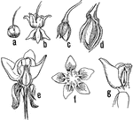
"Milkweed (Asclepias sp.). a, flower-bud; b, flower; c, very young pod; d, older pod in section, showing seeds; e, section of flower; f, top view of flower, showing the 5 hoods f the crown, each with a horn incurving to the stigma; between the horns are the cleft glands to which the pollinia are attached." -Gager, 1916

Milkweed Stages

"Milkweed (Asclepias sp.). a, flower-bud; b, flower; c, very young pod; d, older pod in section, showing…
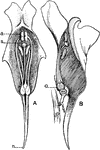
"Sections of flowers of the toad-flax (Linaria vulgaris). A, front view; a, anthers; s, stigma; n, nectar-gland. B, side view; o, ovary." -Gager, 1916

Common Toadflax

"Sections of flowers of the toad-flax (Linaria vulgaris). A, front view; a, anthers; s, stigma; n, nectar-gland.…

Embryo chick (36 hours), viewed from beneath as a transparent object (magnified). Labels:pl, outline of pellucid area; FB, forebrain, or first cerebral vesicle: from its sides project op, the optic vesicles; SO, backward limit of somatopleure fold, "tucked in", under head; A, head-fold of true amnion; a', reflected layer of amnion, sometimes termed "false amnion;" sp, backward limit of splanchnopleure folds, along which run the omphalomesaraic veins uniting to form h, the heart, which is continued forwards into ba, the bulbus arteriosus; d, the foregut, lying behind the heart, and having a wide crescentic opening between the splanchnopleure folds; HB, hindbrain; MB, midbrain; pv, protovertebrae lying behind the foregut; mc, line of junction of medullary folds and of notochord; ch, front end of notochord; vpl, vertebral plated; pr, the primitive groove at its caudal end.

Embryo Chick

Embryo chick (36 hours), viewed from beneath as a transparent object (magnified). Labels:pl, outline…
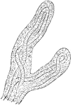
Very soon after the entrance of the ovum into the uterus, in the human subject, the outer surface of the chorion is found beset with fine processes, the so-called chorion villi, which give it a rough and shaggy appearance. Shown is a magnified view of chorion villi.

Magnified View of Chorion Villi

Very soon after the entrance of the ovum into the uterus, in the human subject, the outer surface of…
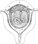
Diagrammatic view of a vertical transverse section of the uterus at the seventh week of pregnancy. Labels: c, c', cavity of uterus, which becomes the cavity of the decidua, opening at c, the cornua, into the Fallopian tubes, and at c' into the cavity of the cervix, which is closed by a plug of mucus; dv, decidua vera; dr, decidua reflexa, with the sparser villi imbedded in its substance; ds, decidua serotina, involving the more developed chorionic villi of the commencing placenta. The fetus is seen lying in the amniotic sac; passing up from the umbilicus is seen the umbilical pedicle of the yolk sac, which lies in the cavity between the amnion and chorion.

Uterus at Seventh Week of Pregnancy

Diagrammatic view of a vertical transverse section of the uterus at the seventh week of pregnancy. Labels:…
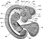
Embryo chick at fourth day, viewed as a transparent object, lying on its left side. CH, cerebral hemispheres; FB, forebrain or vesicle of third ventricle, with Pn, pineal gland projecting from its summit; MB, midbrain; Cb, cerebellum; IV V, fourth ventricle; L, lens; chs, choroidal slit; Cen V, auditory vesicle; s m, superior maxillary process; 1F, 2F, 3F,4F., first, second, third, and fourth visceral folds; V, fifth nerve, sending one branch (ophthalmic) to the eye, and another to the first pharyngeal nerve, passing to the third visceral arch; G.Pg, pneumogastric nerve, passing towards the fourth visceral arch; iv, investing mass; ch, notochord; its front end cannot be seen in the living embryo, and it does not end as shown in the figure, but takes a sudden bend downwards, and then terminated in a point; Ht, heart seen through the walls of the chest; MP, muscle-plates; W, wing, showing commencing differentiation of segments, corresponding to arm, forearm, and hand; H L, hind-limb, as yet a shapeless bud, showing no differentiation. Beneath it is seen the curved tail.

Embryo Chick at Fourth Day

Embryo chick at fourth day, viewed as a transparent object, lying on its left side. CH, cerebral hemispheres;…
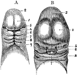
A, Magnified view of the head and neck of a human embryo of three weeks. Labels: 1, anterior cerebral vesicle or cerebrum; 2, middle cerebral vesicle; 3, middle or frontonasal process; 4, superior maxillary process, or first visceral arch, and below it the first cleft; 7, 8, 9, second, third, and fourth arches and clefts. B, Anterior view of the head of a human fetus of about the fifth week. 1, 2, 3, 5, the same parts as in A; 4, the external nasal or lateral frontal process: 6, the superior maxillary process; 7, the lower jaw; X, the tongue; 8, first branchial cleft becoming the meatus auditorius externus.

Head of an Embryo

A, Magnified view of the head and neck of a human embryo of three weeks. Labels: 1, anterior cerebral…
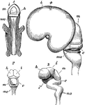
Early stages in development of human brain. 1, 2, 3, are from an embryo about seven weeks old; 4, about three months old. m, middle cerebral vesicle (mesencephalon); c, cerebellum; m o, medulla oblongata; i, thalamencephalon; h, hemispheres; i', infundibulum; Fig. 3 shows the several curves which occur in the course of development; Fig. 4 is a lateral view, showing the great enlargement of the cerebral hemispheres which have covered in the thalami, leaving the optic lobes, m, uncovered.

Development of the Brain

Early stages in development of human brain. 1, 2, 3, are from an embryo about seven weeks old; 4, about…

Side view of fetal brain at six months, showing commencement of formation of the principal fissures and convolutions. F, frontal lobe; P, parietal; O, occipital; T, temporal; a, commencing frontal convolutions; s, Sylvian fissure; s', its anterior division; c, within it the central lobe or island of Reil; r, fissure of Rolando; p, perpendicular fissure.

Fetal Brain at Six Months

Side view of fetal brain at six months, showing commencement of formation of the principal fissures…
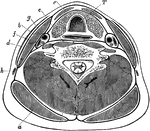
Transverse section through the lower part of the neck, to show the arrangement of the cervical fascia. Labels: a, Trapezius; b, sternomastoid; c, depressors of hyoid bone; d, platysma; e, anterior spinal muscles; f, scalenus anticus; g, carotid artery; h, external jugular vein; i, posterior spinal muscles; T, trachea, with gullet behind and thyroid body in front.

Transverse Section of the Neck

Transverse section through the lower part of the neck, to show the arrangement of the cervical fascia.…

Types of parallel prisms: right triangular prism, isosceles triangular prism, rectangular prism, cylinder.

Parallel Prisms

Types of parallel prisms: right triangular prism, isosceles triangular prism, rectangular prism, cylinder.

Types of oblique prisms: right triangular prism, isosceles triangular prism, rectangular prism, cylinder.
Oblique Prisms

Types of oblique prisms: right triangular prism, isosceles triangular prism, rectangular prism, cylinder.

Types of cylinders: vertical, horizontal, receding, and oblique.

Cylinders

Types of cylinders: vertical, horizontal, receding, and oblique.

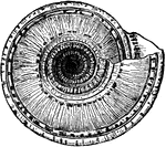
"The Staircase-shell is recognized by its deep umbilicus, wide and funnel-shaped, in the interior of which may be seen the little teeth which follow the edge of every turn of the spiral up to the top. The diameter of this shell is sometimes two and a half inches."

Solarium Perpectivum (Bottom View)

"The Staircase-shell is recognized by its deep umbilicus, wide and funnel-shaped, in the interior of…

"The Staircase-shell is recognized by its deep umbilicus, wide and funnel-shaped, in the interior of which may be seen the little teeth which follow the edge of every turn of the spiral up to the top. The diameter of this shell is sometimes two and a half inches."

Solarium Perpectivum (Side View)

"The Staircase-shell is recognized by its deep umbilicus, wide and funnel-shaped, in the interior of…

This temple is at the southern end of the Great Temple in Philae. The temple was dedicated to the worship of the god Isis.

Egyptian-Front of Temple of Isis at Philae

This temple is at the southern end of the Great Temple in Philae. The temple was dedicated to the worship…
A teacher in front of her classroom.

Teacher

A teacher in front of her classroom.

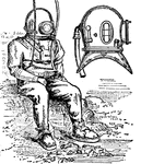
The diving dress in a rubber suit with a metal helmet, having pieces of glass in front to enable to diver to see.

Diving-dress and diving-helmet

The diving dress in a rubber suit with a metal helmet, having pieces of glass in front to enable to…
An upclose view of the arm of a Sepia Tuberculosa.

Arm of Sepia Tuberculosa (Lamarck)

An upclose view of the arm of a Sepia Tuberculosa.

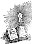
The anti-slavery novel by Harriet Beecher Stowe was published in 1852 and had an effect on the view of slavery.

Uncle Tom's Cabin

The anti-slavery novel by Harriet Beecher Stowe was published in 1852 and had an effect on the view…
A view on upper Saint John's River in Florida.

Saint John's River

A view on upper Saint John's River in Florida.

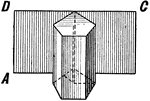
Illustration of a pentagonal prism showing, "If a piece of paper is fitted to cover the convex surface of a prism or a cylinder, and then unrolled, its form will be that of a rectangle, as ABCD."

Surfaces Of A Prism

Illustration of a pentagonal prism showing, "If a piece of paper is fitted to cover the convex surface…

Illustration of a cylinder showing, "If a piece of paper is fitted to cover the convex surface of a prism or a cylinder, and then unrolled, its form will be that of a rectangle, as ABCD."

Surfaces Of A Cylinder

Illustration of a cylinder showing, "If a piece of paper is fitted to cover the convex surface of a…

Illustration used to compare the surfaces of a cylinder and a sphere.

Comparative Surfaces Of A Cylinder And Sphere

Illustration used to compare the surfaces of a cylinder and a sphere.
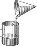
Illustration used to show finding the volume of a pentagonal prism.

Volume Of Cylinder

Illustration used to show finding the volume of a pentagonal prism.

Illustration used to compare the volumes of a cone and a cylinder by emptying sand from the cone into the cylinder.

Comparative Volumes Of A Cone And Cylinder

Illustration used to compare the volumes of a cone and a cylinder by emptying sand from the cone into…

Illustration used to compare the volumes of a cone, a sphere, and a cylinder.

Comparative Volumes Of A Cone, Sphere, And Cylinder

Illustration used to compare the volumes of a cone, a sphere, and a cylinder.
"The front leg of a genera of beetles."

Front Leg of a Zophosis

"The front leg of a genera of beetles."

Various geometric solids, mostly rectangular prisms with a cylinder and a hexagonal prism.

Solids

Various geometric solids, mostly rectangular prisms with a cylinder and a hexagonal prism.

The bushel, a unit of dry measure.

Bushel

The bushel, a unit of dry measure.

The peck, a unit of dry measure.

Peck

The peck, a unit of dry measure.

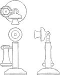
Orthographic projection drawing of a candlestick or upright telephone. An orthographic projection is the method of representing the exact form of an object in two or more views on planes generally at right angles to each other, by dropping perpendiculars fro the object to the planes.

Orthographic Projection of Candlestick Telephone

Orthographic projection drawing of a candlestick or upright telephone. An orthographic projection is…
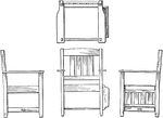
Orthographic projection drawing of an arm-chair. An orthographic projection is the method of representing the exact form of an object in two or more views on planes generally at right angles to each other, by dropping perpendiculars from the object to the planes.

Orthographic Projection of Arm-Chair

Orthographic projection drawing of an arm-chair. An orthographic projection is the method of representing…
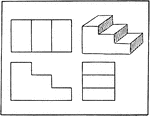
Orthographic projection drawing of stairs. An orthographic projection is the method of representing the exact form of an object in two or more views on planes generally at right angles to each other, by dropping perpendiculars fro the object to the planes.

Orthographic Projection of Stairs

Orthographic projection drawing of stairs. An orthographic projection is the method of representing…

A cylindrical helix is a curve generated by a point moving uniformly around a cylinder and uniformly lengthwise of the cylinder at the some time. The hypotenuse of a right triangle will form one turn of a helix if it is wrapped around a cylinder. The base of the triangle is equal to the circumference of the cylinder and the altitude is the pitch of the helix.

Helix

A cylindrical helix is a curve generated by a point moving uniformly around a cylinder and uniformly…
The trunk showing rib, iliac crest, and pubis.

Side View of the Bones of the Trunk

The trunk showing rib, iliac crest, and pubis.

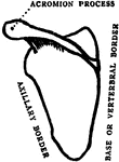
Back view of scapula.

Back View of Scapula

Back view of scapula.

Back view of the trunk.

Back View of the Bones of the Trunk

Back view of the trunk.

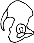
Side view of pelvis.

Side View of Pelvis

Side view of pelvis.

Back view of the femur.

Back View of the Femur

Back view of the femur.

Back view of muscles of the calf.

Back View of the Muscles of the Calf

Back view of muscles of the calf.

Front view of the muscles of the leg.

Front View of the Muscles of the Leg

Front view of the muscles of the leg.

Front view of the bones of the forearm

Front View of the Bones of the Forearm

Front view of the bones of the forearm

Front view of the bones of the forearm

Back View of the Bones of the Forearm

Front view of the bones of the forearm

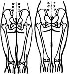
Diagram showing the position of the pelvic and thigh bones in back view in the male and female.

Position of the Pelvic and Thigh Bones in the Male and Female

Diagram showing the position of the pelvic and thigh bones in back view in the male and female.
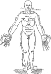
Front view of a man in the anatomical position. On one lateral half the parts are labeled in English, on the other in Latin. The right upper limb is drawn away from the trunk in the order to show the arm more fully than is possible when it hangs perpendicularly.

Front View of the Parts of the Human Body Labeled in English and Latin

Front view of a man in the anatomical position. On one lateral half the parts are labeled in English,…

Back view of a man. On one lateral the names of the parts are given in English, on the other in Latin.
Back View of the Parts of the Human Body Labeled in English and Latin

Back view of a man. On one lateral the names of the parts are given in English, on the other in Latin.

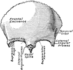
The frontal bone, seen from in front.

Frontal Bone

The frontal bone, seen from in front.

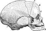
Side view of a fetal skull. The coronal suture extends from the top of the head downwards on either side to the point E; the lambdoidal suture from the back forward, on either side to the point F. The sagittal suture is not shown, but is indicated by the upper margin of the illustration, beginning at the base of the nose, passing backward across the coronal suture (anterior fontanelle), and ending at the lambdoidal suture (posterior fontanelle).

Side View of Fetal Skull

Side view of a fetal skull. The coronal suture extends from the top of the head downwards on either…

The hyoid bone. Viewed from the left and in front.

Hyoid Bone

The hyoid bone. Viewed from the left and in front.

The spinal column, right lateral view and dorsal view.

Lateral and Dorsal View of the Vertebral Column

The spinal column, right lateral view and dorsal view.

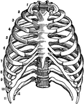
The skeleton of the thorax, front view.

Skeleton of the Thorax

The skeleton of the thorax, front view.

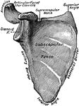
The right scapula, ventral view.

Scapula

The right scapula, ventral view.

The bones of the right forearm, dorsal view.

Dorsal View of the Bones of the Forearm

The bones of the right forearm, dorsal view.

The bones of the right forearm, ventral view.

Ventral View of the Bones of the Forearm

The bones of the right forearm, ventral view.

The right femur, rear view.

Femur

The right femur, rear view.

The right tibia and fibula in their normal relation, rear view.

Bones of the Lower Leg

The right tibia and fibula in their normal relation, rear view.