
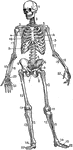
Bones of the Foot
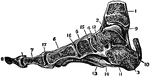
The bones of the foot. Labels: Ca, Calcaneum, or heel bone; Ta, articular surface for tibia on the astragalus;…
Arm Bones
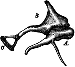
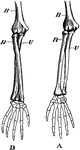
Demonstration of the movement of a pivot joint. Labels: A, arm in supination (palm uppermost); B, arm…
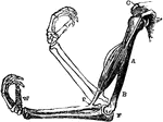
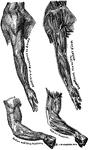
Biceps
The bicep muscle and the arm bones, to illustrate how, under ordinary circumstances, the elbow joint…
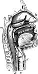
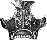
The Mouth, Nose, and Pharynx
The mouth, nose, and pharynx, with the commencement of the gullet (esophagus) and larynx, as exposed…
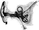
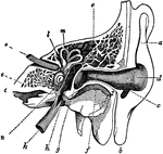
Section Through the Right Ear
Semi-diagrammatic section through the right ear. Labels: M, concha; G, external auditory meatus; T,…
Middle Ear
The middle ear and its bones, considerably magnified. Labels: G, the inner end of the external auditory…
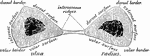
Forearm Bones
Transverse section through the bones of the forearm (radius and ulna), taken at about the middle of…
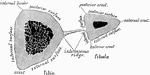
Shank Bones
A section of the bones of the crus (shank of the leg) taken at about the middle of their length (schematized)…
Inferior Turbinal Bones
Interior turbinal bones (or conchae nasales inferiores), which are situated in the nasal fossae.
The Nasal Bones
The nasal fossae (bones), which together form the cavity of the nose and are separated from either other…
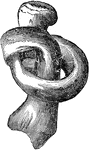
The Tympanic Ossicles
The tympanic ossicles, which are 3 small bones that form a chain across the tympanic cavity, connecting…
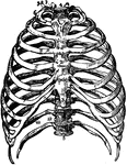
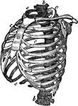
The Bones of the Thorax
Front view of the bones of the thorax, including the ribs, sternum and vertebrae. Labels: 1, first bone…
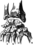
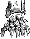
Bones of the Carpus
Articulations of bones of the carpus (wrist area). Labels: 1, ulna; 2, radius; 3, inter-articular fibro-cartilage;…

Upper Surface of the Left Foot
Bones of the upper surface of the left foot. Labels: 1, astragalus; 2, its anterior face; 3, os calcis;…
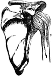
Bones and Ligaments of the Shoulder Articulation
Ligaments of the acromio-clavicular and scapulo-humeral articulations (joints of the shoulder). Labels:…
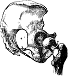
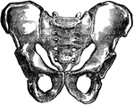
Bones and Ligaments of the Hip and Pelvis
Ligaments and bones of the hip joint and pelvis. Labels: 1, posterior sacro-iliac ligament; 2, greater…
Ankle Joint and Foot
Vertical section of the ankle joint and foot. Labels: 1, tibia; 2, astragalus; 3, os calci; 4, scaphoides;…
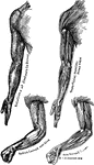
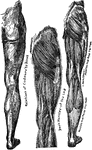
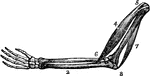
Bones and Muscles of the Arms
Showing relations of the muscles and bones of the arms from the inner side.
Bones and Muscles of the Arms
Showing relations of the muscles and bones of the arms from the outer side
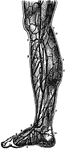
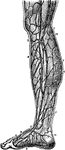
Veins of the Legs
Superficial veins of the legs. Labels: 1, saphena major; 2, collateral branch; 3, anastomosis of veins;…
A Cross-Section of the Ear
Cross-section of the external and internal ear. a, b, and c: External ear. d: Entrance…
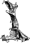
Bone Exposed to Acid and Twisted
This figure shows a thigh bone that has been softened by exposing it to acid, then twisted in a knot…
Structure of the Chest
Structure of the chest, showing the framework of the bones which are connected together chiefly by muscles.…
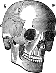
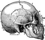
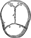
Cranial Sutures
The bones of the top of the head are fastened together by what are called sutures which are locked together…
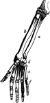
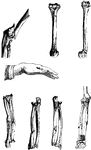
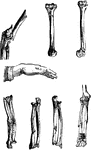
Bones of the Arm and Hand
Bones of the arm and hand. Labels: a, large end of ulna; b, radius; c, small end of the ulna; d, carpal…
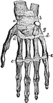
Bones and Ligaments of the Hand
Bones and ligaments of the hand. There are 27 bones in all, including 8 small bones called the carpal…

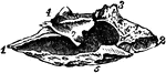
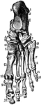
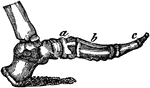
Bones of the Foot
Bones of the foot. At e d f g h are the 7 bones of the tarsus; at a are the 5 bones…
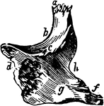
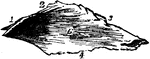
Side View of the Bone of the Foot
Bones of the foot, side view. In this figure the bones of the tarsus extend from the heel to a;…
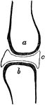
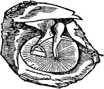
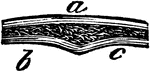
Joint
A joint between two bones (a and b). The ends of all bones are tipped with cartilage so that they may…
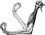
Arm Muscles
Two of the principal muscles f the arm (4 and 7). Between these is the bone of the arm (1) and the bones…
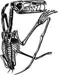
Tendons of a Finger
The arrangement of the tendons of a finger. At a b c are the 3 bones of the finger. At f…
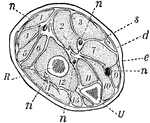
Forearm, Section of
A section across the forearm a short distance below the elbow-joint. R and U, its two supporting bones,…

Bones of the Foot
The bones of the foot. Labels: Ca, calcaneum, or os calcis; Ta, articular surface for tibia on the astragalus;…
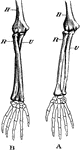
Bones of the Arm
Bones of the arm. Labels: A, arm in supination; B, arm in pronation. H, humerus; R, radius; U, ulna.
The Biceps Muscle and Arm Bones
The biceps muscle and arm bones, to illustrate how, under ordinary circumstances, the elbow-joint is…
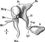
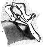
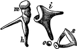
The Auditory Ossicles
The auditory ossicles of the right ear, seen from the front. Labels: M, malleus; J, incus; S, stapes;…
Veins of the Leg
Veins of the leg. Labels: 1, saphenous; 2, collateral branch; 3, anastomosis; 4, internal saphenous;…
Bone Structure
If we divide any of the long bones longitudinally, we find two kinds of structure, the hard or compact,…
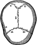
Skull Sutures
Sutures of the skull. Labels: a,a, the coronal suture, from the Latin corona, crown, so called from…
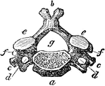
Vertebra of the Neck
A vertebra of the neck. Labels: a, body of the bone; b, the spinal process; c, d, the transverse processes…