The diaphragm, which is the principal muscle that act sin breathing. Here you have the cavity of the chest, <em>Cc</em>, laid open, the ribs being cut away in front, and the heart and lungs taken out; <em>DD</em> is the diaphragm. It is fastened to the spinal column behind, to the breast-bone in front, and to the lower ribs all around its side. It is not flat, but arched upward.

The Diaphragm

The diaphragm, which is the principal muscle that act sin breathing. Here you have the cavity of the…

Here you see the thigh bone on the left and the arm bone on the right. In both, the shaft is hollow, while the large ends are more dense. In the living bone, bone marrow is found in the hollow area which serves in blood cell production.

Thigh Bone and Arm Bone

Here you see the thigh bone on the left and the arm bone on the right. In both, the shaft is hollow,…
The human skull, showing the bones of the head. <em>a</em>is the large bone of the forehead known as the frontal bone; <em>b</em> is known as the parietal bone, and <em>c</em> is the temporal bone.

Skull

The human skull, showing the bones of the head. ais the large bone of the forehead known as…

The bones of the top of the head are fastened together by what are called sutures which are locked together by little teeth of bone that shoot by each other, as shown in the figure. <em>b</em> is the suture at the top of the head between the two parietal bones; <em>a a</em> is that between the two parietal bones and the frontal bone in front; and <em>c c </em> is that between the parietal bones and the bone at the back part of the cranium. These bones are not completely formed in the newborn infant.

Cranial Sutures

The bones of the top of the head are fastened together by what are called sutures which are locked together…

Two views of vertebra. 24 vertebrae make up the spinal column. On the left figure, <em>a</em> is the body of the bone; <em>b</em> is the hole through the vertebra which forms its part of the canal for the spinal marrow; and <em>c</em> is the spinous process. On the right is a side view of the same vertebra.

Two Views of a Vertebra

Two views of vertebra. 24 vertebrae make up the spinal column. On the left figure, a is the…

Rear view of the thigh bone.

Rear View of the Thigh Bone

Rear view of the thigh bone.

Bones of the foot, side view. In this figure the bones of the tarsus extend from the heel to <em>a</em>; the metatarsal bones are at <em>b</em>; and the bones of the toes are at <em>c</em>. In every movement of the foot there is some little motion between all these bones, and it is this that gives ease and grace to its motions.

Side View of the Bone of the Foot

Bones of the foot, side view. In this figure the bones of the tarsus extend from the heel to a;…

A joint between two bones (a and b). The ends of all bones are tipped with cartilage so that they may move easily upon each other. And besides this, the ends are lined with a very fine membrane, so arranged as to make a closed sac (c).

Joint

A joint between two bones (a and b). The ends of all bones are tipped with cartilage so that they may…

The lower part of the large muscle that raises the heel during walking. P, the muscle, makes up most of the bulk of the calf of the leg. The weight of the body rests upon the bone W. In walking, as we raise the body in taking a step, we do it first by raising the heel with the muscle P, the pressure being on the ball of the foot, F. This muscle does here with the weight of the body what the muscles of your arms do to the load in a wheel barrow when you raise it by the handles.

Heel Muscle

The lower part of the large muscle that raises the heel during walking. P, the muscle, makes up most…

Two of the principal muscles f the arm (4 and 7). Between these is the bone of the arm (1) and the bones of the forearm (2).

Arm Muscles

Two of the principal muscles f the arm (4 and 7). Between these is the bone of the arm (1) and the bones…

The arrangement of the tendons of a finger. At <em>a b c </em> are the 3 bones of the finger. At <em>f</em> is the tendon that bends the second bone, <em>b</em>. This is divided into two parts, just at its end, where it is fastened to the bone. Through this division the tendon <em>e</em> passes, to go to the last bone, <em>c</em>.

Tendons of a Finger

The arrangement of the tendons of a finger. At a b c are the 3 bones of the finger. At f

Bones of the ear, including the hammer (m), the anvil (i), and the stirrup (s).

Bones of the Ear

Bones of the ear, including the hammer (m), the anvil (i), and the stirrup (s).

The humerus bisected lengthwise. Labels: a, marrow-cavity; b, hard bone; c, spongy bone; d, articular cartilage.

Bisection of the Humerus

The humerus bisected lengthwise. Labels: a, marrow-cavity; b, hard bone; c, spongy bone; d, articular…
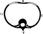
Diagrammatic representation of a segment of the axial skeleton V, a vertebra; C, Cv, ribs articulating above with the body and transverse process of the vertebra; S, the breastbone. The lighter-shaded part between S and C is the costal cartilage.

Segment of the Axial Skeleton

Diagrammatic representation of a segment of the axial skeleton V, a vertebra; C, Cv, ribs articulating…
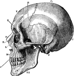
A side view of the skull. Labels: O, occipital bone; T, temporal; Pr, parietal; F, frontal; S, sphenoid; Z, malar; Mx, maxilla; N, nasal; E, ethmoid; L, lachrymal; Md, inferior maxilla.

Side View of the Skull

A side view of the skull. Labels: O, occipital bone; T, temporal; Pr, parietal; F, frontal; S, sphenoid;…

The hyoid bone. Labels: 1, body; 2, great cornua; 3, small cornua.

Hyoid Bone

The hyoid bone. Labels: 1, body; 2, great cornua; 3, small cornua.

The bones of the foot. Labels: Ca, calcaneum, or os calcis; Ta, articular surface for tibia on the astragalus; N, scaphoid bone; CI, CII, first and second cuneiform bones; Cb, cuboid bone; M1, metatarsal bone of great toe.

Bones of the Foot

The bones of the foot. Labels: Ca, calcaneum, or os calcis; Ta, articular surface for tibia on the astragalus;…
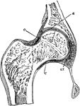
Section through the hip-joint. Labels: a and b, articular cartilages; c, capsular ligament.

Hip Joint Section

Section through the hip-joint. Labels: a and b, articular cartilages; c, capsular ligament.

The thigh bone cut through the middle. Labels: b, hard bone; h, d, spongy bone; ma, marrow.

Thigh Bone

The thigh bone cut through the middle. Labels: b, hard bone; h, d, spongy bone; ma, marrow.

Section through a premolar tooth of a cat still embedded in its socket. Labels: 1, enamel; 2, dentine; 3, cement; 4, the gum; , the bone of the lower jaw; c, the pulp-cavity.

Premolar Tooth

Section through a premolar tooth of a cat still embedded in its socket. Labels: 1, enamel; 2, dentine;…
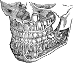
The deciduous and permanent teeth, shown as they are placed in the jaw with portions of bone removed to reveal the roots.

The Deciduous and Permanent Teeth

The deciduous and permanent teeth, shown as they are placed in the jaw with portions of bone removed…
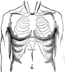
Surface of the breast in a normal condition; contours or cardial torpor to the left of the breast bone. The spaces included in the dotted lines represent the intermediate spaces.

Breast Surface

Surface of the breast in a normal condition; contours or cardial torpor to the left of the breast bone.…

Front view of the upper surface of the tongue; as also of the arch of the bone of the palate.

Upper Surface of the Tongue

Front view of the upper surface of the tongue; as also of the arch of the bone of the palate.

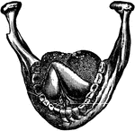
View of the lower jaw bone with the tongue turned up.

Jaw Bone and Tongue

View of the lower jaw bone with the tongue turned up.

Examples of bone fractures.

Bone Fractures

Examples of bone fractures.

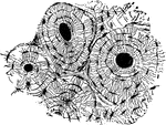
If we divide any of the long bones longitudinally, we find two kinds of structure, the hard or compact, and the alveolar or spongy. Indeed there is no bone that does not exhibit to some extend both of these structures; the compact forming its external, and the spongy its internal part. These two formations are clearly seen in the bones which compose the skull, as in the following cut, with the spongy or cancellated structure between them. This serves to illustrate the structure of all of the flat bones.

Bone Structure

If we divide any of the long bones longitudinally, we find two kinds of structure, the hard or compact,…

The above cut represents a section of the thigh bone. The extremities (a, w) having a shell or thin plate of compact texture, crowded with small cells, diminishing in size, but increasing in number as they approach the articulation (c), the cavity for containing the marrow; the walls of the shaft are very firm and solid (b, b). The compact part is thickest near the middle of the bone where the greatest strength is required.

Thigh Bone Section

The above cut represents a section of the thigh bone. The extremities (a, w) having a shell or thin…

Membranous, or gelatinous portion of bone; the earthy portion being so completely removed (by immersion in nitric acid), that it is capable of being tied in a knot.

Membranous Portion of Bone

Membranous, or gelatinous portion of bone; the earthy portion being so completely removed (by immersion…

Earthy portion of bone, resulting from exposure to a strong heat. The resulting bone is very brittle, as the animal matter has been wholly consumed by the heat or fire, while the earthy part remains unchanged.

Earthy Portion of Bone

Earthy portion of bone, resulting from exposure to a strong heat. The resulting bone is very brittle,…

Bone structure. Labels: a, the external, b,c, the internal table; the intermediate cellular texture, being soft and spongy, and conveying vessels and nerves from one part to another.

Bone Structure

Bone structure. Labels: a, the external, b,c, the internal table; the intermediate cellular texture,…
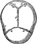
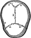
Sutures of the skull. Labels: a,a, the coronal suture, from the Latin corona, crown, so called from its situation on that part of the head, upon which the ancients placed the laurel, or olive crown, given to the victors in their games. It connects the frontal to the parietal bones; b, the sagittal suture, from a Latin word, signifying arrow, from its straight course. It runs from the middle of the frontal to the angle of the occipital bone, connecting the two parietals; c, the lambdoidal suture, extending from the sagittal suture down to the base of the brain on each side; e,e, the scaly overlapping of the temporal upon the parietal bones; hence called squamous suture.

Skull Sutures

Sutures of the skull. Labels: a,a, the coronal suture, from the Latin corona, crown, so called from…
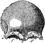
A front view of the frontal bone; a,a, frontal sinuses; b, the temporal arch, beneath which lies the temporal muscle, which closes the lower jaw; d, the supra-orbitary hole for a passage of a branch of the fifth pair of nerves.

Frontal Bone

A front view of the frontal bone; a,a, frontal sinuses; b, the temporal arch, beneath which lies the…
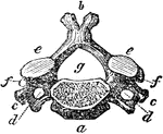
A vertebra of the neck. Labels: a, body of the bone; b, the spinal process; c, d, the transverse processes double, showing circular holes for the passage of the vertebral artery; e,e,. the superior; f,f, the articular of oblique processes; g, the spinal hole for the spinal marrow. The roots of the articular processes are hallowed out above and below into notches; and these, when the bones are fitted together, form apertures on each side of the spine, through which the nerves pass out from the spinal canal.

Vertebra of the Neck

A vertebra of the neck. Labels: a, body of the bone; b, the spinal process; c, d, the transverse processes…

Fourth rib. Labels: a, vertebral extremity, called the head, which is connected with the bodies of the two contiguous dorsal vertebrae. At b, the bone is contracted, forming the neck; c, is the tubercle at the back of the rib, which is articulated with the transverse process of the vertebrae; d, the angle; e, the sternal extremity; f, a groove for the intercostal vessels. This will serve for a general description of the ribs.

Fourth Rib

Fourth rib. Labels: a, vertebral extremity, called the head, which is connected with the bodies of the…

The sternum in this cut consists of two bones. The first is broad and thick above, and contracts as it descends. It is convex before and concave behind. At the upper angle a, the collarbone is articulated; b, the articular surface for the cartilage of the first rib; b, for the second rib; c,d,e,f,g, mark the articular surfaces of the 3rd, 4th, 5th, 6th, and 7th ribs; h, the ensiform cartilage, terminates the lower extremity of the sternum. In older people, this cartilages is often changed into bone.

Sternum

The sternum in this cut consists of two bones. The first is broad and thick above, and contracts as…
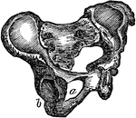
The pelvis.

Pelvis

The pelvis.

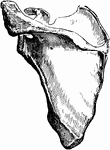
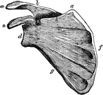
Scapula. Labels: a, superior angle; d, the glenoid cavity, or socket for the round head of the arm bone; m, the aeromion process; n, the caracoid process, which serve to protect the joint; f, the base; g, the costa, or inferior border, and h, the superior border of the triangle; l, the spine; o, the semilunar notch, for the passage of an artery, vein, and nerve.

Scapula

Scapula. Labels: a, superior angle; d, the glenoid cavity, or socket for the round head of the arm bone;…

The radius, a bone of the arm.

Radius

The radius, a bone of the arm.

The ulna, a bone of the arm.

Ulna

The ulna, a bone of the arm.

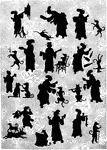
Japanese silhouettes telling the story of Old Mother Hubbard and her dog.

Old Mother Hubbard

Japanese silhouettes telling the story of Old Mother Hubbard and her dog.

A dog watching another dog burying a bone.

Dogs

A dog watching another dog burying a bone.

"Skull of Boa constrictor. a, quadrate bone; b, b, halves of lower jaw." -Cooper, 1887

Boa Constrictor Skull

"Skull of Boa constrictor. a, quadrate bone; b, b, halves of lower jaw." -Cooper, 1887

"1. Lower mandible. 2. Upper mandible. 3. Forehead. 4. Loral space. 5. Crown of the head. 6. Hind part of the head. 7. Scapulars--long feathers frm shoulders over sides of back. 8. Smaller wing coverts. 9. Bend of the wing. 10. Larger wing coverts. 11. Tertials, arising from the second bone of the wing at the elbow-joint. 12. Secondaries, from the second bone of the wing. 13. Primaries, from the first bone of the wing. 14. Tibia, the thigh. 15. Tarsus, the shank. 16. Upper tail coverts. 17. Lower tail coverts. 18. Tail feathers." -Roosevelt, 1884

Bird Parts

"1. Lower mandible. 2. Upper mandible. 3. Forehead. 4. Loral space. 5. Crown of the head. 6. Hind part…
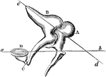
A, the malleus, or mallet, with its long handle running down, to touch with its delicate extremity the membrane of the drum. B, is the incus, or anvil, nicely fitted to the mallet, and showing C, the orbicular bone placed between its termination, and D, the stirrup. The line a,b, represents the center of motion of the malleus, and c,d, the center of motion of the incus. Now this chain of bones acts on the principle of a long level, as a small motion at one end of a long pole serve to move it through a wide space at the other.

Bones of the Ear

A, the malleus, or mallet, with its long handle running down, to touch with its delicate extremity the…

"Fracture of the arm: Apply two splints, one in front, the other behind, if the lower part of the bone is broke; or to the inner and outer sides, if the fracture is in the middle or upper part; support by sling." -Moss, 1914

Splints and Sling

"Fracture of the arm: Apply two splints, one in front, the other behind, if the lower part of the bone…

"Fracture of the thigh: Apply a long splint, reaching from the armpit to beyond the foot on the outside, and a short splint on the inside." -Moss, 1914

Splint

"Fracture of the thigh: Apply a long splint, reaching from the armpit to beyond the foot on the outside,…
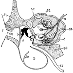
"Diagram showing the ear and related parts in a young cat. P., Pinna; Sq., squamosal: E.A.M., external auditory meatus; T., tympanum; M., malleus; I., incus; St., stapes abutting onforamen ovale; B., bulla of tympanic bone; Se., a septum in the bulla; E.T., eustachian tube leading from the tymmpanic cavity to the back of the mouth; B.O., basi-occipital; C., cochlea; S., sacculus; U., utriculus; D.E., ductus endolymphaticus; N., auditory nerve; S.C., semi-circular canal; PE., periotic bone." -Thomson, 1916

Cat Ear

"Diagram showing the ear and related parts in a young cat. P., Pinna; Sq., squamosal: E.A.M., external…
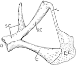
"Pectoral girdle of a Chelonian. G., Glenoid cavity; SC., scapula; P.C., procoracoid fused to the scapula; C., coracoid; E.C., epicoracoid cartilage; L., ligament." -Thomson, 1916

Turtle Pectoral Girdle

"Pectoral girdle of a Chelonian. G., Glenoid cavity; SC., scapula; P.C., procoracoid fused to the scapula;…
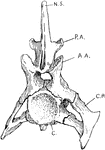
"Cervical vertebra of crocodile. N.S., Neural spine; P.A., posterior articular process; A.A., anterior articular process; C.R., cervical rib; C., procoelous centrum." -Thomson, 1916

Crocodile Vertebra

"Cervical vertebra of crocodile. N.S., Neural spine; P.A., posterior articular process; A.A., anterior…
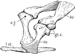
"Pectoral girdle of crocodile. sc., Scapula; glc., glenoid cavity; co., coracoid; c.f., coracoid foramen; i.cl., episternum." -Thomson, 1916

Crocodile Pectoral Girdle

"Pectoral girdle of crocodile. sc., Scapula; glc., glenoid cavity; co., coracoid; c.f., coracoid foramen;…
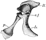
"Half of the pelvic girdle of a young crocodile. Il., Ilium; a.f., acetabulum; Is., ischium; P., pubis or epipubis." -Thomson, 1916

Crocodile Pelvic Girdle

"Half of the pelvic girdle of a young crocodile. Il., Ilium; a.f., acetabulum; Is., ischium; P., pubis…
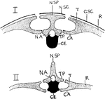
"Vertical section through backbone and ribs of Chelonian (I.) and Mammal (II.). N.SP., neural spine; N.SC., neural scute; T., tubercle of rib; C.SC., costal scute over rib (R.); CA., capitulum of rib; T.P., transverse process; CE., centrum; NA., cavity of neural canal. In the Chelonian the tubercle abuts against the flattened neural spine, and the capitulum against the transverse process. In the Mammal, the tubercle articulates with the transverse process and the capitulum with the centrum." -Thomson, 1916

Mammal and Chelonian Backbones

"Vertical section through backbone and ribs of Chelonian (I.) and Mammal (II.). N.SP., neural spine;…
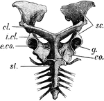
"Pectoral girdle of Echidna. sc., Scapula; cl., clavicle; i.cl., prosternum or "interclavicle"; co., coracoid or metacoracoid; e.co., procoracoid or precoracoid; st., mesosternum." -Thomson, 1916

Echidna Pectoral Girdle

"Pectoral girdle of Echidna. sc., Scapula; cl., clavicle; i.cl., prosternum or "interclavicle"; co.,…
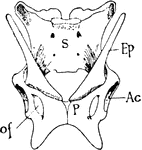
"Pelvis of Echidna. S., Sacrum; Ep., epipubic bones; Ac., acetabulum; o.f., obturator foramen between ischium and pubis (p.)." -Thomson, 1916

Echidna Pelvis

"Pelvis of Echidna. S., Sacrum; Ep., epipubic bones; Ac., acetabulum; o.f., obturator foramen between…

"Bony Tissue. A, portion of cross-section of a bone, the upper portion of the figure representing the outer surface of the bone, just beneath the periosteum. The open spaces, h, are Haversian canals; l, lacunae, occupied in life by bone cells. The minute canals through the bone connecting the lacunae are canaliculi. B, a portion of one Haversian system much magnified. h, Haversian anal, containing arter (a), vein (v), lymphatic spaces, nutritive cells; c, canaliculi; l, lacunae; la, plae of bone intercellular substance." -Galloway, 1915

Bony Tissue

"Bony Tissue. A, portion of cross-section of a bone, the upper portion of the figure representing the…
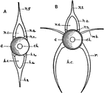
"Diagram of vertebrae of a bony fish. A, caudal; B, trunk. c, centrum or body of the vertebra; ch., the notochord; h.a., haemal arch; h.c., haemal canal; h.s., haemal spine; h.z., haemal zygapophysis, or articulating facet; m.b., inter-muscular bone; n.a., neural arch; n.c., neural canal; n.s., neural spine; n.z., neural zygapophysis; r, rib." -Galloway, 1915

Fish Vertebrae

"Diagram of vertebrae of a bony fish. A, caudal; B, trunk. c, centrum or body of the vertebra; ch.,…
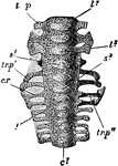
"Columba livia. Sacrum of a nestling (about fourteen days old), ventral aspect. c1, centrum of first sacral vertebra; c1, centrum of fifth caudal; c. r, first sacral rib; l1, centrum of first lumbar; l3, of third lumbar; s1, of fourth lumbar; s3, of sixth lumbar; tr. p, transverse process of first lumbar; tr. p', of fifth lumbar; tr. p'', of first sacral." -Parker, 1900

Rock Pigeon Sacrum

"Columba livia. Sacrum of a nestling (about fourteen days old), ventral aspect. c1, centrum of first…

"Columba livia. Left innominate of a nestling. The cartilage is dotted. ac, acetabulum; a. tr, anti-trochanter; il, pre-acetabular; and il', post-acetabular portion of ilium; is, ischium; i s. f, ischiatic foramen; ob. f, obturator notch; pu, pubis." -Parker, 1900

Rock Pigeon Innominate

"Columba livia. Left innominate of a nestling. The cartilage is dotted. ac, acetabulum; a. tr, anti-trochanter;…
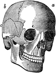
The humerus bone.

The Humerus

The humerus bone.

The scapula bone.

The Scapula

The scapula bone.