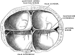
Crucial prolongation of the dura. Frontal section passing through the tentorium cerebelli. The torcular herophili is seen in the center.

Dura of the Brain

Crucial prolongation of the dura. Frontal section passing through the tentorium cerebelli. The torcular…
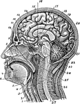
Section of head and neck from front to back. Labels: 1, windpipe; 2, larynx; 3, spinal marrow; 4, pharynx; 5, tongue or hyoid bone; 6, epiglottis; 7, tongue; 8, hard palate; 9, soft palate; 10; bridge of the nose; 11, frontal cavity; 12; sphenoid cavity; 13, nasal cavity; 14, skin of skull; 15, bony skull; 16, hypophisis; 17, corpus callosum; 18, septum lucidum; 19, straight sinus; 20, cerebellum; 21 cerebrum; 22, lobes of medulla; 23, pons varolii; 24, medulla oblongata; 25, zone of the epistropheus; 26, vertebrae; 27, spinal continuation of the vertebrae.

Section of the Head and Neck

Section of head and neck from front to back. Labels: 1, windpipe; 2, larynx; 3, spinal marrow; 4, pharynx;…
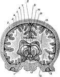
A cross section of the brain from left to right. Labels: 1, thalamus; 2, skull; 3, cerebral membrane; 4, cerebral hemisphere; 5, lateral ventricle; 6, optic lobe; 7, septum lucidum; 8, longitudinal sinus; 9, great longitudinal fissure; 10, corpus callosum; 11, median cerebral cavity; 12, cerebral hemisphere; 13, gray matter; 14, white matter; 15, Corpora Albicantia.

Brain

A cross section of the brain from left to right. Labels: 1, thalamus; 2, skull; 3, cerebral membrane;…
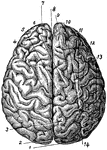
The brain viewed from above. Labels: 1, occipital convolution; 2, occipital lobe; 3, inner parietal convolution; 4, left cerebral hemisphere; 5, inner frontal convolution; 6, right cerebral hemisphere; 7, frontal lobe; 8, longitudinal fissure; 9, median frontal convolution; 10, occipital center convolution; 11, frontal center convolution; 12, outer frontal convolution; 13, outer parietal convolution; 14, median parietal convolution.

Brain Viewed from Above

The brain viewed from above. Labels: 1, occipital convolution; 2, occipital lobe; 3, inner parietal…
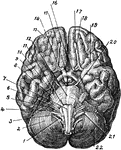
The base of the brain. Labels: 1, eleventh or spinal accessory nerve; 2, right hemisphere of cerebellum; 3, twelfth or hypoglossal nerve; 4, ninth or glosso-pharyngeal nerve; 5, eighth or auditory nerve; 6, seventh or facial nerve; 7, medulla oblongata; 8, fifth or trigemenus; 9, central lobe; 10, fourth or trochlear nerve; 11, sixth or abduceus nerve; 12, pons varolii; 13, right frontal lobe of the cerebrum; 14, lobes of the medulla; 15, optic chiasm; 16, second or optic nerve; 17, left frontal lobe; 18, first or olfactory nerve; 19, sylvian fissure; 20, third or oculamotor nerve; 21, tenth or pneumogastric nerve; 22, left hemisphere of the Cerebellum.

Base of Brain

The base of the brain. Labels: 1, eleventh or spinal accessory nerve; 2, right hemisphere of cerebellum;…

Diagram of a developing ovum, seen in longitudinal section. Labels: a, pericardium; b, bucco-pharyngeal; c, ectoderm; d, entoderm; e,placental area. f, spinal cord; i, brain; g, notochord; k, extra embryonic coelom; h, dorsal wall of alimentary canal.

Developing Ovum

Diagram of a developing ovum, seen in longitudinal section. Labels: a, pericardium; b, bucco-pharyngeal;…

The developmental stages exhibited by a pyramidal cell of the brain. Labels: a, neuroblast with rudimentary axon, but no dendrites; b and c, the dendrites beginning to sprout out; d and e, further development of the dendrites and appearance of collateral branches on the axon.

Pyramidal Cells of the Brain

The developmental stages exhibited by a pyramidal cell of the brain. Labels: a, neuroblast with rudimentary…

Human fetus in the third month of development, with the brain and spinal cord exposed from behind.
Brain and Spinal Cord of Fetus

Human fetus in the third month of development, with the brain and spinal cord exposed from behind.

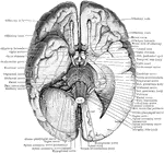
The base of the brain. The under part of the left temporal and occipital lobes has been sliced off so as to open into the lateral ventricle. The left hemisphere of the cerebellum has also been removed.

Base of the Brain

The base of the brain. The under part of the left temporal and occipital lobes has been sliced off so…

Schema showing the connections of the several parts of the brain.

Connection of the Brain Parts

Schema showing the connections of the several parts of the brain.

Two stages in the development of the human brain. A. Brain of an human embryo of the third week. B. Brain of an embryo of five weeks.

Development of Human Brain

Two stages in the development of the human brain. A. Brain of an human embryo of the third week. B.…

Two cross sections through the forebrain. A. Through the forebrain of the early human embryo. B. Through the forebrain and optic vesicles of Lepidosteus embryo of eight days.

Section Through Forebrain of Human and Lepidosteus Embryos

Two cross sections through the forebrain. A. Through the forebrain of the early human embryo. B. Through…
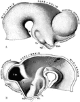
The brain of a human embryo in the fifth week. A, Brain as seen in profile. B, Mesial section through the same brain. Labels: M, mammillary eminence, Tc, tuber cinereum; Hp, Hypophysis (pituitary diverticulum from buccal cavity); Opt, optic stalk; TH, optic thalamus; Tg, tegmental part of mesencephalon; Ps, pars subtalamica; Cs, corpus striatum; FM, foramen of Monro; L, lamina terminalis; RO, recessus opticus; RI, recessus infundibuli.

Brain of Embryo

The brain of a human embryo in the fifth week. A, Brain as seen in profile. B, Mesial section through…
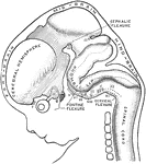
Profile view of brain of a human embryo of ten weeks. The various cranial nerves are indicated by numerals. A, Cerebral diverticulum of pituitary body. B, Buccal diverticulum of pituitary body.

Brain of Embryo

Profile view of brain of a human embryo of ten weeks. The various cranial nerves are indicated by numerals.…
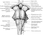
Front view of the medulla, pons, and mesencephalon of a full term human fetus.

Front view of Medulla, Pons, and Mesencephalon

Front view of the medulla, pons, and mesencephalon of a full term human fetus.
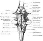
Back view of the medulla, pons, and mesencephalon of a full term human fetus.

Back View of Medulla, Pons, and Mesencephalon

Back view of the medulla, pons, and mesencephalon of a full term human fetus.
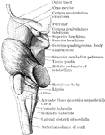
Lateral view of the medulla, pons, and mesencephalon of a full term human fetus.

Lateral View of Medulla, Pons, and Mesencephalon

Lateral view of the medulla, pons, and mesencephalon of a full term human fetus.

Transverse section through the lower end of the medulla of a full term fetus.

Transverse Section Through the the Medulla

Transverse section through the lower end of the medulla of a full term fetus.
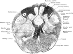
Transverse section through the closed part of the human medulla immediately above the decussation of the pyramids.

Transverse Section Through Closed Part of Medulla

Transverse section through the closed part of the human medulla immediately above the decussation of…

Transverse section through the closed part of the fetal medulla immediately above the decussation of the pyramids.

Transverse Section Through Closed Part of Fetal Medulla

Transverse section through the closed part of the fetal medulla immediately above the decussation of…
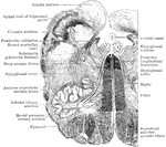
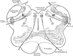
Transverse section through the human medulla in the lower olivary region.

Section Through Medulla in Olivary Region

Transverse section through the human medulla in the lower olivary region.

Transverse section through the the middle of the olivary region of the human medulla or bulb.

Section Through Medulla in Olivary Region

Transverse section through the the middle of the olivary region of the human medulla or bulb.
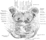
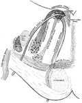
Diagram of the cerebello olivary fibers.

Cerebello Olivary Fibers

Diagram of the cerebello olivary fibers.

Section through the lower part of the human pons varolii immediately above the medulla.

Section Through Pons Varolii

Section through the lower part of the human pons varolii immediately above the medulla.
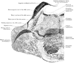
Section through the pons varolii at the level of the nuclei of the trigeminal nerve in an orangoutang.

Section Through Pons Varolii

Section through the pons varolii at the level of the nuclei of the trigeminal nerve in an orangoutang.

Section through the upper part of the pons varolii of the orangoutang above the level of the trigeminal nerve.

Section Through Pons Varolii

Section through the upper part of the pons varolii of the orangoutang above the level of the trigeminal…

Two sections through the tegmentum of the pons at its upper part, close to the mesencephalon. A is at a slightly lower level than B.

Section Through Tegmentum

Two sections through the tegmentum of the pons at its upper part, close to the mesencephalon. A is at…

The upper surface of the cerebellum.

Upper Surface of Cerebellum

The upper surface of the cerebellum.

The lower surface of the cerebellum. The tonsil on the right side has been removed so at to display more fully the inferior medullary velum and the furrowed band.

Lower Surface of Cerebellum

The lower surface of the cerebellum. The tonsil on the right side has been removed so at to display…

Sagittal section through the left lateral hemisphere of the cerebellum. Showing the "arbor vitae" and the corpus dentatum.

Sagittal Section Through Cerebellum

Sagittal section through the left lateral hemisphere of the cerebellum. Showing the "arbor vitae" and…
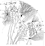
Transverse section through a cerebellar folium. Labels: A, axon of cell Purkinje; F, moss fibers; K and K', fibers from the white core of folium ending in molecular layer in connection with the dendrites of the cells of Purkinje; M, small cell of the molecular layer; GR, granule cell; M1, basket cell; ZK, basket work around the cells of Purkinje; GL, neuroglial cell; N, axon of an association cell.

Sagittal Section Through Cerebellar Folium

Transverse section through a cerebellar folium. Labels: A, axon of cell Purkinje; F, moss fibers; K…
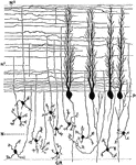
Section through the molecular and granular layers in the long axis of a cerebellar folium. Labels: P, cell of Purkinje; GL, neuroglial cell; N, axon of a granule cell; N1, axons of granule cells in molecular layers.

Sagittal Section Through Cerebellar Folium

Section through the molecular and granular layers in the long axis of a cerebellar folium. Labels: P,…
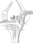
Diagram showing the brain connections of the vagus, glossopharyngeal, auditory, facial, abducent, and trigeminal nerves.

Connection of Brain Nerves

Diagram showing the brain connections of the vagus, glossopharyngeal, auditory, facial, abducent, and…
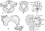
Central connections of the cochlear and vestibular divisions of the auditory nerve.

Auditory Nerves

Central connections of the cochlear and vestibular divisions of the auditory nerve.

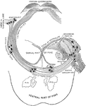
Diagram of the intrapontine course pursued by the facial nerve.

Facial Nerve

Diagram of the intrapontine course pursued by the facial nerve.

Showing the development of the cerebellum. A, Transverse section through the forepart of the cerebellum of a sheep embryo. B, Transverse section through the hinder part of the cerebellum of a sheep embryo. C, Cerebellum of a human fetus 17 cm long. D, Median section through cerebellum of sheep embryo 5 cm long.E, Median section through cerebellum of human fetus 17 cm long. Labels: 1, sulcus primarius; 2, sulcus above and afterwards behind the pyramid; 3, sulcus between pyramid and uvula; 4, great horizontal fissure; r.l., lateral recess ventricle IV; T, transverse groove in roof of ventricle IV.

Development of Cerebellum

Showing the development of the cerebellum. A, Transverse section through the forepart of the cerebellum…
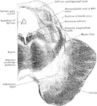
Transverse section through the mesencephalon at the level of the inferior quadrigeminal body.

Section of Mesencephalon at Inferior Quadrigeminal Body

Transverse section through the mesencephalon at the level of the inferior quadrigeminal body.
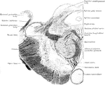
Transverse section through the mesencephalon at the level of the superior quadrigeminal body.

Section of Mesencephalon at Superior Quadrigeminal Body

Transverse section through the mesencephalon at the level of the superior quadrigeminal body.
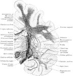
Coronal section through the cerebrum of an orangoutang passing through the subthalamic tegmental region.
Coronal Section Through Cerebrum

Coronal section through the cerebrum of an orangoutang passing through the subthalamic tegmental region.

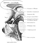
Mesial section through the pituitary region in a child of twelve months old.

Pituitary Region in a Child

Mesial section through the pituitary region in a child of twelve months old.
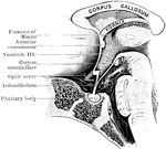
Mesial section through the pituitary region in an adult.

Pituitary Region in an Adult

Mesial section through the pituitary region in an adult.

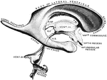
Cast of the ventricles of the brain. Labels" R.SP., recessus suprapinealis; R.P., recessus pinealis inferius; A.S., aqueduct of Sylvius; F.M., foramen of Monro.

Ventricles of the Brain

Cast of the ventricles of the brain. Labels" R.SP., recessus suprapinealis; R.P., recessus pinealis…

Gyri and sulci, on the outer surface of the cerebral hemisphere. Labels: f1, sulcus frontalis superior; f2, sulcus frontalis inferior; f.m., sulcus frontalis medius; p.m., sulcus paramedialis; A, pars basilaris; B, pars triangularis; C, pars orbitalis; S, Sylvian fissure; s1, anterior horizontal limb (Sylvian fissure); s2, posterior horizontal limb (Sylvian fissure); p.c.i., inferior praecentral sulcus; p.c.s., superior praecentral sulcus; r, fissure of Rolando; g.s., superior genu; g.i., inferior genu; d, sulcus diagonalis; t1, superior temporal sulcus (parallel); t2, inferior temporal sulcus; p1, inferior postcentral sulcus; p2. superior postcentral sulcus; p3, ramus horizontalis; p4, ramus occipitalis; s.o.t., sulcus occipitalis transversus; c.m., callosomarginal sulcus; c.t.r., inferior transverse furrow.

Gyri and Sulci on the Brain

Gyri and sulci, on the outer surface of the cerebral hemisphere. Labels: f1, sulcus frontalis superior;…

Three stages in the development of the insula and the insular opercula.

Development of the Insula

Three stages in the development of the insula and the insular opercula.

Diagram to illustrate the development of the opercula which cover the insula. A, Sylian fossa before opercula begin to form. B, Fronto-paretal and temporal opercula wells advances. C, All the four opercula developed but not in opposition. Labels: F.P., fronto-parietal operculum; T, temporal operculum; F.R., frontal wall of fossa; O.R., orbital wall of fossa; F., frontal operculum; O, orbital operculum; s2, anterior horizontal limb of Sylvian fissure; s3, ascending limb; s4, posterior horizontal limb.

Development of the Opercula

Diagram to illustrate the development of the opercula which cover the insula. A, Sylian fossa before…
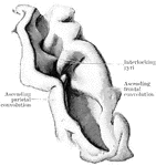
Fissure of Rolando fully opened up, so as to exhibit the interlocking gyri and deep annectant gyrus within it.

Fissure of Rolando

Fissure of Rolando fully opened up, so as to exhibit the interlocking gyri and deep annectant gyrus…

The left cerebral hemisphere, from a fetus in the early part of the seventh month of development. Labels: p.c.s., sulcus praecentralis superior; p.c.i., sulcus praecentralis inferior; r1, lower part of Rolandic fissure; r2, upper part of Rolandic fissure; p1, inferior postcentral sulcus; p3, ramus horizontalis; p4, ramus occipitalis; e.p., external perpendicular fissure; t1, parallel sulcus; S, Sylian fossa; F.P., fronto-parietal wall; F.,frontal wall, O., orbital wall.

Brain of Fetus

The left cerebral hemisphere, from a fetus in the early part of the seventh month of development. Labels:…

The gyri and sulci on the mesial aspect of the cerebral hemisphere. Labels: r, fissure of Rolando; r.o, rostral sulcus; i.t, incisura temporalis.

Gyri and Sulci on the Brain

The gyri and sulci on the mesial aspect of the cerebral hemisphere. Labels: r, fissure of Rolando; r.o,…
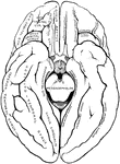
The gyri and sulci on the tentorial and orbital aspects of the cerebral hemispheres.

Gyri and Sulci on the Brain

The gyri and sulci on the tentorial and orbital aspects of the cerebral hemispheres.
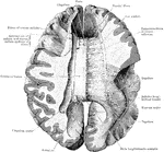
The corpus callosum, exposed from above and the right half dissected to show the course taken by its fibers.

Corpus Collosum

The corpus callosum, exposed from above and the right half dissected to show the course taken by its…
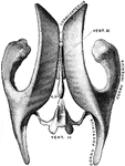
Diagrammatic profile view of the fornix.

Profile View of Fornix

Diagrammatic profile view of the fornix.

Drawing taken from a cast of the ventricular system of the brain, as seen from above. Vent. III, Third ventricle. Vent. IV, Fourth Ventricle. R.SP., recessus suprapinealis.

Ventricles of the Brain

Drawing taken from a cast of the ventricular system of the brain, as seen from above. Vent. III, Third…
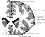
Coronal section through the frontal lobes and the anterior horns of the lateral ventricles.

Section Through Lateral Ventricles

Coronal section through the frontal lobes and the anterior horns of the lateral ventricles.
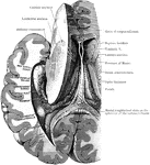
Dissection, to show the fornix and the posterior and descending cornua of the lateral ventricle of the left side.

Dissection to Show Ventricle and Fornix

Dissection, to show the fornix and the posterior and descending cornua of the lateral ventricle of the…
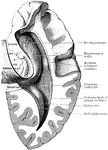
Dissection, to show the posterior and descending cornua of the lateral ventricle. Labels: B.G., Giacomini's band; F.D., gyrus dentatus; F, fimbria; H.C., hippocampal convolution.

Dissection to Show Ventricle

Dissection, to show the posterior and descending cornua of the lateral ventricle. Labels: B.G., Giacomini's…
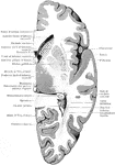
Horizontal section through the right cerebral hemisphere at the level of the widest part of the lenticular nucleus.

Section Through Cerebral Hemisphere

Horizontal section through the right cerebral hemisphere at the level of the widest part of the lenticular…
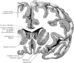
Coronal section through the right cerebral hemispheres as to cut through the anterior part (putamen) of the lenticular nucleus in front of the globus pallidus.

Coronal Section Through Cerebral Hemisphere

Coronal section through the right cerebral hemispheres as to cut through the anterior part (putamen)…
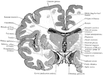
Coronal section through the cerebrum, so as to cut through the three divisions of the lenticular nucleus; posterior surface of the section depicted.

Coronal Section Through the Cerebrum

Coronal section through the cerebrum, so as to cut through the three divisions of the lenticular nucleus;…
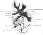
Coronal section through the left side of the cerebrum of an orangoutang. The section passes through the middle of the lenticular nucleus.

Coronal Section Through the Cerebrum

Coronal section through the left side of the cerebrum of an orangoutang. The section passes through…

Diagrammatic representation of the internal capsule (as seen in horizontal section).
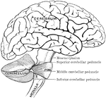
Internal Capsule

Diagrammatic representation of the internal capsule (as seen in horizontal section).