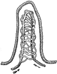
Muscles of the leg
"Muscles of the leg showing how they pass into tendons at the ankle." —Davison, 1910
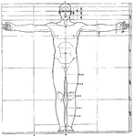
Proportions of human figure
"The proportions of the human figure. As handed down to us by Vitruvius and described by Joseph Bonomi."…
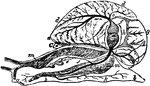
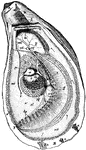
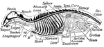
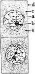
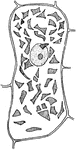
Snail Anatomy
"Anatomy of the Snail: a, the mouth; bb, foot; c, anus; dd, lung; e, stomach, covered above by the salivary…
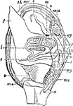
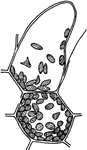
Mollusc Anatomy
"Anatomy of an Acephalous Mollusc (Mactra): s, stomach; ii, intestine; ag, anterior ganglions; pg, posterior…
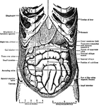
Abdomen
"The Abdominal Viscera in situ, as seen when the abdomin is laid open and the great omentum…
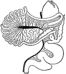
Intestinal Tract of Chauna Chavaria
cc. Colic caeca, d. Duodenum. g. Glandular patch, l.l. Meckel's tract, l.i. Hind-gut, p.v. Cut root…
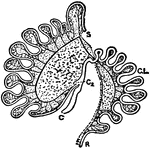
Intestinal Tract of Macropus Bennetti
S, cut end of duodenum; R, cut end of rectum; C, caecum; C2, accessory caecum; C.L., colic loop of hind-gut.
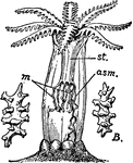
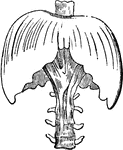
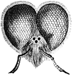
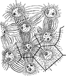
Alcyonarian zooid
"An expanded Alcyonarian Zooid, showing the mouth surrounded by eight pinnate tentacles. st, Stomodaeum…
Diaphragm
"This is the name applied in anatomy to designate the transverse muscle which, in man and the mammalia…
Diaphragm
"This is the name applied in anatomy to designate the transverse muscle which, in man and the mammalia…
Oyster
"Anatomy of the Oyster. A. Hinge or anterior umbonal end of the left valve of an adult oyster, upon…
Organs
Diagram of the chief organs of a mammal. The bones are black. a, opening from the nasal cavity…
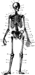
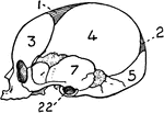
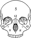
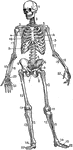
Skeleton
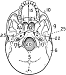
1. Frontal bone 2. Parietal bone 3. Coronal Suture 4. Squamous portion of Temporal bone 5. Mastoid process…
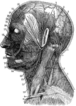
Neck
1. Temporal Artery 2. Artery behind the ear 3. Occipital Artery 4. Greater occipital nerve 5. Smaller…
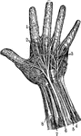
Hand
1. Nerves of the skin 2. Tendons 3. Arteries of the palm of the hand 4. Elbow nerve 5. Elbow artery…
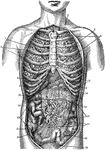
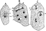
Internal Anatomy
1. Collar bone 2. Left Lung 3. Breast Bone 4. Right Lung 5. Ribs 6. Right lobe of the liver 7. Left…
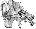
Inner Ear
1. Helix 2. Concha 3. Outer passage 4,5,6. emicircular canals 7. Oval indow 8. Cochlea 9. Eustachian…
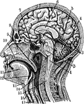
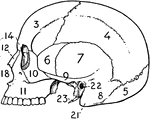
Sectional view of the Head
Section through the Head and Neck on the Median Line. 1. Medulla Oblongata 2. Pons 3. Right lobe of…
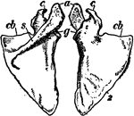
Shoulder blade
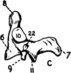
"The Scapula, or shoulder blade, is one of the two bones, the other being the clavicle, which form the…
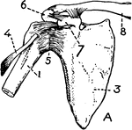
Shoulder bones and ligaments
"Shoulder bones and ligaments. 1. Humerus, 3. Scapula, 4. Tendon of biceps 5. Capsular ligament 6. Acromion…
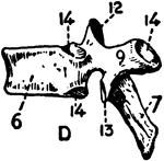
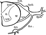
Spinal Nerve Roots
Diagram showing anatomy of the spinal nerve roots and adjacent parts. Labels: G., gray matter of the…
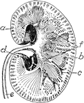
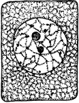
Kidney
Transverse section of the human kidney: "(a) cortex; (b) medulla; (c) small branch of the renal artery;…
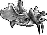
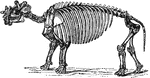
Dinoceras Mirabile
The skull and upper jaw of an early rhinoceros-like mammal from the Cenozoic time.
Onion Cells
"A, embryonic cells from onion root tip; d, plasmatic membrane; c, cytoplasm; a, nuclear membrane enclosing…
Onion Cells
"B, older (onion) cells farther back from the root tip. The cytoplasm is becoming vacuolate; f, vacuole."…
T. Zebrina Cell
In onion cells: "C, a cell from the epidermis of the mid-rib of Tradescantia zebrina, in its natural…
T. Majus Cell
"A, cell from the epidermis of the upper side of the calyx of Tropaeolum majus with crystalline chromoplasts."…
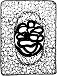
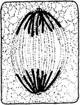
Plant Cell Division 1
First stage in plant cell division: Protophase 1; "Resting cell ready to begin division." -Stevens,…
Plant Cell Division 2
Second stage in plant cell division: Protophase 2; "the nuclear reticulum is assuming the form of a…
Plant Cell Division 3
Third stage in plant cell division: Protophase 3; "The nuclear thread has divided longitudinally throughout…
Plant Cell Division 4
Fourth stage in plant cell division: Protophase 4; "The nuclear membrane and the nucleolus have disappeared,…
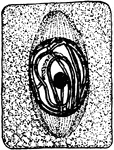
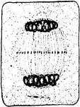
Plant Cell Division 5
Fifth stage in plant cell division: Metaphase; "The metaphase, where the longitudinal halves of the…
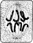
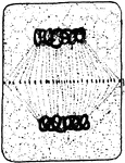
Plant Cell Division 6
Sixth stage in plant cell division: Anaphase; "The anaphase, or movement of the chromosomes toward the…
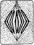
Plant Cell Division 7
Seventh stage in plant cell division: Telophase; "Telophase where the chromosomes have begun to spin…
Plant Cell Division 8
Eighth stage in plant cell division: "The connecting fibers have spread out and come into contact with…
Plant Cell Division 9
Ninth and final stage in plant cell division: "A nuclear membrane has been formed about each daughter…
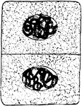
A. Eupatorium Cell
"Formation of endosperm in the embryo-sac of Agrimonia Eupatorium. Cell-walls are being formed between…
E. Communis Cell
"Free cell formation of spores in the ascus of Erysiphe communis. A, ascus with single nucleus; C, cytoplasm;…
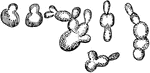
S. Cerevisiae Cell Multiplication
"Various stages of cell multiplication by budding of Saccharomyces cerevisiae." -Stevens, 1916