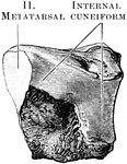
The right internal cuneiform (outer side).

Internal Cuneiform

The right internal cuneiform (outer side).

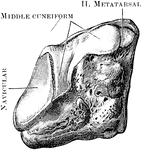
The right middle cuneiform (inner side).

Middle Cuneiform

The right middle cuneiform (inner side).

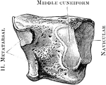
The right middle cuneiform (outer side).

Middle Cuneiform

The right middle cuneiform (outer side).

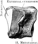
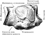
The right external cuneiform (inner side).

External Cuneiform

The right external cuneiform (inner side).

The right external cuneiform (outer side).

External Cuneiform

The right external cuneiform (outer side).

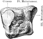
The right cuboid bone (outer side).

Cuboid

The right cuboid bone (outer side).

The right cuboid bone (inner side).

Cuboid

The right cuboid bone (inner side).

The first metatarsal bones of the right foot (plantar aspect).

First Metatarsal Bone

The first metatarsal bones of the right foot (plantar aspect).

View of the bases and shafts of the second, third, and fourth metatarsal bones of the right foot.

Metatarsal Bones

View of the bases and shafts of the second, third, and fourth metatarsal bones of the right foot.
Fifth right metatarsal bone (dorsal aspect).

Fifth Metatarsal

Fifth right metatarsal bone (dorsal aspect).

The phalanges of the toes (dorsal aspect).

Phalanges

The phalanges of the toes (dorsal aspect).

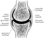
Shown is a a diagram of a diarthrodial joint. In the diarthrodial group the extensive cavity has produced great interruption in the continuity of the uniting structures which originally existed between the bones forming such a joint.

Diarthrodial Joint

Shown is a a diagram of a diarthrodial joint. In the diarthrodial group the extensive cavity has produced…
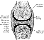
In the diarthrodial group the extensive cavity has produced great interruption in the continuity of the uniting structures which originally existed between the bones forming such a joint. Shown is a a diagram of a diarthrodial joint with an interarticular meniscus dividing the joint cavity into two compartments.

Diarthrodial Joint

In the diarthrodial group the extensive cavity has produced great interruption in the continuity of…
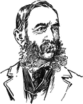
Daniel Coit Gilman (July 6, 1831-October 13, 1908) was an American educator.

Daniel Coit Gilman

Daniel Coit Gilman (July 6, 1831-October 13, 1908) was an American educator.

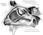
View of the outer wall of the nose. The turbinated bones having been removed. Labels: 1, vestibule, 2, hiatus semilunaris; 3, opening of antrum of highmore; 4, bulla ethmoidalis; 5, agger nasi; 6, opening of anterior ethmoidal cells; 7, cut edge of superior turbinated bone; 8, cut edge of middle turbinated bone; 9, pharyngeal orfice of Eustachian tube.

Outer Wall of Nose

View of the outer wall of the nose. The turbinated bones having been removed. Labels: 1, vestibule,…
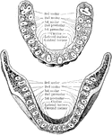
Horizontal section through both the upper and lower jaws to show the roots of the teeth. The sections were carried through the bones a short distance from the edge of their alveolar borders. The upper figure shows the upper teeth, the lower figure the lower teeth. Note the flattened roots of the lower incisors, the two root canals in the anterior root of each lower molar, and the confluence of the three roots of the upper wisdom teeth.

Jaw Showing Roots of Teeth

Horizontal section through both the upper and lower jaws to show the roots of the teeth. The sections…

An illustration of a woodpecker's skull.

Woodpecker Skull

An illustration of a woodpecker's skull.

"Palmar aspect of left fore foot of a black bear (Ursus americanus). scl, scapholunar; c, cuneiform; p, pisiform; tr, trapezium; t, trapezoid; m, magnum; u, unciform. The phalanges show a full series of sesamoid bones (unmarked)." -Whitney, 1911

Bear Foot

"Palmar aspect of left fore foot of a black bear (Ursus americanus). scl, scapholunar; c, cuneiform;…
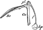
"Right shoulder-girdle or scapular arch of fowl, showing hp, the hypoclidium; f, furculum; Co, coracoid; Sc, scapula; gl, glenoid." -Whitney, 1911

Bird Scapula

"Right shoulder-girdle or scapular arch of fowl, showing hp, the hypoclidium; f, furculum; Co, coracoid;…
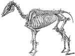
The skeleton of a horse. Axial Skeleton. The Skull. Cranial Bones: a, occipital, 1; b, wormian, 1; c, parietal, 2; d, frontal, 2; e, temporal, 2; sphenoid, 1; ethmoid, 1; auditory ossicles, 8. Facial bones: h, nasal, 2; g, lachrymal, 2; f, malar, 2; i, superior maxilla, 2; k, premaxila, 2; palatine, 2; pterygoid, 2; vomer, 1; tubinals, 4; l, inferior maxilla; 1; hyoid (segments), 5. Teeth: m, incisors, 12; n, canines, 4; o, molars, 24. The trunk: p-v, cervical vertebrae, 7; w, dorsal vertebrae, 18; x, lumbar vertebrae, 6; y, sacrum, 1; z, coccygeal vertebrae (variable), 18, d, d, ribs, 36; sternum (6-7 sternebrae), 7; *,coastal cartilages. Appendicular Skeleton. Pectoral Limb: e', scapula, 2; f', humerus, 2; g', radius, 2; h', ulna, 2. Carpus-i', trapesium, 2; k', cuneiform, 2; l', lunar, 2; m', scaphoid, 2; n', unciform, 2; o', magnum, 2; p', trapezoid, 2; q', pisiform, 2. Metacarpus-r', large bone, 2; s', small bone, 4. Digits- u', proximal phalanges, 2; v, median phalanges; w', distil phalanges, 2; t', large sesamoids, 4; small sesamoids, 2. Pelvic Limbs: Pelvis- b', ilium, 2; d' ischium, 2; c', pubis, 2. The limb- y', femur, 2; z', patella, 2; a', tibia, 2; b', fibula, 2. Tarsus- c', calcaneum, 2; d', astragalus, 2; e'. cuboid, 2; f', cuneiforme magnum, 2; g', cuneiforme medium, 2; h", cuneiforme parvum, 2. Metatarsus: large bone, 2; small bone, 4. Digit: proximal phalanges, 2; median phalanges, 2; distal phalanges, 2; large sesamoids, 4; small sesamoids, 2.

Skeleton of a Horse

The skeleton of a horse. Axial Skeleton. The Skull. Cranial Bones: a, occipital, 1; b, wormian, 1; c,…
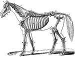
The skeleton of a horse. Showing its relation to the contour of the animal, viewed laterally. Labels: A, temporal fossa; B, inferior maxilla; C, atlas; D, dentata; E, cervical vertebrae; F, dorsal vertebrae; G, lumbar vertebrae; H, sacral vertebrae; I, coccygeal vertebrae; J, scapula; K, humerus; L, radius; L, ulna; M, carpus; N, trapezium; O, metacarpus; P, b. os suffraginis; Q, c. sesamoids; R, d. os coronae; S, e. os pedis; T, ribs; U., ilum; V, femur; X, patella; Y, tibia; y., Fibula; Z, tarsus; a, metatarsus; f, ligamentum nuchae, funicular portion; f' lamellar portion; 1, zygoma; 2, orbital fossa; 3, nasal peak; 4, incisor teeth; 4', canine teeth; 5, molar teeth; 6, external humeral trochanter; 7, scapular fossae; 8, coracoid apophysis; 9, cartilage of prolongation; 10, deltoid ridge, and external tuberosity; 11, olecranon; 12, costal cartilage; 13, anterior iliac spine; 14, ischium; 15, trochanter major; 16, trochanter minor; 18, anterior tibial tuberosity; 19, calcaneum; 20, small metacarpal and metatarsal or splint bones.

Skeleton of a Horse

The skeleton of a horse. Showing its relation to the contour of the animal, viewed laterally. Labels:…

"Schizognathous skull of common fowl. pmx, premaxilla; mxp, maxillopalatine; mx, maxilla; pl, palatine; pt, pterygoid; vo, vomer." -Whitney, 1911

Bird Skull

"Schizognathous skull of common fowl. pmx, premaxilla; mxp, maxillopalatine; mx, maxilla; pl, palatine;…

"Schizorhinal skull of curlew (top view), showing the long cleft, a, between upper and lower forks of each nasal bone." -Whitney, 1911

Curlew Skull

"Schizorhinal skull of curlew (top view), showing the long cleft, a, between upper and lower forks of…

Longitudinal section of a horse's skull. Labels: 1, supraoccipital bone and crest; 2, parietal bone; 3, frontal bone; 4, frontal sinus; 5, nasal bone; 6, superior turbinal; 12, inferior turbinal; 14, superior maxilla; 13 and its palatine plate; 15, premaxilla; 11, palatine bone; 7, ethmoid bone and volutes; 8, basi and presphenoid bones; 9, basioccipital bone; 10, petrosal bone. The cranium is the cavity under 1 and 2.

Longitudinal Section of Horse Skull

Longitudinal section of a horse's skull. Labels: 1, supraoccipital bone and crest; 2, parietal bone;…

The hyoid series is composed of five distinct pieces- a body, or hyoid bone proper, two cornua or horns and two cornicula or lesser horns. Shown us the left aspect of the hyoid series of bones in a horse. Labels: a, proximal, end of left cornu; b, distal end of right cornu; c, proximal end of corniculum; d, hyoid bone. The left heel process points downwards and backwards, the spur process in the opposite direction.

Hyoid Series of Bones in a Horse

The hyoid series is composed of five distinct pieces- a body, or hyoid bone proper, two cornua or horns…

External view of bones of right carpus, metacarpus, and digit of a horse. 1, distal end of radius; 2, groove for extensor tendons; 3, scaphoid; 4, lunar; 5, cuneiform;6, trapezium; 7, magnum; 8, unciform; 9, great metacarpal; 10, external small metacarpal (splint); 11, sesamoids; 12, proximal phalanx; 13, median phalanx; 5, distal phalanx; 16, basilar process.

Horse Leg

External view of bones of right carpus, metacarpus, and digit of a horse. 1, distal end of radius; 2,…

Posterior view of phalanges of a horse disarticulated. Labels: a, os suffraginis; b, os coronae; C, os pedis; D, os naviculare, or third sesamoid; 1, glenoid cavities joined by the groove, 2; 3, rough surface for V-shaped sesamoidean ligament; 4, condyles articulating with cavities, 5; 6, condyles of coronae articulating with glenoid cavities of pedal bone, 7; 8, pyramidal process; 9, basilar process; 10, retrosal processes; 11, articular surfaces between navicular and pedal bones; 12, surface, continues with glenoid cavities, 7; 13, posterior edge; 14, extremities for lateral ligaments of navicular bone.

Phalanges of a Horse

Posterior view of phalanges of a horse disarticulated. Labels: a, os suffraginis; b, os coronae; C,…

Bones of left tarsus of a horse, seen from in front and outside. Labels: 1, calcaneum; 2, astragalus; 3, cuneiforme magnum; 4, cuneiforme medium; 5, cuboid.

Tarsus of a Horse

Bones of left tarsus of a horse, seen from in front and outside. Labels: 1, calcaneum; 2, astragalus;…

External view of bones of left tarsus, metatarsus, and digit of a horse. Labels: 1, distal end of tibia; 2, calcaneum; 3, astragalus; 4, cuboid; 5, cuneiforme magnum; 6, cuneiforme medium; 7, great metatarsal; 8, external small metatarsal (splint); 9, proximal phalanx; 10, sesamoids; 11, median phalanx; 12, distal phlanx; 14, third sesamoid; 15, basilar process.

Horse Leg

External view of bones of left tarsus, metatarsus, and digit of a horse. Labels: 1, distal end of tibia;…
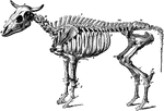
The skeleton of an ox. Axial Skeleton. The skull. Cranial Bones- occipital, 1: b, parietal, 2; a, frontal, 2; c, temporal, 2; sphenoid, 1; ethmoid, 1; auditory ossicles, 8. Facial Bones- h, nasal, 2; e, lachrymal, 2; d, malar, 2; f, maxilla, 2; g, premaxilla, 1; i, inferior maxilla, 2; palatine, 2; pterygoid, 2; vomer, 1; turbinals, 4; hyoid series, 7. Teeth: incisors, 6; canines, 2; molars, 24. The trunk- k, cervical vertebrae, 7; l, dorsal vertebrae, 13; m, lumbar vertebrae, 6; n, sacrum ( five segments), 1; o, coccygal vertebrae (variable), 20; p, ribs, 26; * sternum (seven sternebrae), 1. Appendicular skeleton. Pectoral limb. t, scapula, 2; u, humerus, 2; v, radius, 2; w, ulna, 2. Carpus- x, trapezium, 2; y, cuneiform, 2; z, lunar; a', scaphoid, 2; b', unciform, 2; c', magnum, 2. Metacarpus: d', large bone, 2; e', small bone, 4; f', large sesamoids, 8. Digit- g', proximal phalanges, 4; h', median phalages, 4; i', distal phalanges, 4; k', small sesamoids, 4. Pelvic Limb. Pelvis- q, ilium, 2; s, ischium, 2; r, pubis, 2. The limb- l', femur, 2; m', patella, 2; n', tibia, 2. Tarsus-o', maleolar, 2; p', calcaneum, 2; q', astragalus, 2; r', cubocuneiform, 2; s', cuneiforme medium, 2; t', cuneiforme parvum, 2. Metatarsus: large bone, 2; u', small bone, 2. Large sesamoids, 8. Digit: proximal phalanges, 4; median phalanges, 4; distal phalanges, 4; small sesamoids, 4. Visceral skeleton. Bones of the heart, 2. The separate bones of the ruminant skeleton, as here considered are 251.

Ox Skeleton

The skeleton of an ox. Axial Skeleton. The skull. Cranial Bones- occipital, 1: b, parietal, 2; a, frontal,…
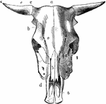
Skull of an ox, superior aspect. Labels: a, frontal crest; b, lateral crest; c, horn core; d, nasal peak; e, supraorbital foramina. Bones: 1, frontal; 2, lachrymal; 3, malar; 4, superior maxillary; 5, nasal; 6, premaxilla, with incisive openings.

Ox Skull

Skull of an ox, superior aspect. Labels: a, frontal crest; b, lateral crest; c, horn core; d, nasal…

Skeleton of the hog. Axial skeleton. The skull. Cranial bones- a, occipital, 1; b, parietal, 2; d, frontal, 2; c, temporal, 2; sphenoid, 1; ethmoid, 1; auditory ossicles, 8. Facial Bones- g, nasal, 2; h, os rostri, 1; f, lachrymal, 2; e, malar, 2; i, maxilla, 2; k, premaxilla, 2; l, inferior maxilla, 1; palatine, 2; 2, pterygoid, 2; vomer, 1; turbinals, 4; hyoid series, 5. Teeth: incisors, 12; canines, 4; molars, 28. The trunk- m, cervicle vertebre (variable), 18; r, ribs, 28; * sternum (seven sternebrae), 1. Appendicular Skeleton. Pectoral Limb. v, scapula, 2; w, humerus, 2; x, radius, 2; y, ulna, 2. carpus: z, trapezium, 2; a', cuneiform, 2; b', lunar, 2; c', scaphoid, 2; d', unciform, 2; e', magnum, 2; f', trapezoid, 2; g', pisiform, phalanges, 8; l', distal phalanges, 8; l", small sesamoids, 8. Pelvic Limb. Pelvis: s, ilium, 2; t, pubis, 2; u, ischium, 2. The limb-m', femur, 2; n', patella, 2; o', tibia, 2; p' fibula, 2. Tarsus- q' calcaneum, 3; r', astragalus, 2; s', cuboid, 2; t', cuneiforme magnum, 2; u', ecto-cuneiforme, 2; v' w', meso and endo cuneiformes, 4. Metatatarsus: large bones, 8; x', small bones 2; y', large sesamoids, 16. Digit: proximal phalanges, 8; median phalanges, 8; distal phalanges, 8; small sesamoids, 8. Visceral Skeleton- None. The separate bones of the skeleton of the hog, as here considered, are 234.

Skeleton of a Hog

Skeleton of the hog. Axial skeleton. The skull. Cranial bones- a, occipital, 1; b, parietal, 2; d, frontal,…
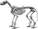
The skeleton of the dog. Axial skeleton. The skull. Cranial bones- a, occipital, 1; b, parietal, 2; d, frontal, 2; c, temporal, 2; sphenoid, 1; ethmoid, 2; auditory ossicles, 8. Facial Bones- f, nasal, 2; e, lachrymal, 2; d, malar, 2; h, maxilla, 2; g, premaxilla, 2. Teeth: incisors, 12; canines, 4; molars, 26. The trunk- l, cervical vertebrae, 7; m, dorsal vertebrea, 13; n, lumbar vertebrae, 7; o, sacrum (three segments), 1; p, coccygeal phalanges (variable), 20; t, ribs, 26; * sternum (eight sternebrae), 1. Appendicular Skeleton. Pectoral Limb. u, scapula, 2; v, humerus, 2; w, radius, 2; x, ulna. Carpus: y, trapezium, 2; z cuneiform, 2; a', scaphoid, 2; b', unciform, 2; c', magnum, 2; d', trapezoid, 2; e', pisiform,2; metacarpal bones, 10; h', anterior sesamoids, 10; g' posterior sesamoids, 20. Digits- i' proximal phalanges, 10; k', median phalanges, 8; l', distal phalanges, 10; small sesamoids wanting. Pelvic Limb. Pelvis: q, ilium, 2; r, pubis, 2; s, ischium, 2. The limb-m', femur, 2; o', fabellae, 4; n', patella, 2; q', tibia, 2; p', tibial sesamoid, 2; r', fibula, 2. Tarsus: s', calcaneum, 2; t', astragalus, 2; u', cuboid, 2; v', superior cuneiform, 2; w', ectocuneiforme, 2; x', mesocuneiforme, 2; y', edocuneiforme, 2. Metatarsus: large bones, 8; z',small bones, 2; anterior sesamoids, 8; posterior sesamoids, 16. Digit: proximal phalanges, 8; median phalanges, 8; distal phalanges, 8; small sesamoids wanting. Visceral Skeleton. Os Penis, 1; Rudimentary clavicle (inconstant), 2. The bones of the Carnivora Skeleton, thus considered, are 345.

Skeleton of a Dog

The skeleton of the dog. Axial skeleton. The skull. Cranial bones- a, occipital, 1; b, parietal, 2;…
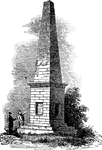
The Monument marks the grave site of the bones of victims of the Wyoming Massacre, which took place on July 3, 1778. Local residents banded together to defend the area against an invasion of British Tories as well as pro-Tory Native Americans. The battle ended in defeat for the colonial fighters and considerable brutality followed the actual Battle. In 1833, the bones were reinterred in a vault under the present Monument. The monument is located in the borough of Wyoming, Pennsylvania.

The Wyoming Monument

The Monument marks the grave site of the bones of victims of the Wyoming Massacre, which took place…
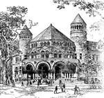
Many buildings stood on the Old Campus which were removed to make way for the current configuration of structures, including The Old Laboratory (1782–1888), The Cabinet (1819–1890), Trumbull Gallery (1832–1901), Alumni Hall (1853–1911, Alexander Jackson Davis; the towers from this building were rescued and rebuilt behind the tomb of Skull and Bones), Old Dwight Hall (1885–1926, J. Cleaveland Cady) and Osborn Hall (1888–1926, Bruce Price).

Osborn Hall, Yale University

Many buildings stood on the Old Campus which were removed to make way for the current configuration…
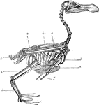
An illustration of a bird skeleton.

Bird Skeleton

An illustration of a bird skeleton.

The skeleton of a bird. Labels: a, radius and ulna; b, dorsal vertebrae; c, sacrum and pelvis; g, ploughshare bone; h, tibia; i, metatarsus; d, furculum; e, carina of the sternum; f, distal phalanx of pectoral limb.

Skeleton of a Bird

The skeleton of a bird. Labels: a, radius and ulna; b, dorsal vertebrae; c, sacrum and pelvis; g, ploughshare…
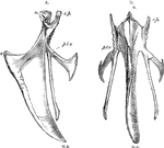
The sternum of a bird. Label: a, lateral aspect; b, inferior aspect; r, rostrum; c.p, costal process; pl.o, pleurosteon; c, carina; m.x, middle xiphoid process.

Sternum of a Bird

The sternum of a bird. Label: a, lateral aspect; b, inferior aspect; r, rostrum; c.p, costal process;…
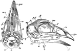
The skull of a bird. Labels: a, inferior aspect, the mandible being removed; b, lateral aspect; px, premaxilla; mx, superior maxilla; p, palatine bone; v, vomer; j, jugal bone; qj, quadrato-jugal bone; lachrymal; me, median ethmoid; f, frontal; pf, post-frontal process; p, parietal; pt, pterygoid; q, os quadratum; sq, sqamosal; eu, process before the sustachian aperture; bo, basi-occipital, and so, supra-occipital bone; ar, articular; a, surangular, and d, dentary portion of the mandible.

Skull of a Bird

The skull of a bird. Labels: a, inferior aspect, the mandible being removed; b, lateral aspect; px,…
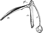
The pectoral arch of a bird. Labels: sc, scapula; co, coracoid bone; f, clavicle, terminating below in the hypocleideum; gl, glenoid cavity.

Pectoral Arch of a Bird

The pectoral arch of a bird. Labels: sc, scapula; co, coracoid bone; f, clavicle, terminating below…
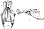
The pelvis of a bird. Labels: a, superior; b, lateral aspect; sm, sacrum; Il, ilium; Is, ischium; Am, acetabulum.

Pelvis of a Bird

The pelvis of a bird. Labels: a, superior; b, lateral aspect; sm, sacrum; Il, ilium; Is, ischium; Am,…

Pogonias chromis, a name of several fish so called from the drumming noise they make, said to be due, in part at least, to the grinding of bones upon each other.

Saltwater Drum

Pogonias chromis, a name of several fish so called from the drumming noise they make, said to be due,…
This Skull Head was a design found on the shield of death, monuments, tombs and often represented over two crossed bones.

Skull Head

This Skull Head was a design found on the shield of death, monuments, tombs and often represented over…
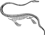
The skeleton of an elasmosaurus, a dinosaur in the order of plesiosaurs.

Elasmosaurus Skeleton

The skeleton of an elasmosaurus, a dinosaur in the order of plesiosaurs.

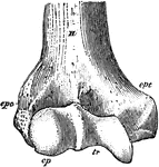
"Anterior View, Distal End, of Right Humerus of a Man. H, humerus; epc, epicondyle, or external supracondyloid protuberance; ept, epitrochlea, or internal supracondyloid protuberance; cp, capitellum, or convex articular suface for head of radius; tr, trochlea, or transversely concave articular surface for the ulna; epc and cp are together the ectocondyle, and ept and tr are together the entocondyle." -Whitney, 1911

Humerus

"Anterior View, Distal End, of Right Humerus of a Man. H, humerus; epc, epicondyle, or external supracondyloid…

"Epipleurae.-- Thorax, scapular arch, and part of pelvic arch of a bobolink (Dolichonyx oryzivorus). ep, four epipleurae or uncinate processes of as many ribs; pl, pleurapophysial parts of seven ribs; hp, hemapopysial parts of six ribs; v, dorsolumbar vertebrae; st, sternum (the letters are on the carina or keel); m, manubrium sterni; cp, costal process of sternum, bearing f, furcula; ec, epiclidium of furcula; h, hypoclidium of fucula; gl, glenoid fossa, formed by coracoid and scapula; il, ilium; is, ischium; p, pubis; a, acetabulum." -Whitney, 1911

Bobolink Epipleurae

"Epipleurae.-- Thorax, scapular arch, and part of pelvic arch of a bobolink (Dolichonyx oryzivorus).…
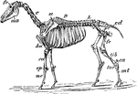
"Skeleton of Horse (Equus caballus). fr, frontal bone; C, cervical vertebrae; D, dorsal vertebrae; L, lumbar vertebrae; cd, caudal vertebrae; sc, scapula; pe, pelvis; ma, mandible; hu, humerus; ra, radius; cp, carpus; mc, metacarpus; fe, femur; tib, tibia; ca, calcaneum; tar, tarsus; mt, metatarsus; p, phalanges." -Whitney, 1911

Horse Skeleton

"Skeleton of Horse (Equus caballus). fr, frontal bone; C, cervical vertebrae; D, dorsal vertebrae; L,…

"Skull of Cat (Felis catus), showing the following bones, viz. : na, nasal; pm, premaxillary; m, maxillary; l, lacrymal; f, frontal; j, jugal; pa, palatine; p, parietal; sq, squamosal; ip, interparietal; so, supra-occipital; eo, exoccipital (the line leads to the occipital condyle); t, tympanic bulla; smf, stylomastoid foramen; mf, mental foramen; c, coronoid process of mandible; ar, ascending ramus of mandible; hr, horizontal ramus of mandible; an, angle of jaw." -Whitney, 1911

Domestic Cat Skull

"Skull of Cat (Felis catus), showing the following bones, viz. : na, nasal; pm, premaxillary; m, maxillary;…

"Bones of Human Foot, or Pes, the third principal segment of the hind limb, consisting of tarsus, metatarsus, and phalanges. a, astralagus; ca, calcaneum; n, navicular, or scaphoid; co, cuboid; c1, c2, c3, entocuneiform, mesocuneiform, and ectocuneiform, or inner, middle, and outer cuneiform bones. The foregoing seven bones constitute the tarsus, and m1 to m5, first to the fifth metatarsal, constitue the metatarsus. The remaining fourteen bones are the phalanges, three to each digit excepting the great toe; h, distal phalanx of the hallux or great toe." -Whitney, 1911

Bones of Human Foot

"Bones of Human Foot, or Pes, the third principal segment of the hind limb, consisting of tarsus, metatarsus,…
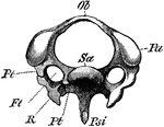
"Third cervical vertebra of Woodpecker (Picus viridis). (Viewed anteriorly.) Ft, vertebrarterial foramen; Ob, upper arch ; Pa, articular process; Psi, haemal spine ; Pt, Pt, the two bars of the transverse process, shewn on one side ancylosed with the cervical rib (R); Sa articular surface of centrum. (From Wieersheim.) A. H. Evans, 1900

The Diagram of a Third Cervical Vertebra of a Woodpecker

"Third cervical vertebra of Woodpecker (Picus viridis). (Viewed anteriorly.) Ft, vertebrarterial foramen;…

"Skeleton of the trunk of a Falcon. Ca, coracoid, which articulates with the sternum (St) at ; Cr, keel of sternum; Fu (Cl), furcula (clavicles); G, glenoid cavity for humerus; S, scapula; Un uncinate process; V, vertebral, and Sp, sternal, portion of rib. (From Wiedersheim.)" A. H. Evans, 1900

The Skeleton of the Trunk of a Falcon

"Skeleton of the trunk of a Falcon. Ca, coracoid, which articulates with the sternum (St) at ; Cr, keel…
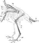
"Skeleton of the Limbs and Tail of a Carinate Bird. (The skeleton of the body is indicated by dotted lines.) F, digits; Fi, fibula; HW, carpus; MF, tarsometatarsus; MH, carpometacarpus; OA, humerus; OS, femur; Py, pygostyle; R, coracoid; Ul, ulna; Sch, scapula; St, sternum, with its keel (Cr); T, tibiotarsus; Rd, radius; Z, Z, digits of foot. (From Wiedersheim.)" A. H. Evans, 1900

Skeleton of the Limbs and Tail of a Carinate Bird

"Skeleton of the Limbs and Tail of a Carinate Bird. (The skeleton of the body is indicated by dotted…

"Pelvis of Apteryx austrlis. Lateral view. a, Acetabulum; il, ilium; is, ischium; p, pectineal process of pubis; p, pubis." (From Wiedersheim.)" A. H. Evans, 1900

Diagram of the Pelvis of a Kiwi

"Pelvis of Apteryx austrlis. Lateral view. a, Acetabulum; il, ilium; is, ischium; p, pectineal process…

"Skull of a Wild Duck (Anus boscas), from the side. ag, Angular; als, alisphenoid; ar, articular; bt, basitemporal; d, dentaryl en, external nostrils, e.o, exoccipital; eth, ethmoid; fr, frontal; j, jugal; lc, lacrymal; mx, maxilla; mx.p, maxillopalatine process; n, nasal, parietal; pg, pterygoid; pl, palatine; ps, presphenoid; px, premaxilla; q, quadrate; q.j, quadratojugal; s.ag, supra-angular; s.o, supraoccipital; sq, squamosal; ty, tympanic cavity; v, vomer; II, foramen for optic nerve; V, for trigeminal." (From Wiedersheim.)" A. H. Evans, 1900

Diagram of the Skull of a Wild Duck

"Skull of a Wild Duck (Anus boscas), from the side. ag, Angular; als, alisphenoid; ar, articular; bt,…
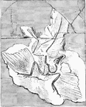
"Archaeornithes is at present represented by but one member, the first undoubted fossil Bird, made known in 1861 by Andreas Wagner form the Jurassic slate formation of Solenhofen in Bavaria, and now preserved in a British Museum. This he described under the name of Griphosaurus; but as Hermann von Meyer had already bestowed the title of Archaeopteryx Lithographica upon a bird, presumably identical, a feather of which had been obtained from the above system." A. H. Evans, 1900 This sample was obtained from the Limestone in Berlin

Archaeopteryx Lithographica

"Archaeornithes is at present represented by but one member, the first undoubted fossil Bird, made known…
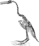
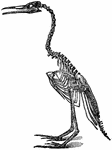
"Hesperornis regalis, (a fossilized restoration) which stood about three feet high, had blunt teeth in the grooves of both maxilla and mandible, the number being thirty or more below, but considerably less above, where they did not reach to the exterior extremity. The bill was long and pointed, the rami of the lower jaw being entirely separate; the head was rather small, the neck long, and the quadrate bone articulated with the skull by one knob only. The sternum was long, broad, and flat, without keel; the furcula was decidedly reduced, the metatarsus, being little more than a humerus; the tail was fairly long and broad, but had no pygostyle." A. H. Evans, 1900

The Restoration of the Hesperornis Regalis

"Hesperornis regalis, (a fossilized restoration) which stood about three feet high, had blunt teeth…

"Ichthyornis victor and I. dispar, ...were small forms of about the size of a Partridge, with the habits and appearance, it is presumed, of Terns or Gulls. The head was extremely large in proportion to the remainder of the skeleton; the beak was long and pointed, with entirely separate rami to the mandible; the sharp teeth, fixed regularly in distinct sockets, were inclined backwards, and occupied the whole of the lower and at least the posterior half of the upper jaw; the keel of the sternum was large and broad; the dorsal and cervico-dorsal vertebrae were biconcave. the quadrate articulated to the skull by one knob,... the metatarsus was short and the whole foot small; a furcula was probably present; the wings were well developed, indicating great powers of flight; while the tail was comparatively short, and ended in a pygostyle." A. H. Evans, 1900

Skeleton Head of a Ichthyornis

"Ichthyornis victor and I. dispar, ...were small forms of about the size of a Partridge, with the habits…

In the Dakotah (Dakota) Territory, the area know as the Bad Lands, is a sunken area thirty miles wide and ninety miles long. It is a type of arid terrain where softer sedimentary rocks and clay-rich soils have been extensively eroded by wind and water. From the bottom of this sunken plain rise domes, pinnacles, monuments, and massive walls. The most impressive feature of this scene is the multitude of fossil bones which appear built into the massive masonry of this mimic architecture.

Bad Lands of Dakota

In the Dakotah (Dakota) Territory, the area know as the Bad Lands, is a sunken area thirty miles wide…

Homology of digits of odd toed mammals, showing gradual reduction in number and consolidation of bones. This figure displays an elephant foot showing five digits.

Bones of Elephant's Foot

Homology of digits of odd toed mammals, showing gradual reduction in number and consolidation of bones.…

Homology of digits of odd toed mammals, showing gradual reduction in number and consolidation of bones. This figure displays a coney showing five digits.

Bones of Coney's Foot

Homology of digits of odd toed mammals, showing gradual reduction in number and consolidation of bones.…