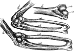
Bones of Rhonoceros' Foot
Homology of digits of odd toed mammals, showing gradual reduction in number and consolidation of bones.…
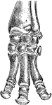
Bones of Horse's Foot
Homology of digits of odd toed mammals, showing gradual reduction in number and consolidation of bones.…
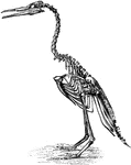
Restoration of Hesperornis regalis
"Hesperornis regalis, (a fossilized restoration) which stood about three feet high, had blunt teeth…
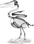
Restoration of Ichthyornis
"Ichthyornis, though the wings are well developed, with fused metacarpals, and the sternum is keeled,…
The Bones of the Right Wing of a Duck
"Fig 27. - Bones of the right wing of a duck, Clangula islandica, A, shoulder, omos; B, elbow, ancon;…
Mechanism of the Elbow-Joint
"Fig. 28. - Mechanism of elbow-joint. ..., where rc and uc show respectively the size, shape, and position…
The Wing Bones of a Young Grouse
"Fig. 29., from a young grouse (Centrocercus urophasianus, six months old), is designed to show the…
Red Shafted Woodpecker Ulna
"Fig. 31. - Ulna of Colaptes mexicanus, showing points of attachment of the secondaries. (Dr. R. W.…
Bird Skulls
"Fig. 114. -Skulls of Turdidae and Sylvicolidae, nat. size; after Shufeldt. A, Oroscoptes montanus;…
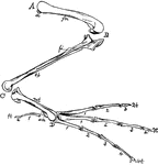
Bones of a Bird's Hind Limb
"Fig 34 - Bones of a bird's hind limb: from a duck, Clangula islandica. A, hip: B, knee: C, heel or…
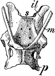
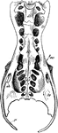
Pelvis of Spiny Anteater
An illustration of the pelvis of a spiny anteater. "m, marsupial bones; il, ilium; p, pubis; s, sacrum."…
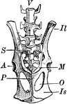
Kangaroo Pelvis
An illustration of a kangaroo pelvis. "M, marsupial bones, borne upon P, pubis; Il, ilium; Is, ischium;…
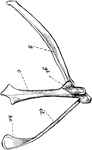
The Right Pectoral Arch of a Bird
"Right pectoral arch of a bird. s, scapula; c, coracoid; gl, glenoid, the cavity for head of humerus;…
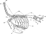
Axial Skeleton
"Fig 56 - Axial skeleton, minus the skull, of an owl, Asio wilsonianus, life size; from nature by Dr.…
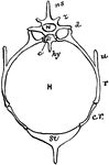
The Pelvis of a Heron
"Pelvis of a heron (ardea herodias), nat. size, viewed from below; from nature by Dr. R.W. Shufeldt,…
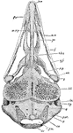
The Pelvis of a Young Grouse
"Pelvis of a young grouse, showing three distinct bones. Il,P, ilium, ischium, pubis. In front of former…
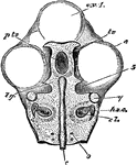
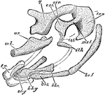
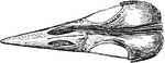
Skull of a Common Fowl
"Fig. 62 Skull of common fowl, enlarged. from nature by Dr. R.W. Shufeldt, U.S.A. The names of bones…
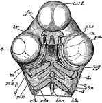
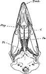
Duck Skull
"Fig 63 - Skull of a duck (Clangula islandica), nat. size; Dr. R.W. Shufeldt, U.S.A. a, premaxillary…
Goose Hyoid
"Fig 72 - Hyoid bones of a goose, nat. size; Dr. R.W. Shufeldt, U.S.A. a, cartilaginous end-piece of…
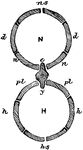
Axial Skeleton
"Ideal plan of the double-ringed body of a vertebrate. N, neural canal; H, haemal canal; the body separating…
The Axial Skeleton
"Fig 55. - Actual section of the body in the thoracic region of a bird. N, neural canal; H, haemal canal;…
Skull of a Chick
"Fig 64 - Skull of chick, fifth day of incubation, x 9 diameters. Seen from above, the membranous roof…
Skull of a Chick Below
"Skull of a chick, but seen from below. cv1, anterior cerebral vesicle; e, eye; m, mouth; pts, pituitary…
Chick Head
"Fig 66 - Head of a chick, second stage, after five days of incubation, section in profile; x6 diameters.…
House Martin Skull
"The post-oral arches of the house martin, at middle of period of incubation, lateral view, X14 diameters.…
The Skull of a Chick Stage Two
"Skull of chick, second stage, in profile, brain and membranes removed to show cartilaginous formations,…
The Skull of a Chick Stage Three
"Skull of chick, third stage, viewed from below, x6 & 2/3 diameters. pn, prenasal cartilage, running…
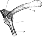
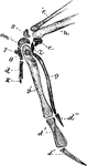
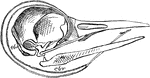
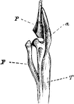
The Knee-joint of a Cormorant
"Phalacrocorax bicristatus. Cormorant. The knee-joint of a Cormorants. F, femur; P, patella; T, tibia;…
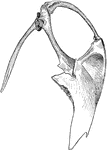
Cormorant Sternum and Shoulder
"Phalacrocorax bicristatus. Red-Faced Cormorant. Sternum and the shoulder from the skeleton of a Cormorant."…
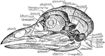
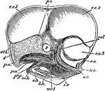
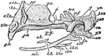
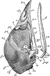
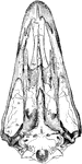
Cormorant Skull
"Phalacrocorax bicristatus. Red-Faced Cormorant. Skull showing sto, occipital style or nuchal bone;…
Ripe Chick's Skull
"Ripe chick's skull, longitudinal section, vied inside, x 3 diameters; after parker. In the mandible…
Ripe Chick's Skull Profile
"Ripe chick's skull, longitudinal section, vied inside, x 3 diameters; after Parker. px, premaxillary;…
The Skull of a Woodpecker
"Side view of a woodpecker's skull, showing the long slender basihyal (bh), bearing slight elements…
Top View of a Woodpecker Skull
"Top view of skull of Cloaptes, (flickers) showing thyrohyals running along the skull and into right…
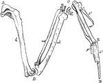
Leg Bones of a Grebe
"F. Fibula; T, tibia, with a, its cnemial process, and P, large patella, of a grebe." Elliot Coues,…
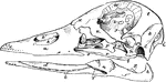
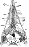
The Skull Structure of an Ostrich
"Dromaeognathous skull of ostrich, nat. size specimen no. 16,629, U.S. Nat Museum, by Dr. R. W. Shufeldt,…
The Skull of a Tinamou
"Dromaeognathous skull of a tinamou (Tinamus robustus); copies by Shufeldt from Huxley. Letters as before;…
Common Fowl Skull
"Schizognathous skull of common fowl, nat. size, from nature, by Dr. R.W. Shufeldt, U.S.A. Letters as…
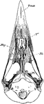
Mallard Duck Skull
"Desmognathous skull of mallard duck, Anas boscas, nat. size, from nature, by Dr. R.W. Shufeldt, U.S.A.…
Woodpecker Skull
"Saurognathous skull of a nesting Picus minor. x4 diameters, after Parker. Px premaxillary: dpx, its…
The Skull of a Raven
"Aegithognathous skull of raven, Corvus corax, nat. size, from nature, by Dr. R. W. Shufeldt, U.S.A.…
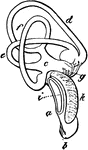
The Ear Bone of Fowl
"Mature stapes of fowl, about x4; after Parker. st, its foot, fitting fenestra ovalis; mst, main shaft,…
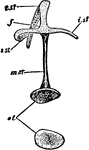
The Inner Ear of an Eagle
"Membranous labyrinth of Haliaetus albicilla (White-tailed Eagle), X2. a,b, cochlea; b, its saccular…
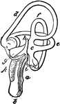
The Inner Ear of a White-tailed Eagle
"Membranous labyrinth of Haliaetus albicilla (White-tailed Eagle), X2. a,b, cochlea; b, its saccular…
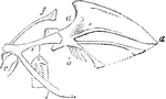
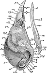
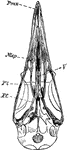
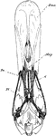
Willow Grouse Sternum
""Views of sternum and pectoral arch of the ptarmigan, Lagapus albus, reduced. Lateral view, with the…
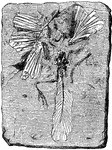
Archaeopteryx Lithographica
"Oldest known ornithological treatise, illustrating also the art of lithography in the Jurassic period,…
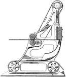
Toothpick Making Machine
This machine is used to make toothpicks. A toothpick is a small stick of wood, plastic, bamboo, metal…
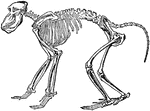
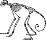
Chacma Baboon Skeleton
"The skeleton, more especially in the higher forms, is in the main similar to that of man, so that only…
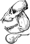
Side View of Howler Monkey
A side view of the howler monkey skull. The monkey have four sharp canines, long teeth on skull, on…

Adult Male Orangutan Skull Viewed from Side
An illustration of an adult male orangutan viewed from the side. The orbit part of the skull is more…
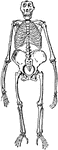
Side View of Skeleton of South American Spider Monkey
"In the other forms the number (vertebrae) varies between twenty and thirty three, the latter being…
Front View of Gorilla Skeleton
"The greatest absolute length of the fore—limb occurs in the gorilla and the orangutan. The humerus…
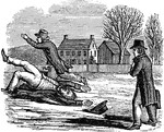
Slow but Sure
"See o'er this icy pathway pictured here, / Three sturdy travelers on foot appear; / One of them slips…
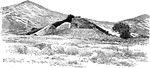
The Mound at Marathon
"Near the southern extremity of the plain of Marathon rises a conical mound, 30 feet high. it covers…
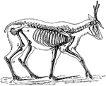
Skeleton of the Deer
"The bones in the extremities of this the fleetest of quadrupeds are inclined very obliquely towards…
Wing of Bird
"Shows how the bones of the arm (a), forearm (b), and hand (c), are twisted, and form a conical screw."—Pettigrew,…
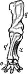
Anterior Extremity of Elephant
"Shows how the bones of the arm (q), forearm (q'x), and foot (o), are twisted to form an osseous screw."—Pettigrew,…
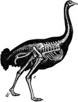
Skeleton of Ostrich
"Shows the powerful legs, small feet, and rudimentary wings of the bird; the obliquity at which the…
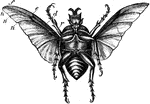
Large Beetle
"Under surface of large beetle, with deeply concave and comparatively small wings, shows that the nervures…