Clipart tagged: ‘duodenum’
Alimentary Canal
Diagram of the abdominal part of the alimentary canal (digestive system). Labels: C, the cardiac, and…
Alimentary Canal
Diagram of the abdominal part of the alimentary canal. Labels: C, the cardiac, and P, the pyloric end…
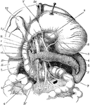
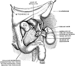
Biliary Apparatus
Portions of liver, duodenum, and pancreas, showing biliary and pancreatic ducts, head of pancreas turned…
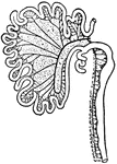
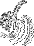
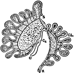
Intestinal Tract from Canis Vulpes
Intestinal tract of Canis vulpes. S, cut end of duodenum; C, caecum; R, cut end of rectum.
Intestinal Tract of Chauna Chavaria
cc. Colic caeca, d. Duodenum. g. Glandular patch, l.l. Meckel's tract, l.i. Hind-gut, p.v. Cut root…
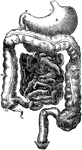
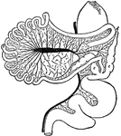
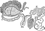
Digestive Organs
The stomach, pancreas, liver, and duodenum, with part of the rest of the small intestine and the mesentery;…
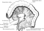
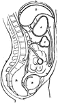
Duodenal Fossae and Folds
The duodenal fossae and folds. The transverse colon and mesocolon have been thrown up, and the mesentery…
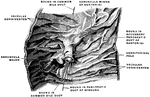
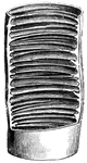
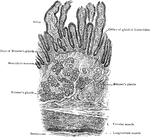
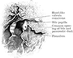
Longitudinal Section of Duodenum
Longitudinal section of duodenum; valvulae conniventes cut across, showing relation of these folds to…

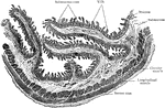
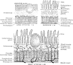
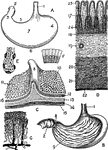
A Magnified Section of the Duodenum
A vertical section of the duodenum, highly magnified. Labels: 1, a fold-like villus; 2, epithelium,…
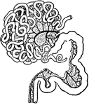
Intestinal Tract of Giraffe
S, cut end of duodenum; R, cut end of rectum; C, caecum; P.C.L., post-caecal loop; S.P., spiral loop;…
Intestinal Tract of a Gorilla
S, cut end of duodenum; R, cut end of rectum; C, vermiform appendix of caecum; X1, X2, X3, cut ends…
Structure of the Intestine
Diagram to show the structure of the small and large intestine and duodenum.
Transverse Section of Small Intestine
Transverse section of small intestine (lower part of duodenum), showing general arrangement of coats.
Intestinal Tract of Macropus Bennetti
S, cut end of duodenum; R, cut end of rectum; C, caecum; C2, accessory caecum; C.L., colic loop of hind-gut.
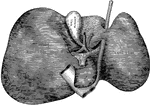
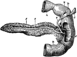
Pancreas
"The pancreas, partly cut away, so as to show the duct, which collects the pancreatic juice, and empties…
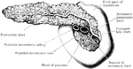
Posterior View of Pancreas
Posterior view of pancreas. Labels: 1, pancreas; 2, pancreatic duct; 6, opening of common duct, formed…
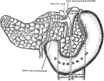
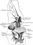
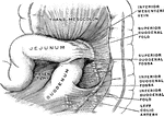
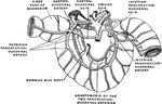
Pancreas and Duodenum from Behind
The pancreas and duodenum from behind, with the pancreatic duct exposed. The superior mesenteric vessels…
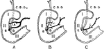
Variations in Termination of the Pancreatic and Bile Ducts
Showing the variations in the manner of termination of the pancreatic and bile ducts. A, Form in which…
The Peritoneum
Diagram of the peritoneum, a serous membrane covering all the contents of the abdominal cavity. Labels:…
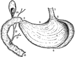
Vertical and Longitudinal Section of Stomach, Gall-Bladder, and Duodenum
Vertical and longitudinal section of stomach, gall-bladder, and duodenum. Labels: 1, esophagus; 2, cardiac…
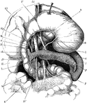
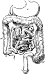
The Stomach, Pancreas, Liver, and Duodenum
The stomach, pancreas, liver, and duodenum, with part of the rest of the small intestine and the mesentery;…
Views of the Stomach
Views of the stomach. Labels: A. stomach (human). B. Same, anterior wall removed. C. Portion of stomach,…