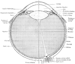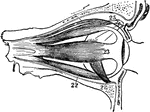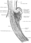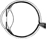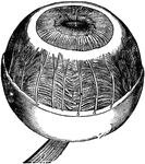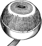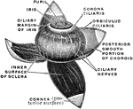
The middle or vascular coat of the eyeball exposed from without. Left eye, seen obliquely from above and before.

Vascular Coat of the Eye

The middle or vascular coat of the eyeball exposed from without. Left eye, seen obliquely from above…
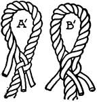
"For making an eye splice, the end of the rope is unlaid and the strands are bent upon the body of the rope so as to form an 'eye' of the size desired."—Finley, 1917

Eye-splice

"For making an eye splice, the end of the rope is unlaid and the strands are bent upon the body of the…
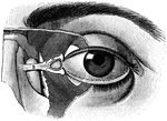
"The Relative Position of the Lachrymal Apparatus, the Eyeball, and the Eyelids. <I>A</em>, lachrymal canals, with the minute orifices represented as two black dots to the right; <em>B</em>, tendon attached to a muscle which surrounds the circumference of the orbit and eyelids; under <em>B</em> is seen the lachrymal sac. The minute openings of the Meibomian glands are seen on the free margins of the eyelids. Below <em>A</em> is seen a small conical elevation, with black dots (the lachrymal papilla, or caruncle)." &mdash; Blaisedell, 1904

Eyeball

"The Relative Position of the Lachrymal Apparatus, the Eyeball, and the Eyelids. A, lachrymal canals,…
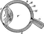
"The most essential parts of human vision are contained in the eyeball, a nearly spherical body, about an inch in diameter, and capable of being turned considerably in its socket by the action of various muscles." — Avery, 1895

Eyeball

"The most essential parts of human vision are contained in the eyeball, a nearly spherical body, about…
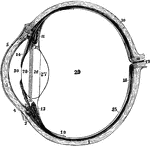
The left eyeball in horizontal section from before back. Labels: 1, sclerotic; 2, junction of sclerotic and cornea; 3, cornea; 4, 5, conjunctival membrane; 7, ciliary muscle; 10, choroid; 11, 13, ciliary processes; 14, iris; 15, retina; 16, optic nerve; 17, artery entering retina; 18, fovea centralis; 19, region where sensory part of retina ends; 26, 27, 28, are placed on the lens; 28 , suspensory ligament placed around lens; 29, vitreous humor; 30, aqueous humor in anterior chamber.

Left Eyeball in Horizontal Section

The left eyeball in horizontal section from before back. Labels: 1, sclerotic; 2, junction of sclerotic…
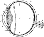
Section of the eyeball. Labels: Con, conjunctiva; C, cornea; A, aqueous humor; I, iris; L, crystalline lens; V, vitreous humor; Sc, sclerotic coat; Ch, choroid coat; R, retina; O, optic nerve, Cm, ciliary muscle.

Section of the Eyeball

Section of the eyeball. Labels: Con, conjunctiva; C, cornea; A, aqueous humor; I, iris; L, crystalline…
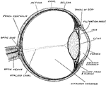
Horizontal section of the eyeball, showing the suspensory ligament of the lens, the aqueous and vitreous chambers, entrance of the optic nerve, and the fovea centralis.

Eyeball

Horizontal section of the eyeball, showing the suspensory ligament of the lens, the aqueous and vitreous…
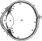
Diagram of a horizontal section through left eyeball and optic nerve.

Eyeball and Optic Nerve

Diagram of a horizontal section through left eyeball and optic nerve.

The left eyeball in horizontal section from before back. Labels: 1, sclerotic; 2, junction of sclerotic and cornea; 3, cornea; 4, 5, conjunctiva; 7, ciliary muscle; 10, choroid; 11, 13, ciliary processes; 14, iris; 15, retina; 16, optic nerve; 17, artery entering retina in optic nerve; 18, fovea centralis; 19, 20, region where sensory part of retina ends; 22, suspensory ligament; 24, the anterior part of the hyaloid membrane; 26, the lends; 29, vitreous humor; 30, aqueous humor.

The Eyeball in Horizontal Section

The left eyeball in horizontal section from before back. Labels: 1, sclerotic; 2, junction of sclerotic…
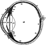
The left eyeball in horizontal section from before back. Labels: 1, sclerotic; 2, junction of sclerotic and cornea; 3, cornea; 4,5, conjunctiva; 6, posterior elastic layer of cornea; 7, ciliary muscle; 10, choroid; 11,13, ciliary processes; 14, iris; 15, retina; 16, optic nerve; 17, artery entering retina in optic nerve; 18, fovea centralis; 19, region where sensory part of retina ends; 22, suspensory ligament; 23 is placed in the canal of Petit, and the line from 25 points to it; 24, the anterior part of the hyaloid membrane; 26, 27, 28, are placed on the lends; 28 points to the line of attachment around it of the suspensory ligament; 29, vitreous humor; 30, anterior chamber of aqueous humor; 31, posterior chamber of aqueous humor.

Section of Left Eyeball

The left eyeball in horizontal section from before back. Labels: 1, sclerotic; 2, junction of sclerotic…

The eyeball with its muscles attached. The upper muscle not attached to the ball belongs to the upper eyelid. The muscle in front is cut away.

The Eyeball with its Muscle

The eyeball with its muscles attached. The upper muscle not attached to the ball belongs to the upper…
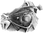
Front and side view of the eyeball.

Front and Side View of the Eyeball

Front and side view of the eyeball.

"<em>A</em>, attachment of tendon connected with the four recti muscles; <em>B</em>, external rectus, divided and turned downward, to expose the internal rectus; <em>C</em>, inferior rectus; <em>D</em>, internal rectus; <em>E</em>, superior rectus; <em>F</em>, superior oblique; <em>H</em>, pulley and reflected portion of the superior oblique; <em>K</em>, inferior oblique; <em>L</em> and <em>M</em>, portions of the muscle which raises the upper eyelid; to the right of <em>D</em> and to the left on the same line are seen cut ends of the optic nerve." &mdash; Blaisedell, 1904

Muscles of the eyeball

"A, attachment of tendon connected with the four recti muscles; B, external rectus,…

Muscles of the eyeball.

Muscles of the Eyeball

Muscles of the eyeball.

Section through ciliary region of eyeball.

Section Through Eyeball

Section through ciliary region of eyeball.

Vertical section of the eyeball.

Vertical Section of the Eyeball

Vertical section of the eyeball.

The tarsi and their ligaments.

Eyelid

The tarsi and their ligaments.

"Showing how the upper eyelid may be everted with a pencil or penholder." — Blaisedell, 1904

Everted eyelid

"Showing how the upper eyelid may be everted with a pencil or penholder." — Blaisedell, 1904

Vertical section through the upper eyelid. Labels: a, Skin; b, Orbicularis palpebrarum; b', Marginal fasciculus of Orbicularis (ciliary bundle); c, Levator palpebrae; d, Conjuctiva; e, Tarsal plate; f, Meibomian gland; g, Sebaceous gland; h, Eyelashes; i, Small hairs of skin; j, Sweat glands; k, Accessory lacrimal glands.

Vertical Section Through Eyelid

Vertical section through the upper eyelid. Labels: a, Skin; b, Orbicularis palpebrarum; b', Marginal…

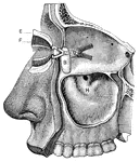
<em>A</em> and <em>C</em>, lachrymal canals; <em>B</em>, lachrymal sac; <em>D</em>, small muscle which serves to compress the lachrymal sac; <em>F</em>, glands upon the inner surface of the eyelids, with ducts opening upon the free margins of the eyelids; <em>H</em>, great openings, or antrum, of the upper jawbone. The oil glands of the nose are plainly shown." &mdash; Blaisedell, 1904

Eyelids and lachrymal apparatus

A and C, lachrymal canals; B, lachrymal sac; D, small muscle which…

"The eye is moved about by six muscles. The back ends of these muscles are attached to the eye sockets. The front ends are attached to the ball of the eye." — Ritchie, 1918

Muscles of the Eyes

"The eye is moved about by six muscles. The back ends of these muscles are attached to the eye sockets.…

"A representation of the manner in which the image is formed in the eye, when the cornea or crystalline lens is flattened. The perfect image would be produced at m m, behind the retina, and, of course, beyond the point necessary to secure perfect vision." —Wells, 1857

Flattened Eye

"A representation of the manner in which the image is formed in the eye, when the cornea or crystalline…

Diagram to illustrate the mechanism of accommodation (focusing); on the right half of the figure for a near, on the left for a distant, object. Labels: rf, ciliary muscle; ch, ciliary process of choroid; sl, suspensory ligament; i, iris.

Focusing of the Eye

Diagram to illustrate the mechanism of accommodation (focusing); on the right half of the figure for…
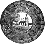
Suppose a person was looking at a church with a tree standing at its side, he would have in each eye an actual inverted panorama of the objects, as seen in this image. The mind then reads this image as right-side up.

Formation of an Image on the Eyeball

Suppose a person was looking at a church with a tree standing at its side, he would have in each eye…

"Galeodes sp., one of the solifugae. Dorsal view. I to VI, Bases of the prosomatic appendages. o, Eyes. a, Lateral region of the cephalic plate to which the first pair of appendages are articulated. b, Cephalic plate with median eye. c, Dorsal element of somites bearing third and fourth pairs of appendages. d, Second plate of the prosoma with fifth pair of appendages. e, Third or hindermost plate of the prosoma beneath which the sixth pair of legs is articulated. 1, 2, 9, 10, First, second, ninth and tenth somites of the opisthosoma. an, Anus." &mdash; The Encyclopedia Britannica, 1910

Galeodes

"Galeodes sp., one of the solifugae. Dorsal view. I to VI, Bases of the prosomatic appendages. o, Eyes.…

"Garypus litoralis, one of the Pseudoscorpiones. Dorsal view. I to VI, The prosomatic appendages. o, Eyes. Prae-gen, Prae-genital somite. 1, Tergite of the genital or first opisthosomatic somite. 10, Tergite of the tenth somite of the opisthosoma. 11, The evanescent eleventh somite of the opisthosoma. an, Anus." &mdash; The Encyclopedia Britannica, 1910

Garypus Litoralis

"Garypus litoralis, one of the Pseudoscorpiones. Dorsal view. I to VI, The prosomatic appendages. o,…

"Garypus litoralis, one of the Pseudoscopions. Lateral view. I to VI, Basal segments of the six prosomatic appendages. o, Eyes. prae-gen, Tergite of the prae-genital somite. I, Genital or first opisthosomatic. 2, 3, 10, The second, third and tenth somites of the opisthosoma. 11, The minute eleventh somite. an, The anus." &mdash; The Encyclopedia Britannica, 1910

Garypus Litoralis

"Garypus litoralis, one of the Pseudoscopions. Lateral view. I to VI, Basal segments of the six prosomatic…
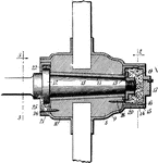
The central part of a car wheel (or fan or propeller etc) through which the shaft or axle passes

Wheel Hub

The central part of a car wheel (or fan or propeller etc) through which the shaft or axle passes
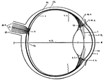
This diagram shows a side view of the right eye of man. a.c., central artery; a.h., aqueous humor; b., blind spot, the entrance of the optic nerve; c, conjunctiva; ch., choroid layer of the eye-ball; c.l., crystalline lens; c.m.c., circular fibers of the ciliary muscles; c.m.r., radial fibers of the ciliary muscles; co., cornea, the transparent portion of the sclerotic; c.p., ciliary process; c.s., canal of Schlemm, a lymphatic vessel; fo., fovea centralis, the point of clearest vision; o.n., optic nerve; o.s., ora serrata, the anterior wavy margin of the visual portion of the retina; r, the retinal layer; sc., sclerotic layer; sh., sheath of theoptic nerve; v.h., vitreous humor.

Human Eye

This diagram shows a side view of the right eye of man. a.c., central artery; a.h., aqueous humor; b.,…

"...where we suppose that the object a, is brought within an inch or two of the eye, and that the rays proceeding from it enter the pupil so obliquely as not to be refracted b the lens, so as to form a distinct image." -Comstock 1850

Indistinct Vision

"...where we suppose that the object a, is brought within an inch or two of the eye, and that the rays…

"The actual position of the vertical object a, as painted on the retina, is therefore such as is represented by the figure." -Comstock 1850

Inversion of Objects by the Eye

"The actual position of the vertical object a, as painted on the retina, is therefore such as is represented…
The iris, the ciliary muscle, and the coroidea membrane.

Iris, Ciliary Muscle and Coroidea Membrane

The iris, the ciliary muscle, and the coroidea membrane.

Iris, ciliary muscle, and choroid of the eye.

Iris, Ciliary Muscle, and Choroid

Iris, ciliary muscle, and choroid of the eye.

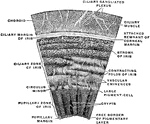
Section of the iris. Anterior surface magnified.

Section of the Iris

Section of the iris. Anterior surface magnified.

A cartoon drawing of Thomas Jefferson.

A Caricature of Jefferson

A cartoon drawing of Thomas Jefferson.

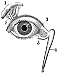
View of the lachrymal gland and nasal duct. Labels: 1, The lachrymal gland. 2, Ducts leading from the lachrymal gland to the upper eyelid. 3, The "tear points". 4, The nasal sac. 5, The termination of the nasal duct.

View of the Lachrymal Gland and Nasal Duct

View of the lachrymal gland and nasal duct. Labels: 1, The lachrymal gland. 2, Ducts leading from the…

Meridional section through the lens of a rabbit. Labels: 1, Lens capsule; 2, epithelium of lens; 3, transition of the epithelium into the fibers; 4, lens fibers.

Lens of a Rabbit

Meridional section through the lens of a rabbit. Labels: 1, Lens capsule; 2, epithelium of lens; 3,…
Flexible crystalline lens of the eye.

Lens of the Eye

Flexible crystalline lens of the eye.

Laminated structure of the crystalline lens. The laminae are split up after hardening in alcohol. Labels: 1, the denser central part or nucleus; 2, the successive external layers.

Crystalline Lens

Laminated structure of the crystalline lens. The laminae are split up after hardening in alcohol. Labels:…

"Diagram showing how the lens changes its form for near and far sight." &mdash; Tracy, 1888

Eye lens

"Diagram showing how the lens changes its form for near and far sight." — Tracy, 1888

Section through the equator of the lens. Showing gradual transition of the epithelium into lens fibers.

Section Through Lens

Section through the equator of the lens. Showing gradual transition of the epithelium into lens fibers.

A convex lens, bends the ray of light which pass through it, so that they meet at a point called a focus. The crystalline lens converges the rays of light which enter the eye, and brings them to a focus on the retina. The healthy lens has a power of changing its convexity so as to adapt itself to near and to distant objects.

The Use of the Crystalline Lens

A convex lens, bends the ray of light which pass through it, so that they meet at a point called a focus.…
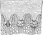
"Section through a portion of the lateral eye of Limulus, showing three ommatidia&mdash;A, B and C. hyp, The epidermic cell-layer (so-called hypodermis), the cells of which increase in volume below each lens, l, and become nerve-end cells or retinula-cells, rl; in A, the letters rh point to a rhabdomere secreted by the cell rl; c, the peculiar central spherical cell; n, nerve fibers; mes, mesoblastic skeletal tissue; ch, chitinous cuticle." &mdash; The Encyclopedia Britannica, 1910

Limulus

"Section through a portion of the lateral eye of Limulus, showing three ommatidia—A, B and C.…
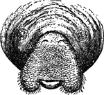
"Front view of head of American Manatee, showing the eyes, nostrils, and mouth with the lobes of the upper lip divaricated." &mdash;The Encyclopedia Britannica, 1903

Manatee Face

"Front view of head of American Manatee, showing the eyes, nostrils, and mouth with the lobes of the…
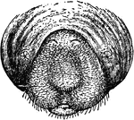
"Front view of head of American Manatee, showing the eyes, nostrils, and mouth with the lip contracted." &mdash;The Encyclopedia Britannica, 1903

Manatee Face

"Front view of head of American Manatee, showing the eyes, nostrils, and mouth with the lip contracted."…

"The horizontal section in the direction of the axis of the telescope. The eye-piece ab consists of two planoconvex lenses a, b, of nearly the same focal length, and with the two convex sides facing each other. They are placed at a distance apart less than the focal length of a, so that the wires of the micrometer, which must be distinctly seen are beyond b. The eye-piece slides into the tube cd, which screws into the brass ring ef, through two openings in which the oblong frame, containing the micrometer slides, passes." &mdash;The Encyclopedia Britannica, 1903

Micrometer

"The horizontal section in the direction of the axis of the telescope. The eye-piece ab consists of…

"The vertical section in the direction of the axis of the telescope. The eye-piece ab consists of two planoconvex lenses a, b, of nearly the same focal length, and with the two convex sides facing each other. They are placed at a distance apart less than the focal length of a, so that the wires of the micrometer, which must be distinctly seen are beyond b. The eye-piece slides into the tube cd, which screws into the brass ring ef, through two openings in which the oblong frame, containing the micrometer slides, passes." &mdash;The Encyclopedia Britannica, 1903

Micrometer

"The vertical section in the direction of the axis of the telescope. The eye-piece ab consists of two…

"The original Merz micrometer of the Cape Observatory, made on Fraunhofer's model. S is the head of the micrometer screw proper, s that of the screw moving the slide to which the so-called "fixed web" is attached, s' that of a screw which moves the eye-piece E. C is the clamp and M the slow motion in position angle, L, L are tubes attached to a larger tube N; the latter fits loosely on a strong hollow cylinder which terminates in the screw V. By this screw the whole apparatus is attached to the telescope." &mdash;The Encyclopedia Britannica, 1903

Micrometer

"The original Merz micrometer of the Cape Observatory, made on Fraunhofer's model. S is the head of…

"A microscope consists of a lens or a combination of lenses used to observe small objects, often so minute as to be invisible to the unaided eye." &mdash; Avery, 1895

Microscope

"A microscope consists of a lens or a combination of lenses used to observe small objects, often so…

"Let a be the distance at which an object can be see distinctly, and b, the distance at which the same object is seen through the lens, and supose tthe distance of a from the eye, be twice that of b. Then because the object is at half the distance that it was before, it will appear twice as large...." -Comstock 1850

Single Microscope

"Let a be the distance at which an object can be see distinctly, and b, the distance at which the same…
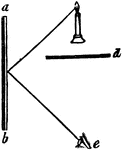
"Suppose the mirror, a b, to be placed on the side of a room, and a lamp to be set in antoher room, but so situated as that its light would shine upon the glass. The lamp itself could not be seen by the eye placed at e, because the partition d is between them; but its image would be visible at e, beacuse the angle of the incident ray, coming from the light, and that of the reflected ray which reaches the eye, are equal." -Comstock 1850

Plane Mirror

"Suppose the mirror, a b, to be placed on the side of a room, and a lamp to be set in antoher room,…

"A representation of the manner in which the image is formed in the eye of a near-sighted person. The curvature of the cornea, s s, and of the crystalline lens, c c, is so great that the image is formed at m m in advance of the retina." —Wells, 1857

Near-sighted

"A representation of the manner in which the image is formed in the eye of a near-sighted person. The…
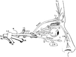
Scheme of the distribution of the ophthalmic nerve. Labels: Vs, trigeminal nerve, afferent root; Mo, efferent root; G.G, gasserian ganglion; M, meningeal branch; I.C, branch to internal carotid artery; Oph, opthalmic nerve; S.M, superior maxillary nerve; I.M, inferior maxillary nerve; III, communication to oculomotor nerve; IV, to trochlear nerve; L, branches to upper eyelid; L.G, long root to lenticular ganglion; Sy, root from sympathetic (on carotid artery); III, short root from motor oculi nerve; C, short ciliary branches; L.C, long ciliary nerves; I.T, infra-trochlear nerve; E.N, external nasal nerve; I.N, internal nasal nerve; O, orbital branch of superior maxillary nerve; L.Gl, lachrymal gland; C, conjuctival branch; L, branch to eyelids and face.

Ophthalmic Nerve

Scheme of the distribution of the ophthalmic nerve. Labels: Vs, trigeminal nerve, afferent root; Mo,…

The terminal portion of the optic nerve and its entrance into the eyeball, in horizontal section.

Optic Nerve

The terminal portion of the optic nerve and its entrance into the eyeball, in horizontal section.

Diagram of the central connection of the optic nerve and optic tract.

Optic Nerve

Diagram of the central connection of the optic nerve and optic tract.

Longitudinal section of the primary optic vesicle in the chick. Labels: A, from an embryo of 65 hours; B, a few hours later; C, of the fourth day; c, the corneous layer or epidermis, presenting in A the open depression from the lends , which is closed in B and C; l, the lens follicle and lens; pr, the primary optic vesicle; in A and B, the pedicle is shown; in C, the section being to the side of the pedicle, the latter is not shown; v, the secondary ocular vesicle and vitreous humour.

Development of the Primary Optic Vesicle in a Chick

Longitudinal section of the primary optic vesicle in the chick. Labels: A, from an embryo of 65 hours;…

An illustration of the position and the size of the image viewed by the eye. The eye approximates the the height of the image. The image's height is the hypotenuse of the triangle, creating an angle with the eye. This image is then reflected upside down in the eye.

Position and Size of Image for Eye Optics

An illustration of the position and the size of the image viewed by the eye. The eye approximates the…

"If y be the object the image appears to a normal eye situated behind the system L with passive accommodation at a very great distance under the angle w'. Since H' P= F O, = y, from the focal length of the simple microscope is the image side focal length." —Encyclopaedia Britannica, 1910

Optical Position and Size of Image Through Magnifying Glasses

"If y be the object the image appears to a normal eye situated behind the system L with passive accommodation…

"The intersection of the principal rays in this case lies in the middle of the entrance pupil or of the exit pupil. By head and eye motion the various parts of the whole field can be viewed one after another." —Encyclopaedia Britannica, 1910

Optical Position of Diaphragms using Lens

"The intersection of the principal rays in this case lies in the middle of the entrance pupil or of…

Diagram to show the action of the orbital muscle. The arrows show the direction of the action of each muscle.

Diagram to Show the Action of the Orbital Muscle

Diagram to show the action of the orbital muscle. The arrows show the direction of the action of each…