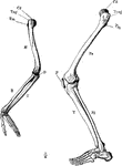
Arm and Leg Skeleton
The skeleton of the arm and leg. Labels: H, the humerus; Cd, its articular head which fits into the…

Leg of Bear
This illustration shows the plantigrade leg of a bear. Plantigrade means that the animal walks flat…
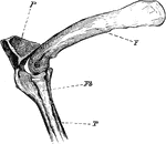
The Knee-joint of a Cormorant
"Phalacrocorax bicristatus. Cormorant. The knee-joint of a Cormorants. F, femur; P, patella; T, tibia;…
Femur
Section of the femur. 1: External view; 2: Cellular portion at end; 3: Hollow in middle; 4: Thick shell…
Femur
Posterior view of the femur (bone of the leg). Labels: 1, depression for round ligament; 2, the head;…
Femur
The thigh bone cut through the middle. Labels: b, hard bone; h and d, spongy bone; ma, marrow.

Ossification of the Femur and the Condition of Coxa Vara
Illustrating the ossification of the upper end of the femur and the condition of coxa vara. Labels:…
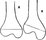
Femur in Knock-Knee
A, Normal Femur. B, Femur in an advanced state of knock-knee, showing the enlargement of the internal…

Calcar Femorale and its Relationship to Fractures of the Femur
The calcar femorale and its relationship to impacted fractures of the neck of the femur.

Displacement of the Femur
Diagram showing the line used by Nelaton to test upward displacement of the femur, and another which…

Femoral Spur of the Femur
In the midst of the cancellous tissue the femoral spur, which commences at the point where the neck…
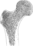
Frontal Section Through Upper End of Femur
Frontal section through upper end of femur, showing arrangement of pressure and tension lamellae.
Human Femur Bone
The Femur (upper leg bone) is the longest, largest, and strongest bone in the skeleton. Labels: b, rounded…
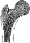
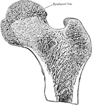
Longitudinal Section of the Femur
The longitudinal section of the extremity of the femur, exhibiting the arrangement of the spongy substance.…
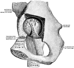
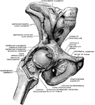
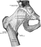
Hip Joint from Mesal Side
Right hip joint, from the mesal side. The bottom of the acetabulum had been chiseled away sufficiently…
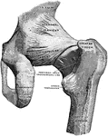
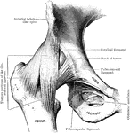
Back View of Hip Joint
Right hip joint, from behind. The joint capsule, except for strengthening ligaments, has been removed.
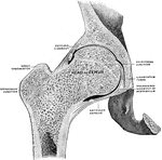
Frontal Section of Hip Joint
Right hip joint. Posterior half, viewed from in front. The joint surfaces have been somewhat pulled…
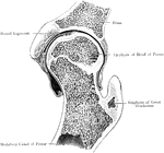
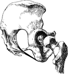
Frontal Section Through Hip Joint
A frontal section through the left hip joint of a boy. Front view.
Human Leg (Front View)
This illustration shows a front view of a human leg. P. Pelvis, FE. Femur, TI. Tibia, FI. Fibula, TA.…

Human Leg (Front View), and Comparative Diagrams showing Modifications of the Leg
This illustration shows a human leg (front view), and comparative diagrams showing modifications of…

Human Leg (Side View)
This illustration shows a side view of a human leg. P. Pelvis, FE. Femur, TI. Tibia, FI. Fibula, TA.…
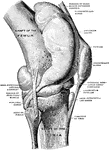
Knee Joint from Lateral Surface
Right knee joint from the lateral surface. The joint cavity and several bursae have been injected with…
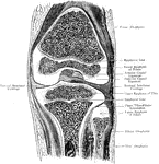
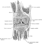
Frontal Section Through Knee Joint
A frontal section through the right knee joint of a boy. Seen from behind.
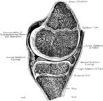
Sagittal Section Through Knee Joint
A sagittal section through the right knee joint of a boy. Seen from the outer side.
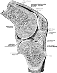
Sagittal Section Through Knee Joint
Right knee joint. Sagittal section through the external condyle of the femur. Mesal half of section,…
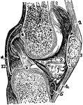
Vertical Section of the Knee Joint
A vertical section of the knee joint. Labels: femur; 3, patella; 2, 4, ligaments of the patella; 5,…
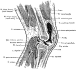
Sagittal Section Through Knee
Sagittal section of the right knee, viewed from the outer side. The joint cavity proper lies to each…

Leg Bones
"Bones of the leg. a, femur; b, tibia; c, fibula; d, tarsal bones; e, metatarsal bones; f, phalanges;…
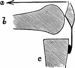
Mechanism of Fracture of the Patella by Muscular Action
Diagram to show mechanism of fracture of the patella by muscular action. a, Line of action of quadriceps…
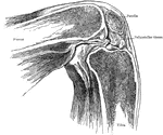
Position of Patella in Relation to Condyles of Femur with Knee Flexed
Showing position of patella in relation to condyles of femur with knee flexed at a right angle.

Position of Patella in Relation to Condyles of Femur with Knee Partially Flexed
Showing position of patella in relation to condyles of femur with knee partially flexed.
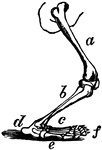
Polar Bear Leg
The polar bear, Plantigrada, is part of the subdivision Carnivora, which includes other carnivorous…
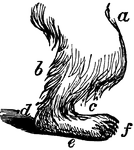
Polar Bear Leg
The anatomy of a polar bear's leg. a, femur (thigh); b, tibia (leg); c, tarsus and metatarsus (foot);…

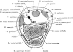
Cross Section Through Thigh Five Inches Above Knee Joint
Section through the lower third of the thigh, five inches above knee joint.


