"The head is represented as having been thrown back, and the tongue drawn forward. <em>A</em>, <em>B</em> and <em>C</em>, cainine; <em>D, E</em> bicuspids; <em>F, H, K,</em> molars; <em>M</em> anterior pillar of the fauces; <em>N</em>, tonsil; <em>L</em> uvula; <em>O</em>, upper part of the pharynx; <em>P</em>, tongue drawn forward; <em>R</em> linear ridge, or raphe</em>." &mdash; Blaisedell, 1904

Back view of the adult mouth

"The head is represented as having been thrown back, and the tongue drawn forward. A, B

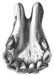
"Skull and teeth of Bennett's Kangaroo (Macropus bennettii). i1, i2, i3, first second and third upper incisors; pm, second or posterior premolar (the first having been already shed); m1, m2, m3, m4, the four true molars. The last, not fully developed, is nearly concealed by the ascending ramus of the jaw." &mdash;The Encyclopedia Britannica, 1910

Bennett's Kangaroo

"Skull and teeth of Bennett's Kangaroo (Macropus bennettii). i1, i2, i3, first second and third upper…
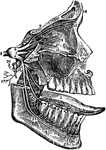
The fifth cranial nerve. Labels: 1, orbit; 2, antrum of Highmore; 3, tongue; 4, lower jaw; 5, Casserian ganglion; 6, first branch of fifth; 7, second; 8, third; 9, frontal branch; 10, lacrymal branch; 11, nasal branch; 122, internal nasal nerve; 13, external nasal; 14, external and internal frontal nerve; 15, infra-orbital; 16, posterior dental branches; 17, middle dental; 18, anterior dental; 19, terminating branches infra-orbital nerve; 20, orbital branch; 21, ptyregoid nerve; 22, five anterior branches; 23, lingual branch; 24, inferior dental; 25, mental branches; 26, superficial temporal nerve; 27, auricular branches; 28, mylo-hyoid.

Fifth Cranial Nerve

The fifth cranial nerve. Labels: 1, orbit; 2, antrum of Highmore; 3, tongue; 4, lower jaw; 5, Casserian…
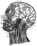
Muscles of the face, jaw and neck. 1, longus colli; 2, rapezius; 3, sterno-hyoid; 4, sterno-mastoid; 5, erico-thyroid; 6, trapezius; 7, constrictor of pharynx; 8, sterno-mastoid; 9, digastric; 10, attrahens aurem; 11, mylo-hyoid; 12, masseter; 13, depressor oof lower lip; 15, orbicularis oris; 17, levator of upper lip; 19, levator of angle of mouth; 21, orbicularis palpebrarum.

Facial Muscles

Muscles of the face, jaw and neck. 1, longus colli; 2, rapezius; 3, sterno-hyoid; 4, sterno-mastoid;…

"Fig. 62 Skull of common fowl, enlarged. from nature by Dr. R.W. Shufeldt, U.S.A. The names of bones and some other parts are printed, requiring no explanation; but observe the following points: The distinction of none of the bones composing the brain-case (the upper back expanded part) can be found in a mature skull. The brain is contained between the occipital, sphenoidals, squamosals, parietals and part of frontal; the ethmoidals belong to the same group of cranial bones proper. All other bones, excepting the three otic ear-bones, are bones of the face and jaws. The lower jaw, of five bones, is drawn detached; it articulates by the black surface marked articular with the prominence just above- the quadratic bone. Observe that from this quadrate a series of bones quadrato-jugal, jugal, maxillary-makes a slender rod running to the premaxillary; this is the zygoma, or jugal bar. Observe from the quadrate also another series, composed of pterygoid and palatine bones, to the premaxillary; this is the pterygo-palatine bar; it slides along a median fixed axis of the skull, the rostrum, which bears the loose vomer at its end. The under mandible, quadrate, pterygoid, and vomer are the only movable bones of this skull. But when the quadrate rocks back and forth, as it does by its upper joint, its lower end pulls and pushes upon the upper mandible, by means of the jugal and pterygo-palatine bars, setting the whole scaffolding of the upper jaw in motion. This motion hinges upon the elasticity of the bones of the forehead, at the thin place just where the reference-lines from the words "lacrymal" and "mesethmoid" cross each other. The dark oval space behind the quadrate is the external orifice of the ear; the parts in it to which the three reference-lines go are diagrammatic, not actual representations; thus, the quadrate articulates with a large pro-otic as well as with the squamosal. The great excavation at the middle of the figure, containing the cirlet of the unshaded bones, is the left orbital cavity, orbit, or socket of the eye. The mesethmoid includes most of the background of this cavity, shaded diagonally. The upper one of the two processes of bone extending into it from behind is post-frontal or sphenotic process; the under one (just over the quadrate) is the squamosal process. A bone not shown, the presphenoid, lies just in front of the oval black space over the end of basisphenoid. This black oval is the optic foramen, through which the nerve of sight passes from the brain-cavity to the eye. The black dot a little behind the optic foramen is the orifice of exit of a part of the trifacial nerve. The black mark under the letters "on" of the word "frontal" is the olfactory foramen, where the nerve of smell emerges from the brain-box to go to the nose. The nasal cavity is the black space behind nasal and covered by that bone, and in the oval blank before it. The parts of the beak covered by horn are only premaxillary, nasal, and dentary. The condyle articulates with the first cervical vertebra; just above it, not shown, is the foramen magnum, or great hole through which the spinal medulla, or main nervous cord, passes from the spinal column. The basioccipital is hidden, excepting its condyle; so is much of the basisphenoid. The prolongation forward of the basisphenoid, marked "rostrum," and bearing the vomer at its end, is the parasphenoid, as far as its thickened under border is concerned. Between the fore end of the pterygoid and the basisphenoidal rostrum, is the site of the basipterygoid process, by which the bones concerned articulate by smooth facets; further forward, the palatines ride freely upon the parasphenoidal rostrum. In any passerine bird , the vomer would be thick in front, and forked behind, riding like the palatine upon the rostrum. The palatine seems to run into the maxillary in this view; but it continues on to premaxillary. The maxillo-palatine is an important bone which cannot be seen in the figure because it extends horizontally into the paper from the maxillary about where the reference life "maxillary" goes to that bone. The general line from the condyle to the end of the vomer is the cranial axis, basis cranii, or base of the cranium. This skull is widest across the post-frontal; next most so across the bulge of the jugal bar." Elliot Coues, 1884

Skull of a Common Fowl

"Fig. 62 Skull of common fowl, enlarged. from nature by Dr. R.W. Shufeldt, U.S.A. The names of bones…

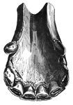
"Skull and teeth of Gray's Rat Kangaroo (Bellongia grayii). c, upper canine tooth. i1, i2, i3, first, second, and third upper incisors; pm, second or posterior premolar (the first having been already shed); m1, m2, m3, m4, the four true molars." &mdash;The Encyclopedia Britannica, 1910

Gray's Rat Kangaroo

"Skull and teeth of Gray's Rat Kangaroo (Bellongia grayii). c, upper canine tooth. i1, i2, i3, first,…
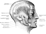
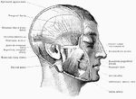
A deep view of the muscles of mastication. The zygoma and masseter muscle are removed.

Head Showing Deep Mastication Muscles

A deep view of the muscles of mastication. The zygoma and masseter muscle are removed.
Superficial view of the muscles of mastication.

Head Showing Mastication Muscles

Superficial view of the muscles of mastication.

Bones of the head.

Bones of the Head

Bones of the head.

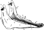
Inferior maxilla of a horse-anterolateral view. Labels: a, body; b, b', rami; c, neck; d, mental foramen; e, buccinator; e', masseter surface; f, f', inner surface of ramus; g, molor alveoli; g', anterior border; g', bar, bounding diastema; h, h', h', posterior border. The angle lies between h' and h'; i, condyles; km coronoid processes; l, maxillary space; m, inferior dental foramen; n, sigmoid notch.

Inferior Maxilla of a Horse

Inferior maxilla of a horse-anterolateral view. Labels: a, body; b, b', rami; c, neck; d, mental foramen;…
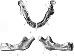
The lower jaw at birth. A, As seen from above. B, As seen from outer side. C, As seen from inner side. Labels: a, mental foramen; b, inferior dental canal; c, lingula; d, sockets fro the dental sacs.

Jaw at Birth

The lower jaw at birth. A, As seen from above. B, As seen from outer side. C, As seen from inner side.…
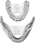
Inferior surface of upper jaw at about birth.

Inferior Surface of Upper Jaw at Birth

Inferior surface of upper jaw at about birth.

Mesial surface of upper jaw at about birth.

Mesial Surface of Upper Jaw at Birth

Mesial surface of upper jaw at about birth.

Horizontal section through both the upper and lower jaws to show the roots of the teeth. The sections were carried through the bones a short distance from the edge of their alveolar borders. The upper figure shows the upper teeth, the lower figure the lower teeth. Note the flattened roots of the lower incisors, the two root canals in the anterior root of each lower molar, and the confluence of the three roots of the upper wisdom teeth.

Jaw Showing Roots of Teeth

Horizontal section through both the upper and lower jaws to show the roots of the teeth. The sections…
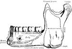
Inner surface of lower jaw, showing various areas.

Areas of Lower Jaw

Inner surface of lower jaw, showing various areas.

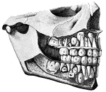
Jaw showing the temporary and permanent teeth.

Human Jaw

Jaw showing the temporary and permanent teeth.

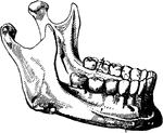
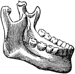
Lower jawbone with the teeth.

The lower jaw

Lower jawbone with the teeth.

Dislocation of the lower jaw.

Dislocation of the Lower Jaw

Dislocation of the lower jaw.

The mandible is the largest and strongest bone of the face. It serves for the reception of the lower teeth. It consists of a curved, horizontal portion, the body, and two perpendicular portions, the rami, which join the back party of the body nearly at right angles.

Mandible

The mandible is the largest and strongest bone of the face. It serves for the reception of the lower…
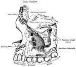
The right half of the mandible.

Mandible

The right half of the mandible.

The largest bones of the face, excepting the mandible, and form, by their union, the whole of the upper jaw.

Maxilla

The largest bones of the face, excepting the mandible, and form, by their union, the whole of the upper…
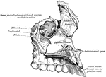
The largest bones of the face, excepting the mandible, and form, by their union, the whole of the upper jaw.

Maxilla

The largest bones of the face, excepting the mandible, and form, by their union, the whole of the upper…
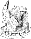
The lower maxillary bone, including the bottom set of teeth.

Maxillary

The lower maxillary bone, including the bottom set of teeth.

Superior maxillary bone. With it's fellow on the opposite side, it forms the whole of the upper jaw. Each bone assists in forming part of the floor of the orbit, the floor and outer wall of the nasal fossae, and the greater part of the roof of the mouth. Labels: 1, orbital surface; 2, facial surface; 3, alveolar process.

Human Maxillary (Upper Jaw) Bone

Superior maxillary bone. With it's fellow on the opposite side, it forms the whole of the upper jaw.…

Inferior Maxillary Bone (lower jaw). It is the largest and strongest bone in the face and serves for the reception of the lower teeth.

Human Maxillary (Upper Jaw) Bone

Inferior Maxillary Bone (lower jaw). It is the largest and strongest bone in the face and serves for…
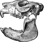
Right superior maxillary bone, inner surface.

Superior Maxillary Bone

Right superior maxillary bone, inner surface.

"Cranium and lower jaw of Mesotherium Cristatum." &mdash;The Encyclopedia Britannica, 1903

Mesotherium Cristatum

"Cranium and lower jaw of Mesotherium Cristatum." —The Encyclopedia Britannica, 1903

"Diagram of a Sectional View of Nasal and Throat Passageways. <em>C</em>, nasal cavities; <em>T</em>, tongue; <em>L</em>, lower jaw; <em>M</em>, Mouth; <em>U</em>, uvula; <em>E</em>, epiglottis; <em>G</em>, larynx; <em>O</em>, gullet, or esophagus." &mdash; Blaisedell, 1904

Nasal and throat passageways

"Diagram of a Sectional View of Nasal and Throat Passageways. C, nasal cavities; T,…
A colt's nippers at five years of age (side view)

Nippers at five years

A colt's nippers at five years of age (side view)

Side view of a colt's nippers at seven years of age.

Nippers at seven years

Side view of a colt's nippers at seven years of age.

Side view of a colt's nippers at six years of age.

Nippers at six years

Side view of a colt's nippers at six years of age.

Side view of the nippers of an old horse.

Nippers of an Old Horse

Side view of the nippers of an old horse.

A colt's lower nippers at eight years.

Lower nippers at eight years

A colt's lower nippers at eight years.

Lower nippers of a colt at five years of age.

Lower nippers at five years

Lower nippers of a colt at five years of age.

The lower nippers of a colt at four years.

Lower nippers at four years

The lower nippers of a colt at four years.

Lower nippers of a colt at six years of age.

Lower nippers at six years

Lower nippers of a colt at six years of age.

The lower nippers of a colt at three years of age.

Lower nippers at three years

The lower nippers of a colt at three years of age.

The lower nippers of a colt two years old.

Lower nippers at two years

The lower nippers of a colt two years old.

Lower nippers of an old horse.

Lower Nippers of an Old Horse

Lower nippers of an old horse.

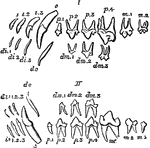
"Milkand permanent dentition of upper (I) and lower (II) jaw of the dog, with the symbols by which the different teeth are commonly designated. The third upper molar (m3) is the only tooh wanting in this animal to complete the typical heterodont mammalian dentition." &mdash;The Encyclopedia Britannica, 1903

Permanent Dentition

"Milkand permanent dentition of upper (I) and lower (II) jaw of the dog, with the symbols by which the…
A moving jaw is spring urged, against a fixed jaw, o capture picks.

Lasting Pinchers

A moving jaw is spring urged, against a fixed jaw, o capture picks.

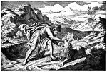
"Then went Samson down, and his father and his mother, to Timnah, and came to the vineyards of Timnah: and, behold, a young lion roared against him. And the Spirit of Jehovah came mightily upon him, and he rent him as he would have rent a kid; and he had nothing in his hand: but he told not his father or his mother what he had done." Judges 14:5-6 ASV
<p>Illustration of Samson tearing apart the jaw of a lion he is killing. He is wearing a tunic and cloak. He has long hair and a thick beard. The lion appears to be alive still. The terrain is rocky with some foliage.

Samson Tears a Lion Apart with His Bare Hands

"Then went Samson down, and his father and his mother, to Timnah, and came to the vineyards of Timnah:…
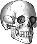
The human skull. Labels: 1, frontal lobe; 2, parietal lobe; 3, temporal lobe; 4, the sphenoid bone; 5, ethmoid bone; 6, superior maxillary (upper jaw) bone; 7, malar bone; 8, lachrymal bone; 9, nasal bone; 10, inferior maxillary (lower jaw) bone.

The Skull

The human skull. Labels: 1, frontal lobe; 2, parietal lobe; 3, temporal lobe; 4, the sphenoid bone;…
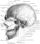
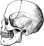
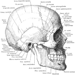
Skull and mandible, lateral view.

Skull and Mandible

Skull and mandible, lateral view.

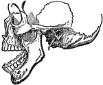
Median section of the skull and mandible, viewed from the left.

Median Section of the Skull and Mandible

Median section of the skull and mandible, viewed from the left.
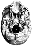
"A, Palate process of upper jawbone; B, zygoma, forming zygomatic arch; C, condyle, for forming articulation with atlas; D, foramen magnum; E, occipital bone" &mdash; Blaisedell, 1904

Base of skull

"A, Palate process of upper jawbone; B, zygoma, forming zygomatic arch; C, condyle, for forming articulation…
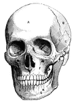
"A, frontal bone; B, parietal bone; C, temporal bone; D, sphenoid bone; E, malar bone; F, upper jawbone; G, lower jawbone" &mdash; Blaisedell, 1904

Human skull

"A, frontal bone; B, parietal bone; C, temporal bone; D, sphenoid bone; E, malar bone; F, upper jawbone;…

The backward and forward movement of the jaws and the great size and strength of the lower jaw, adapt rodents and gnawers to their mode of life, as in the squirrel. Labels: 1, The enamel of the gnawing tooth. 2, The ivory. 3, The lateral furrows of the molar teeth.

The Lower Jaw of a Squirrel

The backward and forward movement of the jaws and the great size and strength of the lower jaw, adapt…
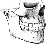
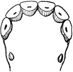
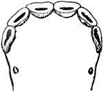
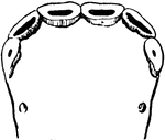
To show the relation of the upper to the lower teeth when the mouth is closed. The manner in which a tooth of one row usually strikes against two teeth of the opposite row, and the resulting interlocking of the teeth, is to be noted.

Teeth

To show the relation of the upper to the lower teeth when the mouth is closed. The manner in which a…
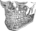
The deciduous and permanent teeth, shown as they are placed in the jaw with portions of bone removed to reveal the roots.

The Deciduous and Permanent Teeth

The deciduous and permanent teeth, shown as they are placed in the jaw with portions of bone removed…
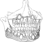
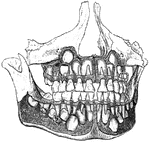
Well formed jaws, from which the alveolar plate has been removed to expose the developing permanent teeth in their crypt in the jaws.

Development of Permanent Teeth

Well formed jaws, from which the alveolar plate has been removed to expose the developing permanent…
A horse's mouth at eight years old.

Eight Year Old Horse Teeth

A horse's mouth at eight years old.

A horse's mouth at five years old.

Five Year Old Horse Teeth

A horse's mouth at five years old.

A horse's mouth at five years old.

Five Year Old Horse Teeth

A horse's mouth at five years old.

A horse's mouth at four and a half years old.

Four and a Half Year Old Horse Teeth

A horse's mouth at four and a half years old.

Teeth from a very old horse. The right tush has fallen out.

Old Horse Teeth

Teeth from a very old horse. The right tush has fallen out.

A horse's mouth at six years old.

Six Year Old Horse Teeth

A horse's mouth at six years old.

"<em>Temporary Teeth:</em> <em>A</em>, central incisors; <em>B</em>, lateral incisors; <em>C</em>, canines; <em>D</em>, anterior molars; <em>E</em>, posterior molars, <em>F</em>, central incisors; <em>H</em>, lateral incisors; <em>K</em>, canines; <em>L</em>, first bicuspids; <em>M</em>second bicuspids; <em>N</em>, first molars." &mdash; Blaisedell, 1904

Temporary and permanent teeth

"Temporary Teeth: A, central incisors; B, lateral incisors; C, canines;…
A horse's mouth at three years old.

Three Year Old Horse Teeth

A horse's mouth at three years old.

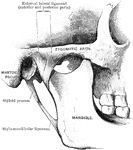
Temporo mandible joint.

Temporo Mandible Joint

Temporo mandible joint.