Clipart tagged: ‘nerves’
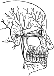
Arteries and Nerves
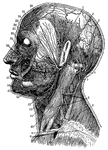
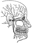
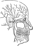
"Superficial arteries and nerves of the face and neck. 1, Temporal artery; 2, artery behind the ear;…
Brain and Spinal Cord
Anterior view of the brain and spinal marrow. Labels: 1, 1, hemispheres of the cerebrum; 2, great middle…
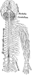
Brain and Spinal Cord
The brain and spinal cord. Labels: 1, 1, hemispheres of cerebrum; 2, great middle fissure; 3, cerebellum;…
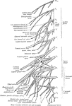
Cervical and Brachial Nerve Plexuses
The cervical and brachial nerve plexuses of the left side of the body.
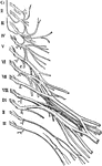
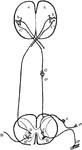
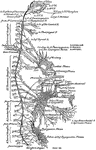
Cervical and Brachial Plexuses (Nerves)
The plan of the cervical and brachial plexuses (nerves), somewhat simplified.
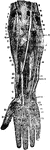
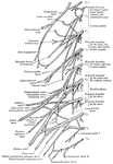
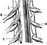
Dissection of Forearm
Deep dissection of front of the forearm. Labels: 1, supinator longus; 2, ulnar nerve; 3, brachialis;…
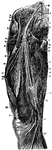
Dissection of the Forearm
Deep dissection of the front of the forearm. Labels: 1, supinator longus; 2, ulnar nerve; 3, brachialis…
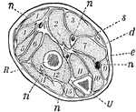
Forearm, Section of
A section across the forearm a short distance below the elbow-joint. R and U, its two supporting bones,…
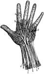
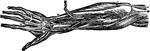
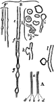
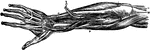
Hand Nerves
"Nerves of the had. 1, Nerves of the skin; 2, tendons; 3, arteries of the palm of the hand; 4, elbow…
Striated Muscle
Striped, or striated, muscle which quickly contracts causing the alternating black and white lines.…
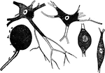
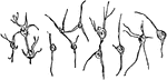
Nerve Cells
Forms of nerve cells. They each have a nucleus and nucleolus and are connected to each other by means…
Nerve Fibers
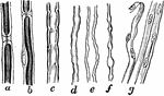
Nerve fibers. Labels: a, nerve-fiber, showing complete interruption of the white substance; b, another…
Nerve Fibers
To illustrate the structure of nerve fibers. Labels: A, nerve fiber examined fresh; n, node. B, nerve…
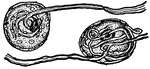
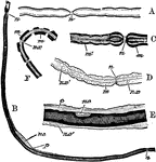
Nerve Tubules
A diagram of nerve tubules A nerve tube consists of a white portion which is fatty, and which protects…
Nerves
The cord-like structures composed of delicate filaments by which sensation or stimulative impulses are…
Nervous System
Element of Nervous System. a: Sensory fiber; b: Motor fiber; c: Center; f: End of sensory fiber; m:…
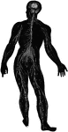
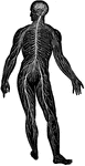
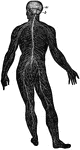
Diagram of the Human Nervous System
Diagram illustrating the general arrangement of the cerebrospinal nervous system.
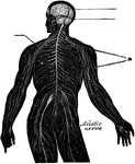
The Nervous System
View of the nervous system of man, showing the nerve centers (brain and spinal cord) giving off nerves…
Nervous System Diagram
Diagram of nervous system. Labels: a, a, cortex of cerebral hemispheres; b, b, cell body and dendrites…
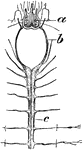
Scale Worm Nervous System
"Esophageal Ring. Anterior end of nervous system of Polynoë, a polychaetous annelid, showing, a,…
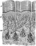
Section of the Skin
A thin slice through the skin. Labels: a, dead part; d, live part of the epidermis; ar, artery; e, sweat…
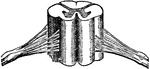
Spinal Cord
Portion of the spinal cord. 1: Body of cord; 2: A spinal nerve from left side of cord; 3: Anterior roots…
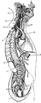
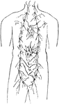
General View of the Sympathetic Nervous System
General view of the sympathetic nervous. Labels: 1,2,3, cervical ganglia; 4, 1st thoracic ganglion;…
Nerves of the Thigh
Nerves of the thigh. Labels: 1, gangliated cord of sympathetic; 2, third lumbar nerve; 3, branches to…