"Nervous System of the Aplysia, a Gasteropodous Mollusc: c, cerebral ganglia; g, thoracle or sub-aesophageal ganglia; o, nervous collar surrounding the aesophagus; l, labial ganglia; v, visceral ganglion." — Chambers, 1881

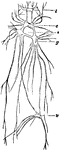
Aplysia Nervous System

"Nervous System of the Aplysia, a Gasteropodous Mollusc: c, cerebral ganglia; g, thoracle or sub-aesophageal…
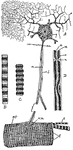
"Some nerves of the face." —Davison, 1910

Nerves of the Face

"Some nerves of the face." —Davison, 1910

This illustration shows a diagram of nervous and cross-striate muscular tissue, showing the mode of connection between nerve fibres and muscle fibres. A, nerve cell (g) connected with muscle fibre (mf.) by nerve fibre (n.f.). The muscle fibre (m.f.) is composed of numerous fibrils (f) which are made up lengthwise of alternating discs of ligter and darker substance. These fibrils are shown more highly magnified in B and C. In B the fibril is uncontracted; in C it is contracted. D, nerve fibre more highly magnified showing a, axis; m, medullary sheath; and s, Schwann's sheath; ax., axon; d, dendron; n, node; n.m., nerve-muscle plate.

Muscular Tissue

This illustration shows a diagram of nervous and cross-striate muscular tissue, showing the mode of…
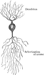
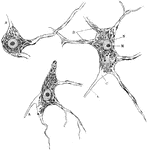
"Nervous tissue is either gray, which is a mass of tailed cells suppoted by a fine connective tissue." — Richardson, 1906

Nerve Cells

"Nervous tissue is either gray, which is a mass of tailed cells suppoted by a fine connective tissue."…

"The Main Nerve Trunks of the Right Forearm, showing the Accompanying Radial and Ulnar Arteries. (Anterior view.)" — Blaisedell, 1904

Nerve trunks

"The Main Nerve Trunks of the Right Forearm, showing the Accompanying Radial and Ulnar Arteries. (Anterior…
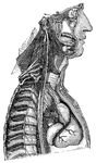
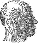
"The Cervical and Thoracic Portions of the Sympathetic Nerve and their Main Branches. In the center of the figure, running almost vertically, is shown the right pneumogastrix nerve. To the left may be seen a chain of ganglia of the sympathetic nerve, running along the vertebrae. The distribution of some smaller ganglia and nerve branches in the neck and chest regions are also shown." — Blaisedell, 1904

Sympathetic nerve

"The Cervical and Thoracic Portions of the Sympathetic Nerve and their Main Branches. In the center…
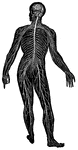
"Scheme showing the essential relations of the parts of a nervous system: 1, the sensory end organ (epithelial); 2, afferent nerve tract; 3, central nervous cells (ganglia); 4, efferent nerves, leading to 5, muscle, gland, etc. g, ganglion cells; gl., gland; m, muscle fibre; n.f., nerve fibre; s.e., sensory epithelium." — Galloway

Nervous System

"Scheme showing the essential relations of the parts of a nervous system: 1, the sensory end organ (epithelial);…
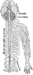
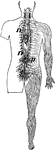
"Part of the nervous system viewed in front." —Davison, 1910

Nervous System

"Part of the nervous system viewed in front." —Davison, 1910

"Diagram illustrating the General Arrangement of the Nervous System. (posterior view.)" — Blaisedell, 1904

Nervous System

"Diagram illustrating the General Arrangement of the Nervous System. (posterior view.)" — Blaisedell,…

"Part of the sympathetic nervous system seen from in front, n, one of the two chief cords, t, i, and p, branches to organs." —Davison, 1910

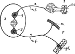
Sympathetic Nervous System

"Part of the sympathetic nervous system seen from in front, n, one of the two chief cords, t, i, and…

"A nerve cell from the brain." — Ritchie, 1918

Neuron

"A nerve cell from the brain." — Ritchie, 1918

Diagram of typical neuron.

Neuron

Diagram of typical neuron.

Diagram of nerve cell of type II, in which axone is not prolonged as nerve fiber.

Neuron

Diagram of nerve cell of type II, in which axone is not prolonged as nerve fiber.

Nerve cells of human spinal cord stained to show Nissl bodies. Labels: D, dendrites; A, axons; C, implantation cone; N, nucleus; M, nucleolus.

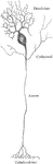
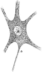
Neuron of Spinal Cord

Nerve cells of human spinal cord stained to show Nissl bodies. Labels: D, dendrites; A, axons; C, implantation…

Semidiagrammatic representation of structure of a neuron; a, axon.

Structure of a Neuron

Semidiagrammatic representation of structure of a neuron; a, axon.

Multipolar nerve cells of various forms. Labels: A, from spinal cord; B, from cerebral cortex; C, from cerebellar cortex (Purkinje cell); a, axon; c, implantation cone.

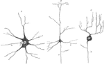
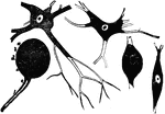
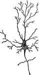
Various Forms of Neurons

Multipolar nerve cells of various forms. Labels: A, from spinal cord; B, from cerebral cortex; C, from…

Diagrammatic view of the Sympathetic cord of the right side, showing its connections with the principal cerebrospinal nerves and the main prae-aortic plexuses. Cerebrospinal nerves. VI, a portion of the sixth cranial as it passes through the cavernous sinus, receiving two twigs from the carotid plexus of the sympathetic nerve; O, opthalmic ganglion connected by a twig with the carotid plexus; M, connection of the spheno-palatine ganglion by the Vidian nerve with the carotid plexus; C, cervical plexus; Br, brachial plexus; D 6, sixth intercostal nerve; D 12, twelfth; L 3, third lumbar nerve; S 5, fifth; Cr, anterior crural nerve winding round the subclavian artery. Sympathetic Cord. c, superior cervical ganglion; c', second or middle; c", inferior; from each of these ganglia cardiac nerves (all deep on this side) are seen descending to the cardiac plexus; d 1, placed immediately below the first dorsal sympathetic ganglion; d 6, is opposite he sixth; l 1, first lumbar ganglion. Preaortic and Visceral Plexuses. p, pharyngeal, and, lower down, laryngeal plexus; pl, post-pulmonary plexus spreading from the vagus on the back of the right bronchus; c a,on the aorta , the cardiac plexus, towards which , in addition to the cardiac nerves from the three cervical sympathetic ganglia, other branches are seen descending from the vagus and recurrent nerves; co, right or posterior and co1, left or an. coronary plexus; o, esophageal plexus in long meshes on the gullet; sp, great splanchnic nerve formed by branched from the fifth, sixth, seventh, eight, and ninth dorsal ganglia; +, small splanchnic from the ninth and tenth; ++, smallest or third splanchnic from the eleventh: the first and second of theses are shown joining the solar plexus, s o; the third descending to the renal plexus, r e; connecting branches between the solar plexus and the vagi are also represented; pn', above the place where the right vagus passes to the lower or posterior surface of the stomach; pn", the left distribution on the anterior side or upper surface of the cardiac portion of the organ: from the solar plexus large branches are seen surrounding the arteries of the coeliac axis, and descending from the solar and sup. mesenteric plexuses and from the lumbar ganglia; mi, the inf. mesenteric plexus surrounding the corresponding artery; hy, hypogastric plexus; pl, the right pelvic or inf. hypogastric plexuses; pl, the right pelvic plexus; from this the nerves descending are joined by those from the plexus on the sup. hemorrhoidal vessels, mi', by nerves from the third and fourth sacral spinal nerves, and there are thus formed the rectal, vesical, and other plexuses, which ramify upon the viscera, ,as towards ir, and v, the rectum and bladder.

Sympathetic System

Diagrammatic view of the Sympathetic cord of the right side, showing its connections with the principal…