Clipart tagged: ‘palate’
Mouth
"The mouth, nose and pharynx, with the commencement of the gullet and larynx, as exposed by a section,…

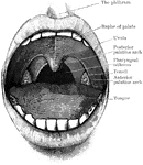
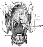
Mouth Showing Palate and Tonsils
Open mouth showing palate and tonsils. It also shows the two palatine arches, and the pharyngeal isthmus…
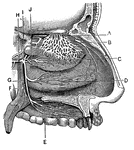
Mouth, Cavity of the
Antero-inferior surface of the soft palate. The tongue has been removed, so that the pharyngeal isthmus…
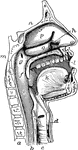
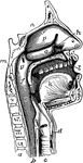
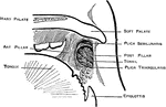
The Mouth, Nose, and Pharynx
The mouth, nose, and pharynx, with the larynx and commencement of gullet (esophagus), seen in section.…
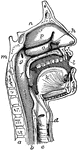
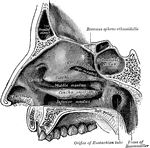
The Mouth, Nose, and Pharynx
The mouth, nose, and pharynx, with the commencement of the gullet (esophagus) and larynx, as exposed…
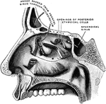
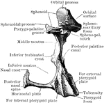
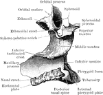
External Wall of Nasal Cavity
External wall of right nasal fossa, parts of the turbinates having been cut away to show the orifices…
Nerves of the Nostril
"A, branches of the nerves of smell; B, nerves of touch to the nostrils; E, F,…

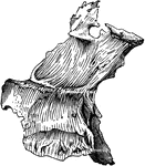
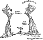
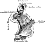
Human Palate Bone
Palate bone. Palate bones form the back part of the roof of the mouth; part of the floor and outer wall…

Muscles of the Soft Palate
Muscles of the soft palate, the pharynx being laid open from behind and mucous membrane removed.
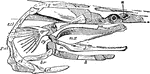
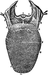
Polyodon
"Skull of Polyodon. n, nasal cavity; sq, squamosal; mh, hyomandibular; sy, symplectic; pa, palate-pterygold;…
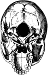
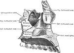
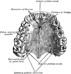
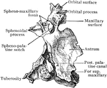
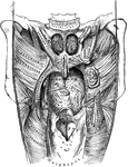
Base of the Skull
The base of the skull. "The lower jaw has been removed. At the lower part of the figure is the hard…
Upper Surface of the Tongue
Front view of the upper surface of the tongue; as also of the arch of the bone of the palate.
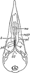
Woodpecker Skull
"Saurognathous skull of woodpecker (Colaptes auratus). v, v, the posterior parts of the abortive vomer;…