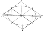
Conjugate Diameters of an Ellipse
Illustration showing that if one diameter is conjugate to a second, the second is conjugate to the first.
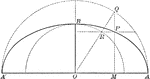
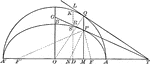
Construction of an Ellipse
Illustration of half of an ellipse and its auxiliary circle used to construct an ellipse by points,…
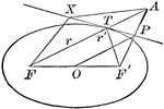
Focal Radii of an Ellipse
Illustration of half of an ellipse. "If d denotes the abscissa of a point of an ellipse, r and r' its…
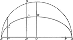
Line Bisecting Angle Between Focal Radii on Ellipse
Illustration of half of an ellipse. "If through a point P of an ellipse a line is drawn bisecting the…
Ordinate and Major Axis of Ellipse
Illustration of half of an ellipse. The square of the ordinate of a point in an ellipse is to the product…
Parallel Tangents to an Ellipse
Illustration showing that tangents drawn at the ends of any diameter are parallel to each other.
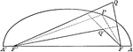
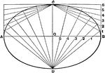
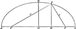
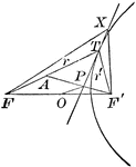
Point Distances to Foci on Ellipse
Illustration of half of an ellipse. "The sum of the distances of any point from the foci of an ellipse…
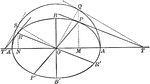
Tangent From External Point to an Ellipse
Illustration of how to draw a tangent to an ellipse from an external point.
Tangent to an Ellipse
Diagram an ellipse with a tangent line that illustrates "A line through a point on the ellipse and bisecting…
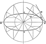
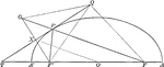
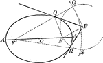
Tangents to an Ellipse
Illustration showing the tangents drawn at two corresponding points of an ellipse and its auxiliary…
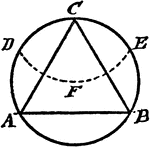
Construction Of An Equilateral Triangle Inscribed In A Circle
An illustration showing how to construct an equilateral triangle inscribed in a circle. "With the radius…
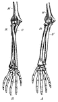
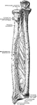
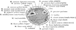
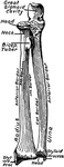
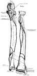
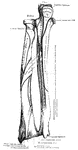
Forearm Bones
This diagram shows the bones of the right fore-arm. H, the humerus; R, the radius; and U, the ulna.
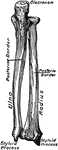
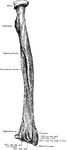
Forearm Bones
Transverse section through the bones of the forearm (radius and ulna), taken at about the middle of…
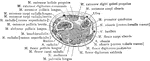
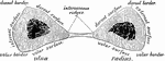
Cross Section One Inch above the Styloid Process of the Forearm
Section one inch above the styloid process of the right radius.
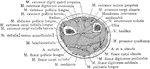
Cross Section Three Inches above the Styloid Process of the Forearm
Section three inches above the styloid process of the right radius.
Cross Section Through the Styloid Process of the Forearm
Section through the styloid process of the right radius.
Cross Section Two Inches above the Styloid Process of the Forearm
Section two inches above the styloid process of the right radius.
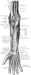
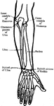
Muscles and Nerves of the Forearm
The muscles and nerves on the front of the forearm and hand. The pronator radii teres, flexor carpi…
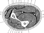
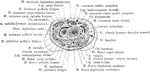
Section Across Forearm
Section across the forearm in the middle third. Labels: A, pronator radii teres; B, flexor carpi radialis;…
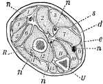
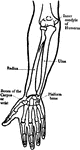
Forearm, Section of
A section across the forearm a short distance below the elbow-joint. R and U, its two supporting bones,…

Geometry
Two students drawing geometrical shapes on the chalkboard. A student is also using a scale while students…
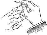
Guiding Needle Point of Compass to Little Finger
The little finger on the left hand can guide the needle point of the compass to the center.
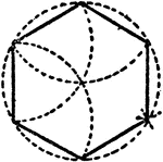
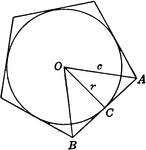
Construction Of A Hexagon In A Circle
An illustration showing how to construct a hexagon in a given circle. "The radius of the circle is equal…
Tangent to a Hyperbola
Diagram part of a hyperbola with a tangent line that illustrates "A line through a point on the hyperbola…
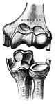
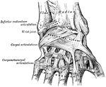
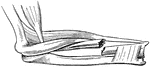
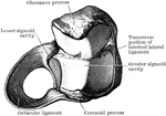
Elbow Joint
"Showing how the Ends of the Bones are shaped to form the Elbow Joint. The cut ends of a few ligaments…
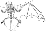
Noctule Bat
"Skeleton and volar Membranes of the Noctule Bat. c, clavicle; h, humerus; r, radius; u, ulna; d1, first…
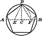
Construction Of A Pentagon Inscribed In A Circle
An illustration showing how to construct a pentagon inscribed in a circle. "Draw the diameter AB, and…
Construction Of A Pentagon On A Line
An illustration showing how to construct a pentagon on a given line. "From B erect BC perpendicular…
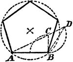
Regular Pentagon With Circle Inscribed
Illustration showing a circle inscribed in a regular pentagon. Or, a regular pentagon circumscribed…
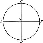
Circle With 2 Perpendicular Diameters
Illustration of a circle with center O and diameters AB and CD perpendicular to each other.
Construction of Radii of a Circle
Illustration used to draw lines which are radii of a circle where the center is inaccessible.
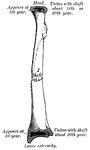
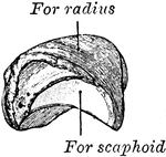
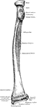
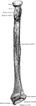
Radius
Anterior view of radius (bone of the arm) of the right side. Labels: 1, cylindrical head; 2, surface…
Broken Radius
"When a bone is broken, blood trickles out between the injured parts, and afterwards gives place to…
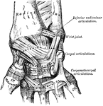
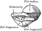
Carpal Articular Surface of the Radius
Carpal articular surface of the radius, and triangular fibro cartilage of the wrist.
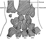
Colle's Fraction of the Radius
Showing the situation of Colle's fracture of the radius, with fracture of the styloid of the ulna. The…

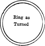
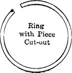
Ring With Piece Cut Out
Illustration of ring (small circle in larger concentric circle) with piece cut out.