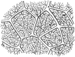
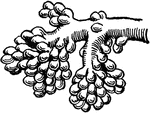
"Diagram showing the capillary network of the air sacs and origin of the pulmonary veins.. <em>A</em>, small branch of pulmonary artery; <em>B</em>, twigs of pulmonary artery; <em>C</em>, capillary network around the walls of the air sacs; <em>D</em>, branches of network converging to form the veinlets of the pulmonary veins." — Blaisedell, 1904

Capillaries of the Air Sac

"Diagram showing the capillary network of the air sacs and origin of the pulmonary veins.. A,…
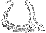
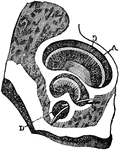
"<em>A</em>, epithelial lining wall; <em>B</em>, partition between two adjacent sacs, in which run capillaries; <em>C</em>, fibers of elastic tissues." &mdash; Blaisedell, 1904

Diagrammatic view of an air sac

"A, epithelial lining wall; B, partition between two adjacent sacs, in which run capillaries;…
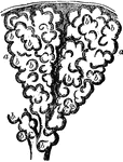
Two alveoli of the lung, highly magnified. Alveoli are cavities which are honeycombed with bulgings in order to increase the surface area exposed to air and covered by capillaries. Labels: b,b, bulgings of the alveoli, a, a.

Two Alveoli of the Lung

Two alveoli of the lung, highly magnified. Alveoli are cavities which are honeycombed with bulgings…

This illustration shows a representation of the circulation of the blood, in its essential features. The arrows indicate the course of the blood. a, arteries; aur., auricle or receiving portion of the heart; d, digestive tract; c. d., capillaries of the digestive tract; c.r., capillaries of the respiratory organs; c.s., capillaries of the system; va., valves; ve, veins; vt, ventricle.

Blood Circulation

This illustration shows a representation of the circulation of the blood, in its essential features.…
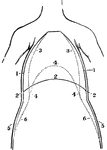
A front view of the chest and abdomen in respiration. Labels: 1, The position of the walls of the chest in inspiration. 2, The position of the diaphragm in inspiration. 3, The position of the walls of the chest in expiration. 4, The position of the diaphragm in expiration. 5, The position of the walls of the abdomen in inspiration. 6, The position of the abdominal walls in expiration.

A Front View of the Chest and Abdomen in Respiration

A front view of the chest and abdomen in respiration. Labels: 1, The position of the walls of the chest…

A side view of the chest and abdomen in respiration. Labels: 1, The cavity of the chest. 2, The cavity of the abdomen. 3, The line of direction for the diaphragm when relaxed in expiration. 4, The line of direction for the diaphragm when contracted in inspiration. 5, 6, The position of the front walls of the chest and abdomen in inspiration. 7, 8, The position of the front walls of the abdomen and chest in expiration.

A Side View of the Chest and Abdomen in Respiration

A side view of the chest and abdomen in respiration. Labels: 1, The cavity of the chest. 2, The cavity…

The left figure represents the natural shape of the chest, and that upon the right, the contracted state of it, owing to tight lacing.

Natural and Contracted Chest

The left figure represents the natural shape of the chest, and that upon the right, the contracted state…
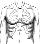
Surface of normal chest; outlines of cardiac dulness to the left of the sternum. The spaces enclosed in the dotted lines represent the interspaces.

Surface of Normal Chest

Surface of normal chest; outlines of cardiac dulness to the left of the sternum. The spaces enclosed…
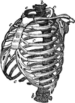
Structure of the chest, showing the framework of the bones which are connected together chiefly by muscles. It is important to understand the structure of the chest in order to understand how the movements of the chest are made in inspiration and expiration. The spinal column <em>b,b</em> is the grand pillar that supports this barrel-shaped framework. The ribs <em>c, c, c</em> are fastened very strongly by ligaments to the spinal column. They are 2 in number, 12 on each side and extend round towards the breast bone (sternum) <em>a</em> in front.

Structure of the Chest

Structure of the chest, showing the framework of the bones which are connected together chiefly by muscles.…

Torso of the Statue known as Venus of Melos (left) and New York Fashion, 1898 (right). "Since abdomen and chest alternately expand and contract in healthy breathing, anything which impedes their free movement is to be avoided. The tight lacing which is still indulged [in 1900] by those who think a distorted form beautiful, seriously impedes one of the most important functions of the body, and leads not only to shortness of breath and an incapacity for muscular exertion, but in many cases to actual deformity or disease." — Newell, 1900.

Effect of Corset Use on Respiration

Torso of the Statue known as Venus of Melos (left) and New York Fashion, 1898 (right). "Since abdomen…
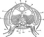

"Diagrammatic cross-section of Cray-fish in the thoracic region, to show relation of circulation and respiration. a, appendage; c, carapace; c.f., flap of carapace overhanging the gills; d, digestive tube; g, gill; h, heart; l, liver; m, body muscles; m', muscles of the appendages; n.c., nerve cord; p.s., paricardial sinus; r, reproductive glands; st, sternal artery; v.a., ventral artery; v.s., ventral blood sinus in which the nerve cord lies." &mdash; Galloway

Cray-fish

"Diagrammatic cross-section of Cray-fish in the thoracic region, to show relation of circulation and…

A diagram of how respiration works. A balloon is attached to the end of a small oil lamp chimney. The container is sealed tight with a sealing wax. As the chimney fills with air, the balloon expands, showing a representation of what a lung looks like while it is expanded. As expected, air leaves the balloon when the chimney is not covered, forcing air to leave the "lung". The piece on the right helps to control the air pressure within the container.

Respiration Diagram

A diagram of how respiration works. A balloon is attached to the end of a small oil lamp chimney. The…

The diaphragm, which is the principal muscle that act sin breathing. Here you have the cavity of the chest, <em>Cc</em>, laid open, the ribs being cut away in front, and the heart and lungs taken out; <em>DD</em> is the diaphragm. It is fastened to the spinal column behind, to the breast-bone in front, and to the lower ribs all around its side. It is not flat, but arched upward.

The Diaphragm

The diaphragm, which is the principal muscle that act sin breathing. Here you have the cavity of the…

Diaphragm of the diaphragm and it's placement in the chest. Let <em>a</em> represent the spinal column, <em>b</em> the front wall of the chest, <em>Cc</em> the cavity of the chest, and <em>Ca</em> the cavity of the abdomen. At <em>d</em> is represented the diaphragm. As you can see from the figure, as the fibers of the diaphragm are shortened the space in the chest increases which is what takes place during inspiration. On the contrary, during expiration the diaphragm is pushed upward as shown in this figure so the room in the chest is lessened and air is forced out.

The Diaphragm

Diaphragm of the diaphragm and it's placement in the chest. Let a represent the spinal column,…

This illustration shows the gills (breathing apparatus) of a perch, as well as the specific veins and arteries running along the branchial arch.

Gills (Branchial Arch of Perch)

This illustration shows the gills (breathing apparatus) of a perch, as well as the specific veins and…

This illustration shows the thorax of a crayfish with a portion of the carapace removed to show the gills.

Gills (Crayfish)

This illustration shows the thorax of a crayfish with a portion of the carapace removed to show the…

This illustration shows a transverse section of a worm with external gills. Gills are the breathing organs of aquatic animals.

Gills (worm)

This illustration shows a transverse section of a worm with external gills. Gills are the breathing…

"Showing the structure of a lobule of the lung. The lobule has been injected with mercury, afterwards dried and cut open. A large bronchial tube with its various branches is well shown." &mdash; Blaisedell, 1904

Lobule of a Lung

"Showing the structure of a lobule of the lung. The lobule has been injected with mercury, afterwards…
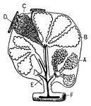
"<em>A</em>, an air sac; <em>B</em>, an air sac cut open; <em>C</em>, capillary network over an air sac; <em>D</em>, branch of pulmonary artery; <em>E</em>, branch of pulmonary vein; <em>F</em>, bronchial tube." &mdash; Blaisedell, 1904

Lobule of the lung

"A, an air sac; B, an air sac cut open; C, capillary network over an air…

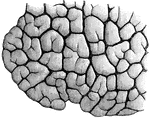
"Lobules of various sizes are well shown." &mdash; Blaisedell, 1904

Portion of a child's lung

"Lobules of various sizes are well shown." — Blaisedell, 1904

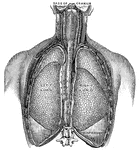
"Relative Postion of the Lungs, the Heart, and Some of the Great Vessels belonging to the latter. <em>A</em>, left common carotid artery; <em>B</em>, external carotid artery; <em>C</em>, internal carotid artery; <em>D</em>, left jugular vein; <em>E</em>, sterno-cleido muscle; <em>F</em>, right innominate artery with branches to head and arm; <em>G</em>, left subclavian artery; <em>H</em>, great azygos vein; <em>K</em>, thoracic duct." &mdash; Blaisedell, 1904

Lungs

"Relative Postion of the Lungs, the Heart, and Some of the Great Vessels belonging to the latter. A,…

The lungs and air passages seen from the front. On the left of the figure the pulmonary tissue has been dissected away to show the ramifications of the bronchial tubes. Labels: a, larynx; b, trachea; d, right bronchus. The left bronchus is seen entering the root of its lung.

Lungs and Air Passages

The lungs and air passages seen from the front. On the left of the figure the pulmonary tissue has been…

Diagram showing the structure of the lungs. At <em>d</em> is the left lung, and at <em>c</em> are represented the main branch of the windpipe that go to the right lung, separated by the lung itself. At the lower part, at <em>e</em>, are represented the very minute branches as they go to the air-cells (alveoli). At <em>b</em> is the windpipe (trachea), and at <em>a</em> is the larynx (or Adam's apple). It is through a chink in this that air passes in and out as we breathe.

Structure of the Lungs

Diagram showing the structure of the lungs. At d is the left lung, and at c are represented…
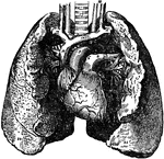
The lung, which are the two essential organs of respiration contained in the cavity of the thorax.

The Lungs

The lung, which are the two essential organs of respiration contained in the cavity of the thorax.

"Anatomy of the Oyster. A. Hinge or anterior umbonal end of the left valve of an adult oyster, upon which the soft parts of the animal are represented as they lie in situ, but with the greater part of the mantle of the right side removed. a u. The auricle of the right side of the heart contracted. B. Posterior or ventral end of the left valve, which in life is usually directed upward more or less, and during the act of feeding and respiration is separated slightly from the margin of its fellow of the opposite side to admit the water for respiration, and which also contains the animal's food in suspension. b m. Body-mass, traversed superficially by the generative ducts g e. b j. The organ of Bojanus, or 'renal' organ, of the right side of the oyster. (The ducts which it sends into the manle are not shown, nor is its connection with the genito-urinary sinus s indicated.)" &mdash; Winston's Encyclopedia, 1919

Oyster

"Anatomy of the Oyster. A. Hinge or anterior umbonal end of the left valve of an adult oyster, upon…

"The external air penetrates into every part of their body by the respiratory tubes, which ramify the whole cellular tissue. Wings alone would fail to support the bird."

Lungs of a Pigeon

"The external air penetrates into every part of their body by the respiratory tubes, which ramify the…
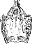
"The external air penetrates into every part of their body by the respiratory tubes, which ramify the whole cellular tissue. Wings alone would fail to support the bird."

Respiratory Organs of a Pigeon

"The external air penetrates into every part of their body by the respiratory tubes, which ramify the…
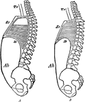
Diagram to show the changes in the sternum, diaphragm, and abdominal wall in respiration. Labels: A, inspiration; b, expiration; Tr, trachea; St, sternum; D, diaphragm; Ab, abdominal wall. The shaded part is to indicate the stationary air

Changes during Respiration

Diagram to show the changes in the sternum, diaphragm, and abdominal wall in respiration. Labels: A,…
A shaft in a building which serves as an air passage in a building.

Ventilation Shaft

A shaft in a building which serves as an air passage in a building.

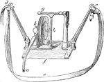
The stethograph or pneumograph consists of a thick rubber of elliptical shape about three inches long, to one end of which a rigid gutta-percha tube is attached. Labels: h, tambour fixed at right angles to plate of steel f; c and d, arms by which instrument is attached to chest by belt e. When the chest expands, the arms are pulled asunder, which bends the steel plate, and the tambour is affected by the pressure of b, which is attached to it on the one hand, and to the upright in connection with horizontal screw g.

Stethograph or Pneumograph

The stethograph or pneumograph consists of a thick rubber of elliptical shape about three inches long,…
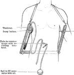
The stethometer consists of a frame formed of two parallel steel bars joined by a third at one end. At the free end of the bars is attached a leather strap, by means of which the apparatus may be suspended from the neck. Attached to the inner end of one bar is a tambour and ivory button, to the end of the other an ivory button. When in use, the apparatus is suspended with the transverse bar posteriorly, the button of the tambour is placed on the part of the chest the movement of which it is desire to record, and the other button is made to press upon the corresponding side of the chest, so that the chest is held between a pair of callipers.

Stethometer

The stethometer consists of a frame formed of two parallel steel bars joined by a third at one end.…
Thorax, or chest; a, the sternum; b,b, the spine; c,c, the ribs.

Thorax

Thorax, or chest; a, the sternum; b,b, the spine; c,c, the ribs.

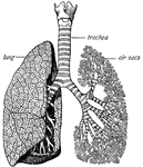
"The <em>trachea</em> has in its walls stiff rings of cartilage that hold it open so that the air can pass freely through it to and from the lungs. At its base the trachea divides and sends a great branch to each lung." &mdash; Ritchie, 1918

Trachea and lungs

"The trachea has in its walls stiff rings of cartilage that hold it open so that the air can…
"A small bronchial tube and the air sacs in which it ends." &mdash; Ritchie, 1918

Bronchial tube

"A small bronchial tube and the air sacs in which it ends." — Ritchie, 1918

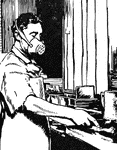
"A workman wearing a mouthpiece to protect himself from dust." — Ritchie, 1918
Worker with Mouthpiece

"A workman wearing a mouthpiece to protect himself from dust." — Ritchie, 1918