The Human Sensory Systems: Hearing ClipArt gallery offers 133 illustrations related to the human auditory sense. The structures within the ear also contribute to the sense of balance or equilibrium.
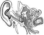
Left Ear
Diagramatic section of the Left Ear. 1: Concha; 2: External canal; 3: Middle ear, crossed by the ossicles;…
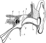
Meatus and Eustachian Tube of the Ear
Section through the external meatus, middle ear, and Eustachian tube. Labels: a, External auditory meatus;…
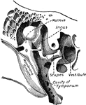
Middle Ear
The middle ear and its bones, considerably magnified. Labels: G, the inner end of the external auditory…
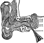
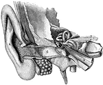
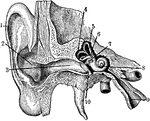
Parts of the Ear
A view of all the parts of the ear, Labels: 1, The tube that leads to the internal ear. 2, The membrana…
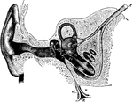
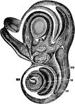
Section of the Ear
Semidiagrammatic section through the right ear. Labels: M, concha; G, external auditory meatus; T, tympanic…
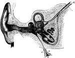
Section Through the Right Ear
Semi-diagrammatic section through the right ear. Labels: M, concha; G, external auditory meatus; T,…
Sectional View of the Ear
General sectional view of the structure of the ear. Labels: a, the meatus auditorius externus; b, the…
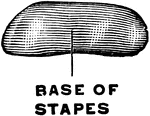
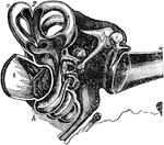
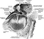
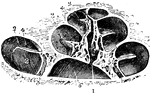
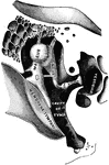
Small Bones and Ligaments of the Ear
Chains of small bones and their ligaments, seen from the front in a vertical, transverse section of…
Vertical Section of Ear
Vertical transverse section of right ear; anterior half of section, viewed from behind.
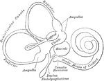
Equilibrium, Sense of
Diagram of epithelium in nervous region of ampulla of a semicircular canal of the ear. These canals…
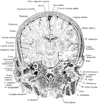
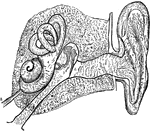
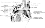
Frontal Section of Head
Frontal section of the head, passing through external and internal auditory meatus, as seen from in…
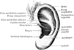
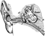
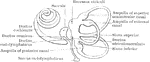
General view of organ of hearing
"A, pinna; B, cavity of the concha, showing the openings of a great number of sebaceous…
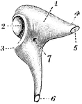
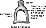
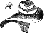
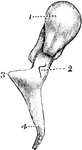
Incus
The incus, or anvil-bone. Labels: 1, body; 2, ridged articulation from the malleus; 4, processus brevis,…
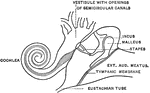
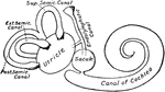
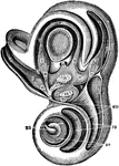
Inner Ear
1. Helix 2. Concha 3. Outer passage 4,5,6. emicircular canals 7. Oval indow 8. Cochlea 9. Eustachian…
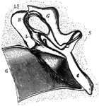
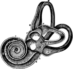
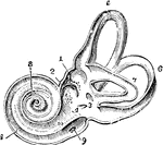
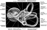
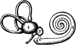
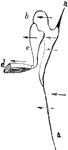
View of the Labyrinth Laid Open
A view of the labyrinth laid open. Labels: 1, The cochlea. 2, 3, Two channels that wind two and half…
Labyrinth and Vestibule of the Ear
View, very much enlarged, of the external phase of the bony labyrinth of the left side, open, exposing…
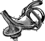
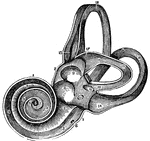
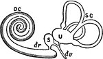
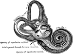
Labyrinth of the Ear
View of the labyrinth in a straight position, open to show the distribution of the nerves.
Labyrinth of the Ear on the Left Side
View of labyrinth on the left side, open throughout, in order to show its structure -- enlarged.

The Labyrinth of the Inner Ear
The labyrinth of the middle ear, which is composed of a system of fluid passages.
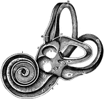
The Labyrinth of the Inner Ear
A view of the labyrinth of the left side laid open in its whole extend, so as to show its structure…
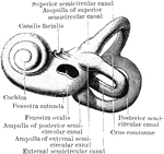
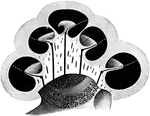
Bony Labyrinth of the Inner Ear
A highly magnified view of the external face of the bony labyrinth of the left side, opened so as to…
Development of Labyrinth
A, Left labyrinth of a human embryo of about four weeks; B, left labyrinth of a human embryo of about…
Interior of the Left Labyrinth
View of the interior of the left labyrinth. The bony wall of the labyrinth is removed superiorly and…
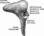
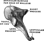
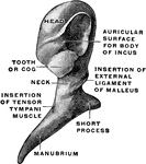
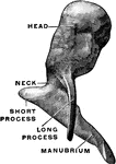
Malleus
The hammer-bone or malleus, seen from the front. 1, the head; 2, neck; 3, short process; 4, long process.
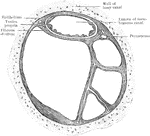
Membrana Tympani
The membrana tympani separates the cavity of the tympanum from the bottom on the external canal. Shown…
Membrana Tympani
The membrana tympani separates the cavity of the tympanum from the bottom on the external canal. Shown…
Membranous Labyrinth
The right membranous labyrinth of an adult, isolated, medial and posterior view.
Membranous Labyrinth
The membranous labyrinth is lodged within the bony labyrinth and has the same general form; it is, however,…
The Membranous Labyrinth
The membranous labyrinth of the ear. It is situated within the osseous labyrinth and consists of two…
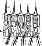
The Organ of Corti
The Organ of Corti (organon spirale), also known as the Papilla Spiralis. It is situated on the inner…
Osseous Cochlea
View of the osseous cochlea divided through the middle. Labels: 1, central canal of the modiolus; 2,…
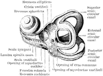
Osseous Labyrinth in Vertical Section
The osseous labyrinth in vertical section. The broken, white lines indicate the position of the basilar…
Ossicles of the Middle Ear
Diagram to illustrate the action of the ossicles of the middle ear in the conduction of sound to the…
Ossicles of the Tympanum
Chain of ossicles and their ligaments, seen from the front in a vertical transverse section of the tympanum.
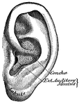
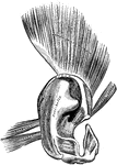
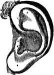
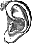
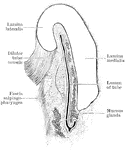
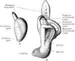
Pinna
"The outer ear consists of a plate of gristle, shaped somewhat like a shell, known as the pinna, or…
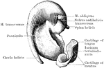
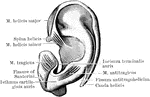
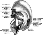
Cartilage of the Pinna
The cartilage of the right pinna, isolated, with muscles, viewed from the inside.