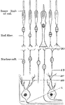
Nerve Cells
The nerve cells that compose the ganglia are generally unipolar, and seldom bipolar; sometimes two cells…
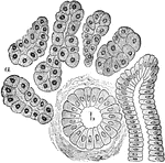
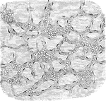
Air Cells of a Monkey
Terminal branch of a bronchial tube, with its infundibula and air cells from the margin of the lung…
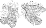
Infundibula
Two small infundibula or groups of air cells, a, with air cells, b, and the ultimate bronchial tubes,…
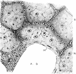
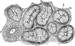
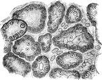
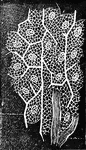
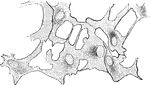
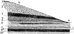
Air Cells from a Cat's Lung
From a section of the lung of a cat, stained with silver nitrate. Labels: A. D., alveolar duct or intercellular…
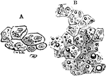
Gland Cells of a Dog
A section of the submaxillary gland of a dog. Showing gland cells, b, and a duct, a, in section.
True Salivary Gland
From a section through a true salivary gland. Labels: a, the gland alveoli, lined with albuminous "salivary…
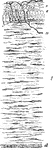
Section of a Mucous Gland
From the section through a mucous gland in a quiescent state. The alveoli are lined with transparent…
Section of a Mucous Gland After Stimulation
From the section through a mucous gland after prolonged electrical stimulation. The alveoli are lined…
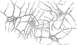
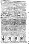
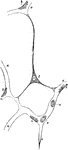
Auerbach's Nerve Plexus
Auerbach's nerve plexus in the small intestine. The plexus consist of fibrillated substance, and is…
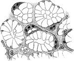
Portion of a Lobule of Liver
Portion of a lobule of liver. Labels: a, bile capillaries between liver cells, the network which is…
Hepatic Cells and Bile Capilaries
Hepatic cells and bile capillaries, from the liver of a child three months old. Both figures represent…
Epithelial Elements of a Malpighian Capsule
Epithelial elements of a Malpighian capsule and tuft, with the commencement of a urinary tubule showing…
Epithelium of the Bladder
Epithelium of the bladder. Labels: a, one of the cells of the first row; b, a cell of the second row;…
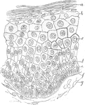
Epidermis
Vertical section of epidermis of the prepuce. Labels: a, stratum corneum, of very few layers, the stratum…
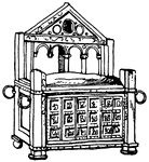
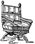
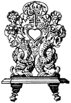
Chair of St. Peter Arm-Chair
The chair of St. Peter was made out of wood with ivory reliefs illustrating the story of Hercules in…
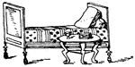
Roman Bedstead
This Roman bedstead had a Pompeian vase-painting. It included a head and foot board. It was made out…
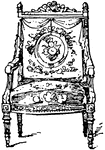
Italian 16th Century Chair
The Italian 16th century chair was made out of wood and supported by perforated carved boards.
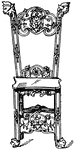
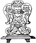
German 17th Century Chair
The German 17th century chair had openings for the hand that were carved into the wooden back of the…
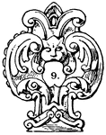
German 17th Century Chair
The German 17th century chair had openings for the hand that were carved into the wooden back of the…
German 17th Century Chair
The German 17th century chair had openings for the hand that were carved into the wooden back of the…
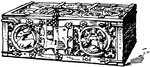
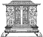
Gothic Chest
The Gothic chest of the 15th century was carved out of chestnut wood and had iron mounts and handles.
Renaissance Chest
This Renaissance chest of Dutch origin was richly decorated with carving, intarsia or wood inlaying,…
Renaissance Chest
This Renaissance chest of Italian origin was richly decorated with carving, intarsia or wood inlaying,…
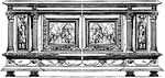
Renaissance Chest
This Renaissance chest of Flemish origin was from the 17th century. It was richly decorated with carving,…
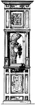
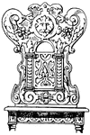
Toilet-Stand Clock-Case
This Toilet-stand clock-case was made during the German Renaissance. Made of various colored wood, it…

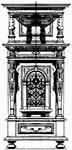
Gothic Lectern
The Gothic lectern of the 15th century had a base made out of wood and the slope was made out of wrought-iron.…
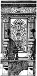
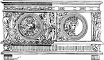
St. Maria Novella Stall
The St. Maria Novella Stall was decorated with intarsias or wood inlaying. It was designed by Baccio…
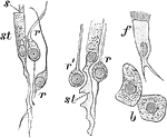
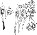
Cells from the Olfactory Region of a Rabbit
Cells from the olfactory region of the rabbit. Labels: st, supporting cells; r, r', varieties of rod-cells;…
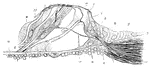
Corti from the Dog
Vertical section of the organ of Corti from the dog. Labels: 1 to 2, homogeneous layer of the so-called…
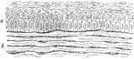
Magnified Rabbit's Cornea
Vertical section of rabbit's cornea. Labels: anterior epithelium, showing the different shapes of the…
Magnified Frog Cornea Showing Branched Corpuscles
Horizontal preparation of cornea of frog; showing the network of branched cornea-corpuscles. The ground…
Lamella of Kitten's Cornea
Surface view of part lamella of kitten's cornea, prepared first with caustic potash and then with nitrate…
Section of Rabbit's Cornea
Vertical section of rabbit's cornea, stained with gold chloride. Labels: e, Laminated anterior epithelium.…
Structure of the Retina
A section of the retina, choroid, and part of the sclerotic. Labels: a, membrana limitans interna; b,…
Nervous Elements of the Retina
Diagram showing the nervous elements of retina. Labels: 1, nerve fiber to ganglion cell; 2, processes…
Layers of the Retina
Section through the macula lutea and fovea centralis of the human retina. Labels: a, fovea; b, descent…
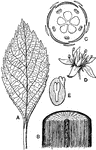
Dicotyledonous Morphology
"Morphology of a typical dicotyledonous plant. A, leaf, pinnately-netted veined; B, portion of stem,…
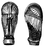
Pholas Dactylus (Linnaeus)
This species is able to hollow out a home in a solid block of gneiss, one of the hardest rocks. To excavate…
Pholas Crispata (Linnaeus)
This species is able to hollow out a home in a solid block of gneiss, one of the hardest rocks. To excavate…
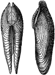
Pholas Papyracea (Solander)
This species is able to hollow out a home in a solid block of gneiss, one of the hardest rocks. To excavate…
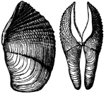
Pholas Melanoura (Sowerby)
This species is able to hollow out a home in a solid block of gneiss, one of the hardest rocks. To excavate…
Capillary Blood Vessels of Larval Frog
Capillary blood vessel of the tail of a young larval frog. a, capillaries permeable to blood; b, fat…
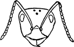
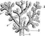
Red Wood Ant Worker's Head
Red Wood Ant worker's head. "Workers supply all the food and are the builders of their wonderful colonies."
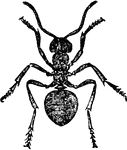
Red Wood Ant Worker
The workers supply all the food and are the builders of their wonderful colonies.
Red Wood Ant Pupae (bottom)
"The cocoons represent the pupa stage; they are commonly called ant-eggs, and are carefully tended by…
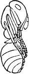
Red Wood Ant Pupae (side)
"The cocoons represent the pupa stage; they are commonly called ant-eggs, and are carefully tended by…
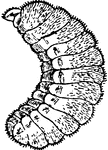
Red Wood Ant Larva
The larva is the stage right after the eggs are laid. In this stage, the larva is white and legless.
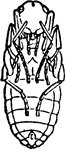
Red Wood Ant Shelter of Pupae, so-called ant-house
While the pupae are in the cocoon, they are incased in a shelter, often called an ant-house.
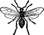
Red Wood Ant Male
The male red wood ant has wings, and after pairing is allowed to stray away and soon die.
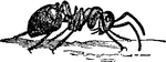
Red Wood Ant Worker (side)
The workers supply all the food and are the builders of their wonderful colonies.
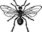
Red Wood Ant Female
"In a colony are the females, which are the largest in size." The females are very long-lived.
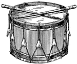
Snare Drum
"The snare drum, having two heads, the upper one only being played upon by two sticks of wood; strings…
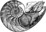
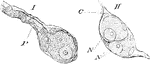
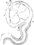
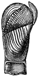
Nautilus Pompilius (Linnaeus), showing the interior of the lower cell, to which the animal is fixed.
"In the last partition of the shell is the animal, covered by its mantle, which lines to walls of the…