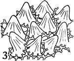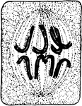
Fourth stage in plant cell division: Protophase 4; "The nuclear membrane and the nucleolus have disappeared, and the nuclear thread has become segmented into chromosomes which are assembling at the equator of the cell." -Stevens, 1916

Plant Cell Division 4

Fourth stage in plant cell division: Protophase 4; "The nuclear membrane and the nucleolus have disappeared,…
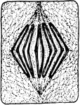
Fifth stage in plant cell division: Metaphase; "The metaphase, where the longitudinal halves of the chromosomes are being drawn apart preparatory to their journey toward opposite poles." -Stevens, 1916

Plant Cell Division 5

Fifth stage in plant cell division: Metaphase; "The metaphase, where the longitudinal halves of the…
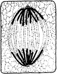
Sixth stage in plant cell division: Anaphase; "The anaphase, or movement of the chromosomes toward the poles, is about completed, connecting fibers extend from pole to pole." -Stevens, 1916

Plant Cell Division 6

Sixth stage in plant cell division: Anaphase; "The anaphase, or movement of the chromosomes toward the…
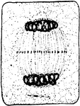
Seventh stage in plant cell division: Telophase; "Telophase where the chromosomes have begun to spin out in the form of a nuclear reticulum. The connecting fibers have begun to thicken in the equatorial plane." -Stevens, 1916

Plant Cell Division 7

Seventh stage in plant cell division: Telophase; "Telophase where the chromosomes have begun to spin…
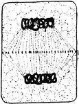
Eighth stage in plant cell division: "The connecting fibers have spread out and come into contact with the wall of the mother cell in the equatorial plane, and the thickening of the fibers throughout this plane has made a complete cell plate within which the dividing wall will be produced." -Stevens, 1916

Plant Cell Division 8

Eighth stage in plant cell division: "The connecting fibers have spread out and come into contact with…
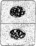
Ninth and final stage in plant cell division: "A nuclear membrane has been formed about each daughter nucleus, and the dividing cell-wall is complete." -Stevens, 1916

Plant Cell Division 9

Ninth and final stage in plant cell division: "A nuclear membrane has been formed about each daughter…
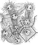
"Formation of endosperm in the embryo-sac of Agrimonia Eupatorium. Cell-walls are being formed between the nuclei." -Stevens, 1916

A. Eupatorium Cell

"Formation of endosperm in the embryo-sac of Agrimonia Eupatorium. Cell-walls are being formed between…
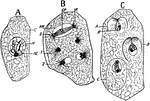
"Free cell formation of spores in the ascus of Erysiphe communis. A, ascus with single nucleus; C, cytoplasm; N, nucleus; NL, nucleolus; B, successive stages in nuclear division with the ascus; at X, early anaphase, nuclear membrane, NM, still persisting; R, kinoplasmic radiations from the poles; at Y, telophase, new nuclear membrane not yet formed; Z, a later stage where the nuclear membranes demark the daughter nuclei." -Stevens, 1916

E. Communis Cell

"Free cell formation of spores in the ascus of Erysiphe communis. A, ascus with single nucleus; C, cytoplasm;…

"Various stages of cell multiplication by budding of Saccharomyces cerevisiae." -Stevens, 1916

S. Cerevisiae Cell Multiplication

"Various stages of cell multiplication by budding of Saccharomyces cerevisiae." -Stevens, 1916

"Nucleus dividing by simple constriction. From the lining of the embryo-sac of Vicia faba." -Stevens, 1916

V. Faba Nucleus Division

"Nucleus dividing by simple constriction. From the lining of the embryo-sac of Vicia faba." -Stevens,…
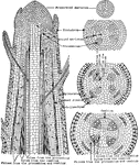
"Diagram showing the evolution of tissues from the primordial meristem down to the beginning of cambial activity." -Stevens, 1916

Plant Tissues

"Diagram showing the evolution of tissues from the primordial meristem down to the beginning of cambial…

The epidermis of an oak leaf.

Plant Epidermis

The epidermis of an oak leaf.

The epidermis of an iris leaf.

Plant Epidermis

The epidermis of an iris leaf.

"Group of cells from petal of Viola tricolor." -Stevens, 1916

Plant Epidermis

"Group of cells from petal of Viola tricolor." -Stevens, 1916

"Two epidermal cells in cross section showing thickened outer wall differentiated into three layers, namely, an outer cuticle, cutinized layer (shaded), and an inner cellulose layer." -Stevens, 1916

Plant Epidermis

"Two epidermal cells in cross section showing thickened outer wall differentiated into three layers,…

"Epidermal outgrowths in the form of scales and hairs." -Stevens, 1916

Plant Epidermis

"Epidermal outgrowths in the form of scales and hairs." -Stevens, 1916

"Epidermal outgrowths in the form of scales and hairs." -Stevens, 1916

Plant Epidermis

"Epidermal outgrowths in the form of scales and hairs." -Stevens, 1916

"Cross section of a portion of leaf of Ficus elastica showing the multiple epidermis from e to a inclusive; c, cystolith; b, palisade parenchyma; d, spongy parenchyma." -Stevens, 1916

F. Elastica Epidermis

"Cross section of a portion of leaf of Ficus elastica showing the multiple epidermis from e to a inclusive;…

"Diagram to show the topography and character of the tissues that evolved from the primary meristems. Cambial activity has not yet begun." -Stevens, 1916

Plant Tissues

"Diagram to show the topography and character of the tissues that evolved from the primary meristems.…

Developmental stages of collenchyma: "1 and 2, cross and longitudinal section of a collenchyma cell in its primary meristem condition." -Stevens, 1916

Collenchyma 1 and 2

Developmental stages of collenchyma: "1 and 2, cross and longitudinal section of a collenchyma cell…

Developmental stages of collenchyma: "3 and 4, longitudinal and cross sections of the same cell at a later stage, the walls in 4 have commenced to thicken at the angles." -Stevens, 1916

Collenchyma 3 and 4

Developmental stages of collenchyma: "3 and 4, longitudinal and cross sections of the same cell at a…

Developmental stages of collenchyma: "5 and 6, longitudinal and cross sections of a mature cell. The arrows indicate the planes through which the longitudinal sections were cut. The stippling inside the walls indicates the protoplasts." -Stevens, 1916

Collenchyma 5 and 6

Developmental stages of collenchyma: "5 and 6, longitudinal and cross sections of a mature cell. The…
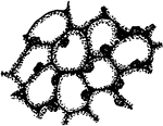
First developmental stage of stone cells: "1, in the primary meristem condition." -Stevens, 1916

Stone Cells 1

First developmental stage of stone cells: "1, in the primary meristem condition." -Stevens, 1916
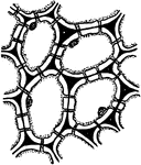
Second developmental stage of stone cells: "2, the cells have enlarged and the walls have begun to thicken and become pitted." -Stevens, 1916

Stone Cells 2

Second developmental stage of stone cells: "2, the cells have enlarged and the walls have begun to thicken…
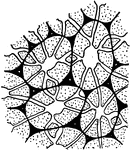
Third and final developmental stage of stone cells: "3, The walls are completed. The primary wall is black, cellulose additions white, and the lignified walls in 3 are stippled. Notice that the protoplasts have disappeared in 3, and the pits in some instances are branched." -Stevens, 1916

Stone Cells 3

Third and final developmental stage of stone cells: "3, The walls are completed. The primary wall is…

Developmental stages of bast fibers: "1 and 2, cross and longitudinal sections of primary meristem cells that are to become bast fibers." -Stevens, 1916

Bast Fibers 1 and 2

Developmental stages of bast fibers: "1 and 2, cross and longitudinal sections of primary meristem cells…

Developmental stages of bast fibers: 3 and 4, cross and longitudinal sections of meristem cells becoming bast fibers.

Bast Fibers 3 and 4

Developmental stages of bast fibers: 3 and 4, cross and longitudinal sections of meristem cells becoming…

Developmental stages of bast fibers: "5, longitudinal section of completed bast fibers. In 5 the stippling of the wall indicates lignification. Note that the walls have become pitted and the protoplasts have disappeared from the fibers." -Stevens, 1916

Bast Fibers 5

Developmental stages of bast fibers: "5, longitudinal section of completed bast fibers. In 5 the stippling…
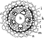
"Portion of a cross section of a root of Allium ascalonicum. h, large central, tracheal tube; i, xylem, and l, phloem portion of the vascular bundle; n, cortex cells; 1, endodermis with thin-walled cell at k to admit passage of materials; m, pericycle or pericambium." -Stevens, 1916

A. Ascalonicum Root

"Portion of a cross section of a root of Allium ascalonicum. h, large central, tracheal tube; i, xylem,…

"Diagram to show different plans in the distribution of bast fibers. A, bast a continuous cylinder in the pericycle; B, isolated strands of bast in the cortex and in the pericycle in front of each vascular bundle; C, a combination of A and B." -Stevens, 1916

Bast Fibers

"Diagram to show different plans in the distribution of bast fibers. A, bast a continuous cylinder in…

First developmental stage of sieve tubes, companion cells, and phloem parenchyma. "A, a and b, two rows of procambial cells; in c and d, a has divided longitudinal and c is to become companion cells; d, a sieve tube, and b, phloem parenchyma." -Stevens, 1916

Plant Cell Development 1

First developmental stage of sieve tubes, companion cells, and phloem parenchyma. "A, a and b, two rows…

Second developmental stage of sieve tubes, companion cells, and phloem parenchyma. "B, c, companion cells, and d, a beginning sieve tube from c and d, respectively in A. The cross-walls in d are pitted; b, phloem parenchyma grown larger." -Stevens, 1916

Plant Cell Development 2

Second developmental stage of sieve tubes, companion cells, and phloem parenchyma. "B, c, companion…

Third developmental stage of sieve tubes, companion cells, and phloem parenchyma. "The pits in the cross-walls of the sieve tubes become perforations, and the nuclei gone from the cells composing the tube." -Stevens, 1916

Plant Cell Development 3

Third developmental stage of sieve tubes, companion cells, and phloem parenchyma. "The pits in the cross-walls…

"Stages in the development of the elements of the xylem. A, progressive steps in the development of a tracheal tube. 1, row of procambial or cambial cells that are to take part in the formation of a tube; 2, the same at a later stage enlarged in all dimensions; 3, the cells in 2 have grown larger, their cross-walls have been dissolved out, and the wall has become thickened and pitted; 4, the walls in 3 have become more thickened, the pits have an overhanging border, the walls have become lignified as indicated by the stippling, and finally the protoplasts have disappeared, and the tube is mature and dead." -Stevens, 1916

Xylem Development 1

"Stages in the development of the elements of the xylem. A, progressive steps in the development of…

Stages in the development of the elements of the xylem. "B, stages in the formation of tracheids from procambial or cambial cells. The steps are the same as in A, excepting that the cross-walls remain and become pitted." -Stevens, 1916

Xylem Development 2

Stages in the development of the elements of the xylem. "B, stages in the formation of tracheids from…

Stages in the development of the elements of the xylem. "C, steps in the development of wood fibers from cambial cells; 1, cambial cells; 2, the same grown larger in all dimensions with cells shoving past each other as they elongate; 3, a later stage with cells longer and more pointed and walls becoming thickened and pitted; 4, complete wood fibers with walls more thickened than in the previous stage and lignified, as shown by the stippling. The protoplasts in this last stage have disappeared and the fibers are dead." -Stevens, 1916

Xylem Development 3

Stages in the development of the elements of the xylem. "C, steps in the development of wood fibers…

Stages in the development of the elements of the xylem. "D, steps in the formation of wood parenchyma from cambial or procambial cells. 1, group of cambial or procambial cells; 2, the same enlarged in all dimensions; 3, the same with walls thickened and pitted; 4 and 5 show the same stages as 2 and 3, but here the cells have enlarged radially or tangentially more than they have vertically. The walls of these cells are apt to become lignified, but the cells are longer lived than the wood fibers." -Stevens, 1916

Xylem Development 4

Stages in the development of the elements of the xylem. "D, steps in the formation of wood parenchyma…

"Diagram showing how the vascular bundles anastomose around the medullary rays. The gaps represent the rays." -Stevens, 1916

Vascular Bundles

"Diagram showing how the vascular bundles anastomose around the medullary rays. The gaps represent the…
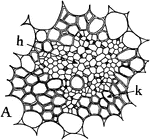
Types of vascular bundles: "A, the concentric type, with xylem, k, surrounding the phloem, h." -Stevens, 1916

Concentric Vascular Bundle

Types of vascular bundles: "A, the concentric type, with xylem, k, surrounding the phloem, h." -Stevens,…
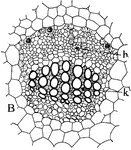
Types of vascular bundles: "B, the collateral type, with phloem, h, standing in front of the xylem, k." -Stevens, 1916

Collateral Vascular Bundle

Types of vascular bundles: "B, the collateral type, with phloem, h, standing in front of the xylem,…

Types of vascular bundles: "C, a portion of the radial type, shown complete in D, where the part outlined at a, corresponds to C. Corresponding parts are lettered the same in both figures; c, xylem; b, phloem; f, cambium ring; e, pericycle; d, endodermis. C and D are from the tap root of Vicia faba." -Stevens, 1916

Radial Vascular Bundle

Types of vascular bundles: "C, a portion of the radial type, shown complete in D, where the part outlined…
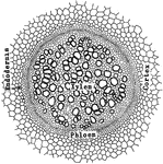
"Gleichenia pubescens: transverse section of stem, showing the protostele." -Stevens, 1916

G. Pubescens Stem

"Gleichenia pubescens: transverse section of stem, showing the protostele." -Stevens, 1916
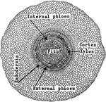
"Adianium pedatum: transverse section of stem, showing the amphiphloic siphonostele." -Stevens, 1916
A. Pedaium Stem

"Adianium pedatum: transverse section of stem, showing the amphiphloic siphonostele." -Stevens, 1916

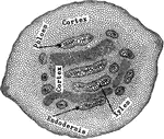
"Pteris aquilina: transverse section of stem, showing the polystele." -Stevens, 1916

P. Aquilina Stem

"Pteris aquilina: transverse section of stem, showing the polystele." -Stevens, 1916
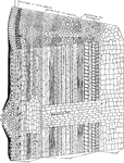
"Diagram showing additions to the primary tissues through the activity of the cambium and phellogen or cork cambium." -Stevens, 1916

Plant Tissues

"Diagram showing additions to the primary tissues through the activity of the cambium and phellogen…

"Diagram showing the relation of this year's leaves to the wood of the current year." -Stevens, 1916
Plant Tissues

"Diagram showing the relation of this year's leaves to the wood of the current year." -Stevens, 1916

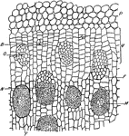
"Portion of a cross section throughout the stem of Dracaena marginata. P, parenchyma of cortex. V, meristematic zone of the pericycle by the activity of which the stem increases in diameter, with the addition of new vascular bundles. M, mature vascular bundle. N, nearly mature vascular bundle. O, newly formed procambium strand from which a vascular bundle is to arise. B, beginning of a procambium strand by the division of cells in the meristematic zone. F, parenchyma of the fundamental tissue." -Stevens, 1916

D. Marginata Stem

"Portion of a cross section throughout the stem of Dracaena marginata. P, parenchyma of cortex. V, meristematic…
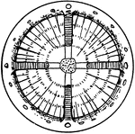
"Diagram showing some types of unusual growth in thickness. A, cross section through a four-year-old stem of Anisostichus capreolata." -Stevens, 1916

A. Capreolata Unusual Stem Growth

"Diagram showing some types of unusual growth in thickness. A, cross section through a four-year-old…
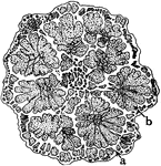
"Diagram showing some types of unusual growth in thickness...B, cross section of stem of species of Bauhinia; the xylem strands, b, are stippled while the surrounding parenchyma and bark tissues are left white." -Stevens, 1916

Bauhinia Unusual Stem Growth

"Diagram showing some types of unusual growth in thickness...B, cross section of stem of species of…
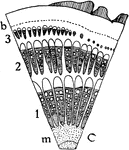
"Diagram showing some types of unusual growth in thickness...C, portion of a cross section of stem of Gnetum scandens; 1, 2, and 3 are successive rings of growth; m, is the pith; b, is a sclerenchyma ring. The xylem portions with the exception of the larger tracheal tubes are shaded, while the medullary rays, phloem and tissues intervening between the rings of growth and the outer cortes tissues are left white." -Stevens, 1916

G. Scandens Unusual Stem Growth

"Diagram showing some types of unusual growth in thickness...C, portion of a cross section of stem of…

"A, cross section through upper half of leaf of Pyrus Japonica, showing cutinized layer of the outer wall at f, and cellulose layer at g." -Stevens, 1916

P. Japonica Epidermis

"A, cross section through upper half of leaf of Pyrus Japonica, showing cutinized layer of the outer…

A cross section through the upper half of a Russian olive leaf, showing a "cutinized layer of the outer wall at f, and cellulose layer at g." -Stevens, 1916

Russian Olive Leaf Epidermis

A cross section through the upper half of a Russian olive leaf, showing a "cutinized layer of the outer…

"C, portion of cross section of submerged stem of Nymphaea odorata, where there is no cutinized layer, and the cuticle is a hardly distinguishable film." -Stevens, 1916

N. Odorata Epidermis

"C, portion of cross section of submerged stem of Nymphaea odorata, where there is no cutinized layer,…
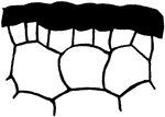
"A, portion of cross section of leaf of Avicennia growing in salty soil; outer wall of epidermis very thick." -Stevens, 1916

Avicennia Epidermis

"A, portion of cross section of leaf of Avicennia growing in salty soil; outer wall of epidermis very…

"B, cross section through skin of apple." -Stevens, 1916

Apple Epidermis

"B, cross section through skin of apple." -Stevens, 1916

"C, cross section through upper half of petal of Japan Quince." -Stevens, 1916

Japan Quince Epidermis

"C, cross section through upper half of petal of Japan Quince." -Stevens, 1916
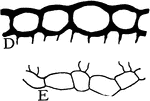
"D, upper, and E, lower epidermis of leaf of Hibiscus moscheutos." -Stevens, 1916

H. Moscheutos Epidermis

"D, upper, and E, lower epidermis of leaf of Hibiscus moscheutos." -Stevens, 1916

"Different forms of epidermal outgrowths. 1, hooked hair from Phaseolus multiflorus; 2, climbing hair from stem of Humulus Lupulus; 3, rod-like wax coating from the stem of Saccharum offininarum; 4, climbing hair of Lossa hispida; 5, stinging hair of Urtica urens." -Stevens, 1916

Epidermal Outgrowths

"Different forms of epidermal outgrowths. 1, hooked hair from Phaseolus multiflorus; 2, climbing hair…

"Multiple epidermis of leaf of mangrove in cross section. This serves as a water reservoir, and the relatively thick walls of the inner cells reenforce the protective power of the outer layer." -Stevens, 1916

Multiple Epidermis

"Multiple epidermis of leaf of mangrove in cross section. This serves as a water reservoir, and the…
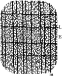
"Cross section of yellow poplar wood. E, early; L, late growth; m, medullary ray." -Stevens, 1916

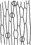
Yellow Poplar Wood

"Cross section of yellow poplar wood. E, early; L, late growth; m, medullary ray." -Stevens, 1916