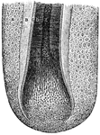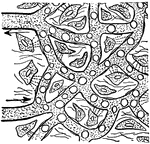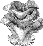
"These animals are generally called <em>Tree-corals</em>, on account of the forms of the polypidons which they build and in which they live. They are very small, and possess twelve short tentacles placed in a circle around the mouth; they occupy the cells in the polypidom, which is of a porus nature, the openings of the cells being placed at the summits of tubercular prominences of greater or lesser elevations. " &mdash; Goodrich, 1859

Tree-coral

"These animals are generally called Tree-corals, on account of the forms of the polypidons…
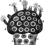
"a a, expanded polypes; b b, polypes withdrawn into their cells; c c, coral uncovered by flesh, showing the cells. In this the corals usually form thick stony masses; the stony rays of the cells are exceedingly numerous, and the cells themselves penetrate deeply into the mass of coral, although they are generally partially divided by imperfect transverse partitions." &mdash; Goodrich, 1859

Astrae virdis

"a a, expanded polypes; b b, polypes withdrawn into their cells; c c, coral uncovered by flesh, showing…

"It is to this family more especially that the formation of the coral reeds is to be attributed. In this the corals usually form thick stony masses; the stony rays of the cells are exceedingly numerous, and the cels themselves penetrate deeply into the mass of coral, although they are generally partially divided by imperfect transverse paritions." &mdash; Goodrich, 1859

Astraea rotulosa

"It is to this family more especially that the formation of the coral reeds is to be attributed. In…
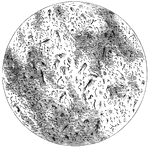
"Their bodies consist either of aa simple elementary cell, with its contents, or of an aggregation of several of these cells; each, however, still appearing to retain its independent existence. They are generally of a very minute size, and only to be observed with the microscope." &mdash; Goodrich, 1859

Protozoans

"Their bodies consist either of aa simple elementary cell, with its contents, or of an aggregation of…

"A, columnar cells found lining various parts of the intestines (called <em>columnar epthelium</em>); B, cells of a fusiform or spindle shape found in the loose tissue under the skin and in other parts (called <em>connective-tissue cells</em>); C, cell having many processes or projections - such are found in some kinds of connective tissue; D, primitive cells composed of protoplasm with nucleus, and having no cell wall." &mdash; Blaisedell, 1904

Various forms of cells

"A, columnar cells found lining various parts of the intestines (called columnar epthelium);…
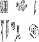
"A, columnar cells of intestine; B, polyhedral cells of the conjuctiva; C, ciliated conical cells of the trachea; D, ciliated cell of frog's mothl E, inverted conical cell of the trachea; F, squamous cell of the cavity of the mouth, seen on its broad surface; G, squamous cell, seen edgeways." &mdash; Blaisedell, 1904

Various kinds of epithelial cells

"A, columnar cells of intestine; B, polyhedral cells of the conjuctiva; C, ciliated conical cells of…

"One of the simplest of the tissues in the body is called the <em>epithelium</em>, and its cells are called epithelial cells. It serves as a lining of various cavities, a covering for the surface of the body, and the essential part of various organs called glands." — Blaisedell, 1904

Cross-Section of the Epithelium

"One of the simplest of the tissues in the body is called the epithelium, and its cells are…

"Consisting of a very fine network of fibrils, around which are cells of various sizes." — Blaisedell, 1904

Connective Tissue from a Lymphatic Gland

"Consisting of a very fine network of fibrils, around which are cells of various sizes." — Blaisedell,…

"Showing (1) cartilage with martrix and cells; (2) cartilage with matrix containing cells and white fibers." &mdash; Blaisedell, 1904

Longitudinal section of cartilage

"Showing (1) cartilage with martrix and cells; (2) cartilage with matrix containing cells and white…

Growing Yeast Cells, showing Method of budding and forming Groups of Cells. Each bud appears as a little swelling on the side of the larger cell, as seen in <em>a</em> and <em>b</em>. In <em>c</em> the little bud has grown to be nearly as large as the parent cell. The little buds grow one after another, making irregularly shaped groups, as shown in <em>d</em>." &mdash; Blaisedell, 1904

Growing yeast cells

Growing Yeast Cells, showing Method of budding and forming Groups of Cells. Each bud appears as a little…
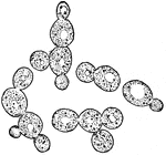
"Yeast Cells, found in the Juice of Apples, which cause the fermentation of Cider." &mdash; Blaisedell, 1904

Yeast cells

"Yeast Cells, found in the Juice of Apples, which cause the fermentation of Cider." — Blaisedell,…

"Hydrozoon is a name given to the great class of the sub-kingdom C&oelig;lenterata, of which hydra is the type. They exhibit a definite histological structure, their tissues having a cellular organization. These tissues are two, an outer or ectoderm, and an inner or endoderm. In most the prey is seized by tentacles surrounding the mouth and furnished with offensive weapons called thread cells, The hydrozoa are all aquatic, and nearly all marine. Their distribution is world-wide. [Pictured] Hydra fusca, with a young bud at b, and a more advanced bud at c."&mdash;(Charles Leonard-Stuart, 1911)

Hydrozoon

"Hydrozoon is a name given to the great class of the sub-kingdom Cœlenterata, of which hydra is…

"<em>A</em>, outer layer of cuticle; <em>B</em>, deeper layer of cuticle; <em>C</em>, duct of sweat gland; <em>D</em>, true skin; <em>E</em>, sublayer of true skin, with columnar cells. The blood vessels are injected to show black." — Blaisedell, 1904

Cross-Section of the Skin

"A, outer layer of cuticle; B, deeper layer of cuticle; C, duct of sweat…

"Cross-section of One Half of a Human Hair. A hair is made up of horny cells of the outer layer of the skin altered in shape and structure." &mdash; Blaisedell, 1904

Cross-section of a human hair

"Cross-section of One Half of a Human Hair. A hair is made up of horny cells of the outer layer of the…

"<em>A</em>, membrane of hair follicle, showing cells with nuclei and pigmentary granules; <em>B</em>, external lining of root sheath; <em>C</em>, internal lining of root sheath; <em>D</em>, cortical or fibrous portion of hairshaft; <em>E</em>, medullary portion (pith) of shaft; <em>F</em>, hair bulb, showing its development from cells." — Blaisedell, 1904

Hair Follicle

"A, membrane of hair follicle, showing cells with nuclei and pigmentary granules; B,…

"The convoluted gland is seen surrounded by fat cells and may be traced through the true skin to its outlet in the horny layers of the outer skin." &mdash; Blaisedell, 1904

Sweat gland

"The convoluted gland is seen surrounded by fat cells and may be traced through the true skin to its…

"Nerve tissue is really made up of a great number of distinctive units called <em>nerve cells</em>. Each cell usually contains a large nucleus and gives off one or more tiny branches, or processes. Thse cells vary more in shape and size than any other cell in the body. Each nerve cell has a number of short branches, and many have also one long branch which can be traced for some distance from the cell body." — Blaisedell, 1904

Nerve Cells

"Nerve tissue is really made up of a great number of distinctive units called nerve cells.…
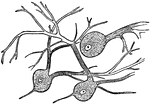
"Wherever nerve cells are abundant, the nerve tissue has a gray color; in other places, it looks white. Most of the gray matter of the brain is on the surface." — Blaisedell, 1904

Nerve Cells of the Brain

"Wherever nerve cells are abundant, the nerve tissue has a gray color; in other places, it looks white.…
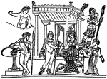
"What in this letter is contained, what here, Is written, all I will repeat to thee, That thou mayst bear my message to my friends, 'Gainst danger thus I guard: if thou preserve, The letter, that though silent will declare, My purport; if it perish in the sea, Saving thyself, my words too thou wilt save." &mdash; The Delphian Society, 1913

Iphegnia delivers letter to Pylades

"What in this letter is contained, what here, Is written, all I will repeat to thee, That thou mayst…
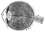
1, the sclerotic thicker behind than in front; 2, the cornea; 3, the choriod; 6, the iris; 7, the pupil; 8, the retina; 10, the anterior chamber of the eye; 11, the posterior chamber; 12, the crystalline lens, enclosed in its capsule; 13, the vitreous humour, enclosed in the hyaloid membrane and in cells formed in its interior by that membrae; 15, the sheath; and 16, the interior of the optic nerve, in the centre of which is a small artery.

Human Eye

1, the sclerotic thicker behind than in front; 2, the cornea; 3, the choriod; 6, the iris; 7, the pupil;…

A genus of plants of natural order Scropulariace&aelig;. having a tubular calyx, the upper lip of the corolla divided, the lower of three nearly equal lobes, the cells of the athers spurred at the base, a two-celled capsule and striated seeds.

Eyebright

A genus of plants of natural order Scropulariaceæ. having a tubular calyx, the upper lip of the…
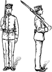
"Without changing the grasp of the right hand, place the piece on the right shoulder, barrel up and inclined at an angle of about 45 degrees from the horizontal trigger guard in the hollow of teh shoulder, right elbow near the side, the piece in a vertical plane perpendicular to the front." — Moss, 1914

Right Shoulder, Arms

"Without changing the grasp of the right hand, place the piece on the right shoulder, barrel up and…

"Carry the piece with the right hand and place it on the left shoulder, barrel up, trigger guard in the hollow of the shoulder, at the same time grasp the butt with the left hand, heel between first and second fingeres, thumb and fingers closed on the stock." — Moss, 1914

Left Shoulder, Arms

"Carry the piece with the right hand and place it on the left shoulder, barrel up, trigger guard in…

"The bayonet is a cutting and thrusting weapon consisting of three principal parts, viz, the <em>blade</em>, <em>guard</em>, <em>grip</em>." &mdash; Moss, 1914

Bayonet

"The bayonet is a cutting and thrusting weapon consisting of three principal parts, viz, the blade,…
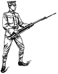
"At the second command sake the position of guard; at the same time throw the rifle smartly to the front, grasp the rifle with the left hand just below the lower band, fingers between the stock and gun sling, barrel turned slightly to the left, the right hand grasping the small of the stock about 6 inches in front of the right hip, elbows free from the body, bayonet point at the height of the chin." — Moss, 1914

Bayonet Guard

"At the second command sake the position of guard; at the same time throw the rifle smartly to the front,…

"Executed in the same manner as the thrust, except that the left foot is carried forward about twice its length. The left heel must always be in rear of the left knee. Guard is resumed immediately without command. Guard may also be resumed by advancing the right foot if, for any reason, it is desired to gain the ground held in lunging. In the latter case, the preparatory command <em>forward</em> will be given." &mdash; Moss, 1914

Lunge

"Executed in the same manner as the thrust, except that the left foot is carried forward about twice…

"Straighten right arm and right leg vigorously and swing butt of rifle against point of attack, pivoting the rifle in the left hand at about the height of the left shoulder, allowing the bayonet to pass to the rear on the left side of the head. Guard is resumed without command." — Moss, 1914

Butt Strike

"Straighten right arm and right leg vigorously and swing butt of rifle against point of attack, pivoting…
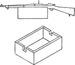
"Take an empty pistol ammunition box or a similar well-made box, remove the top and cut notches in the ends to fit the rifle closely. Place the rifle in these notches with the trigger guard close to and outside one end." &mdash; Moss, 1914

Sighting rest for rifle

"Take an empty pistol ammunition box or a similar well-made box, remove the top and cut notches in the…

"By use of an instrument called a map measurer, set the hand on the face to read zero, roll the small wheel over the distance; now roll the wheel in an opposite direction along the graphical scale, noting the number of yards passed over. A map measurer is valuable for use in solving map problems in patrolling, advance guard, outpost, etc." &mdash; Moss, 1914

Map measurer

"By use of an instrument called a map measurer, set the hand on the face to read zero, roll the small…

"The parts are the butt, A; small, B; magazine well, C; barrel bed, D; air chamber, E, which reduces the charring effect og a heater barrel on the stock; small butt plate screw hole and seat for the butt plate tang, F; butt swivel plate seat, G; mortise for receiver tang lug and hole for rear guard screw, H; mortise for sear and slot for trigger, I; cut-off thumb-piece recess, J; mortise for recoil on the receiver, K; bed for fixed base, L; grasping grooves, N; shoulder for lower band, O; bed for band spring, P; shoulder for upper band, Q; channels for decreasing weight, R; upper band screw hole, S; and the stock screw hole, T. The large hole in butt is for decreasing weight, and the smaller one is a pocket for the combination oiler and thong case." &mdash; Moss, 1914

Stock, top view

"The parts are the butt, A; small, B; magazine well, C; barrel bed, D; air chamber, E, which reduces…

"The parts are the butt, A; small, B; magazine well, C; barrel bed, D; air chamber, E, which reduces the charring effect og a heater barrel on the stock; small butt plate screw hole and seat for the butt plate tang, F; butt swivel plate seat, G; mortise for receiver tang lug and hole for rear guard screw, H; mortise for sear and slot for trigger, I; cut-off thumb-piece recess, J; mortise for recoil on the receiver, K; bed for fixed base, L; grasping grooves, N; shoulder for lower band, O; bed for band spring, P; shoulder for upper band, Q; channels for decreasing weight, R; upper band screw hole, S; and the stock screw hole, T. The large hole in butt is for decreasing weight, and the smaller one is a pocket for the combination oiler and thong case." &mdash; Moss, 1914

Stock, right side view

"The parts are the butt, A; small, B; magazine well, C; barrel bed, D; air chamber, E, which reduces…

A large, circular Chinese encampment.

Chinese encampment

A large, circular Chinese encampment.

"Among the cells of the body there is, besides the blood capillaries, a system of fine, thin-walled lymphatic capillaries." — Ritchie, 1918

Lymphatic Vessels in the Fingers

"Among the cells of the body there is, besides the blood capillaries, a system of fine, thin-walled…
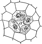
"Young resin gland of fir: <em>a</em>, duct, an intercellular space formed by the separation of the four nucleate cells." &mdash; Coulter, 1910

Resin gland of a fir

"Young resin gland of fir: a, duct, an intercellular space formed by the separation of the…
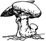
"Many cells, but without differentiation into stem and leaf; growing horizontally in spreading shoots or fronds, as the algae, or sea-weeds; fungi, or toad-stools; and the lichens.

Toad-stool

"Many cells, but without differentiation into stem and leaf; growing horizontally in spreading shoots…
"Blood plasma passing out of capillary to feed the body cells." &mdash;Davison, 1910

Capillaries

"Blood plasma passing out of capillary to feed the body cells." —Davison, 1910

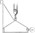
This metal stirrup and guard-rail device which is designed to take the place of rope stirrups. Built of wrought iron to comply with state, city and labor union safety regulations. These are now very generally used in place of the old-fashioned rope rigs.

Swing Stage Safety Stirrup

This metal stirrup and guard-rail device which is designed to take the place of rope stirrups. Built…
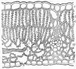
This illustration shows a stem of a plant. e; epidermis; s, stoma; p, palisade mesophyll; ch, chloroplast; sp, spongy mesophyll; i, intercellular spaces; v, small vein cut across; l, end of vein seen from the side, sonsisting of elongated and banded cells.

Epidermis

This illustration shows a stem of a plant. e; epidermis; s, stoma; p, palisade mesophyll; ch, chloroplast;…
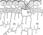
This image shows the cross-section of the outer cells of a leaf of pine. S, stoma; E, epidermis; C, cuticle; ST, stereome; and M, Mesophyll Cells.

Pine Leaf

This image shows the cross-section of the outer cells of a leaf of pine. S, stoma; E, epidermis; C,…
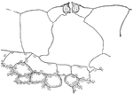
This illustration shows a close up of the plant tradescantia. It shows the delicate character of the cells and the raised stoma of this shade plant.

Tradescantia

This illustration shows a close up of the plant tradescantia. It shows the delicate character of the…
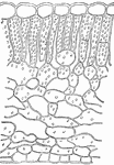
This illustration shows a section of the leaf of skunk cabbage, Spathyema. Note the poorly developed palisade tissue and the loose arrangement of the cells of this plant which lives in moist, shaded places.

Spathyema

This illustration shows a section of the leaf of skunk cabbage, Spathyema. Note the poorly developed…
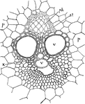
This illustration shows the cross-section of a single vascular bundle of corn stem: ph, phloem; x, small cells of the xylem; v, vessels of xylem; st, stereome that forms a sheath about the bundle; p, parenchyma of the stem.

Corn Stem

This illustration shows the cross-section of a single vascular bundle of corn stem: ph, phloem; x, small…
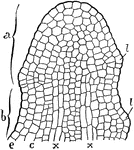
This illustration shows the longitudinal section of the tip of a growing stem: e, epidermis extending over surface of the entire tip; a, formative region; b, upper portion of the zone of elongation; c, cortex; x, cells of the central region that by further growth form the vascular bundles; l, first appearance of the leaves.

Growing Stem

This illustration shows the longitudinal section of the tip of a growing stem: e, epidermis extending…
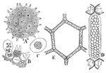
This illustration shows the colonial forms of unicellular green algae: A, Pediastrum, the plants of the colony being arranged in a flat plate. B, a view of the outer cells of the colony showing the formation of a new colony. C, one of these new colonies. D, a plant of the water net containing a young colony. E, enlarged view of one of the meshes of a net showing the geometrical arrangement of the plants.

Green Algae

This illustration shows the colonial forms of unicellular green algae: A, Pediastrum, the plants of…
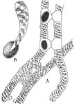
This illustration shows the sexual reproduction of Spirogyra: A, in lower portion of figure formation of the tubes between the opposite cells of the filaments is shown. Above the contents of two cells have united with two cells of another filament, forming two gametospores. B, germination of a gametospore.

Spirogyra

This illustration shows the sexual reproduction of Spirogyra: A, in lower portion of figure formation…
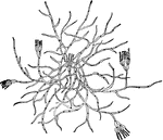
The green mold, Penicillium, one of the most common of the Sac Fungi. The hyphae of the branching mycelium is composed of cells and the spores or conidia are formed in chains that are arranged in brushlike clusters at the ends of the erect hyphae.

Mold

The green mold, Penicillium, one of the most common of the Sac Fungi. The hyphae of the branching mycelium…
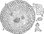
This illustration shows further development of the ascocarp: A, sectional view, showing the branches, s, derived from the germinating gametospore, that are forming numerous lateral branchlets. B, one of the branchlets enlarged, showing how it divides into cells which round off, forming the asci, as. C, ascospore. D, germinating ascspore.

Ascocarp

This illustration shows further development of the ascocarp: A, sectional view, showing the branches,…

This illustration shows the structure of the thallus of Ricciocarpus: A, section of the thallus, showing the apical cell, x, forming cells that by further division develop into plates of cells separated by air spaces, j. At the left the plates thus formed are seen curving over the apical cell. B, an olderp ortion of the upper part of the thallus. The air spaces, j, are greatly enlarged and the upper cells of the vertical plates have divided, arching over the air spaces but leaving small openings which permit the entrance of air for photosynthesis.

Ricciocarpus

This illustration shows the structure of the thallus of Ricciocarpus: A, section of the thallus, showing…
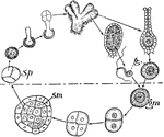
This illustration shows a diagram of the life history of Ricciocarpus. The upper portion of the figure represents the sexual generation and the lower portion, the asexual. The former generation begins with the formation of the spores, sp, from the mother-cell and ends with the formation of the gametes, g. The asexual generation begins with the gametospore, gm, and ends with the spore mother cells, sm.

Ricciocarpus

This illustration shows a diagram of the life history of Ricciocarpus. The upper portion of the figure…
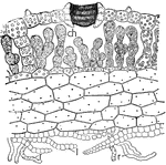
This illustration shows a section through the center of the thallus of Marchantia, showing one of the air chambers and chimney-like pores in the epidermis-ch, palisade-like chlorenchyma arising from bottom of air chamber. The lower cells of the thallus are nearly colorless and filled with watery solutions or mucilage. r, rhizoids; l, leaf-like plates of cells.

Marchantia

This illustration shows a section through the center of the thallus of Marchantia, showing one of the…
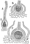
This illustration shows the germination of the gametospore: A, section of a mature archegonium with canal cells dissolved, thus forming a passageway to the large female gamete, g. B, sectional view of base of archegonium, showing the germinating gametospore in two-cell stage. The perianth, p, is seen growing up about the archegonium. C, later stage in growth of the gametospore. The lower cell shown in B is forming stalk cells, while the outer cell has produced densely granular cells that will later by further division form spore mother cells and elaters.

Gametospore

This illustration shows the germination of the gametospore: A, section of a mature archegonium with…

This illustration shows the structure of capsule of Funaria: 5, capsule with calyptra, 5A, removed. 6, section of nearly mature capsule-sp, spore-forming cells surrounded on outside by loosely arranged chlorophyll-bearing cells; o, operculum; r, annulus; p, peristome; a, apophysis. 8, magnified view of a portion of the capsule, showing the annulus, r, and the thick-walled cells of the peristome, p, which are attached at their base to the epidermis by a double row of cells; sp, spore-forming cells, the doted line should run to the four roundish cells at the left. 7, the cells shown in 8, p, have split apart, thus orming the inner and outer teeth-like segments of the peristome.

Funaria

This illustration shows the structure of capsule of Funaria: 5, capsule with calyptra, 5A, removed.…
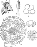
This illustration shows the flower and sporophylls of Angiosperms: 1, flower of Sedum with leaf-like perianth, p; microsporophylls, s; megasporophylls, c. 2, microsporophyll of the buttercup, showing four-lobed anther and filament. 3, diagram of a cross-section of anther, showing the breaking down of the tissue about the four sporangia and the beginning of the opening of the anther. 4, one of the sporangia from a young anther, as seen in cross-section-m, spore mother cells. The large cells surrounding the mother cells are nourishing cells, known as the tapetum, and disorganize as the spores mature. At the right a mother cell forming four microspores, the upper one being characterisitic of dicotyledons and the lowr of monocotyledons.

Angiosperm

This illustration shows the flower and sporophylls of Angiosperms: 1, flower of Sedum with leaf-like…

This illustration shows stages in the germination of the gametospore of Lepidium, sectional view: A, micropylar end of embryo sac, showing the enlarging gametospore provided with cell wall and attached to wall of sac. B, later growth-s, suspensor; e, embryo cell; en, endosperm cells. C, pro-embryo after first division of embryo cell. D, further divisions of embryo cell, showing formation of an epidermis and a central stem region. E, later growth, two growing regions, the cotyledons, appearing on the sides of the stem. F, plant or embryo, consisting of two cotyledons, c; stem, st, which terminates in the root, r; the endosperm cells, en, are ebing absorbed by the enlarging embryo; s, suspensor.

Lepidium

This illustration shows stages in the germination of the gametospore of Lepidium, sectional view: A,…
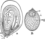
This illustration shows the structure of dicotyledonous seeds: A, nearly mature seed of Lepidium. The embryo consists of the hypocotyl, hy, ending below in the root, r, and the root cap and above the epicotly, pl. Two cotyledons, c; arise laterall from the stem; f, funiculus; mi, micropyle; in, integuments, en, remains of endosperm. B, section of seed of water lily-e, embryo with two cotyledons attached laterally to the minute stem of the embryo and surrounded by a layer of endosperm cells; mg, sporangial cells or perisperm; i, integument.

Dicotylendonous

This illustration shows the structure of dicotyledonous seeds: A, nearly mature seed of Lepidium. The…
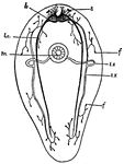
This is a diagram of a Turbellarian, showing the general arrangement of the nervous structures and one of the modes of occurrence of the excretory tubules, which in this case open seperately into the pharynx, on the ventral side of the animal. b., brain; e, eye-spots; ex, excretory canals consisting of a transverse portion passing from the mouth toward the dorsal side, and longitudinal tubes which branch into the capillary vessels terminating in f, the flame cells; lc., lateral nerve cords; m, mouth.

Turbellarian

This is a diagram of a Turbellarian, showing the general arrangement of the nervous structures and one…

This is a diagram of transverse section of a Turbellarian through the region of the mouth. d.m., dermo-muscular wall containing longitudinal fibres; ex, excretory system; f, flame cells; g, gut; l.c., lateral nerve cord; m, mouth; m.f., muscle fibres; ph., pharynx; t, testis; u, uterus; y, yolk glands.

Turbellarian

This is a diagram of transverse section of a Turbellarian through the region of the mouth. d.m., dermo-muscular…

This diagram shows a sagittal section of a Rotifer. b, brain; bl., excretory bladder; c, cloaca, the common opening of digestive and reproductive organs; co, caelom; e, eyespot; ex, excretory canal; f, flame cells; f.g., foot gland; ft., foot; g, gut; m, mouth; m.f., longitudinal muscle fibres; mx, mastax; o, ovary; pf., pharynx; s.g., salivary gland; t, tentacle; tr, trochus, or cilia-bearing disc.

Rotifer

This diagram shows a sagittal section of a Rotifer. b, brain; bl., excretory bladder; c, cloaca, the…

This diagram shows the longitudinal section of the anterior end of the annelid. A, sagittal section; B, frontal section to show anterior portion of nervous system. b, brain; co., nervous collar about the mouth; c.v., contractile blood vessels; d, dissepiment; d.m., dermo-muscular wall; d.v., dorsal blood vessel; m, mouth; n.c., nerve cells; n.f., nerve fibers; np., nephridia; p, prostomium; ph., pharynx; s, setae; sn., segmental nerves; v.g, ventral chain of fanglia; v.v., ventral blood vessel. Only a portion of the blood vascular system is shown, and this appears unsectioned in the figure.

Annelid

This diagram shows the longitudinal section of the anterior end of the annelid. A, sagittal section;…
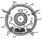
This diagram shows a transverse section of dero. c., caelom; c.l., cells of the so-called "lateral line"; d.m., dermo-muscular wall including muscles and skin; d.v., dorsal blood vessel; ect, ectoderm; ent, entoderm; g, gut; g.f., giant nerve fibers; gl, glandular cells assisting in digestion; m.c., circular muscle fibers; m.l., longitudinal muscle fibers; n, nephridium; n.v., ventral nerve chain, made up of nerve cells and nerve fibers; s, setae; v.v., ventral blood vessel.

Annelid

This diagram shows a transverse section of dero. c., caelom; c.l., cells of the so-called "lateral line";…