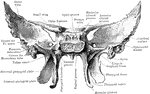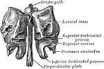
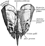
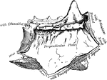
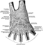
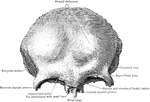
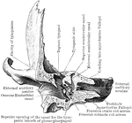
Perpendicular Plate of Ethmoid
Perpendicular plate of ethmoid, shown by removing the right lateral mass.
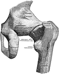
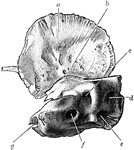
Back View of Hip Joint
Right hip joint, from behind. The joint capsule, except for strengthening ligaments, has been removed.
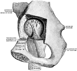
Hip Joint from Mesal Side
Right hip joint, from the mesal side. The bottom of the acetabulum had been chiseled away sufficiently…
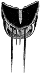
Bristles at the Extremity of the Abdomen of the Hydrophilus Piceus
"The female is sometimes seen clinging to aquatic plants head downward, forming her cocoon, terminated…

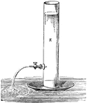
Backward Movement of Discharging Vessel
This experiment illustrates the law of action and reaction, which asserts that momentum cannot be imparted…
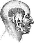
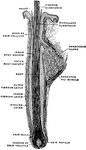
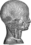
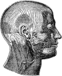
Temporal Muscle
The temporal muscle is a broad muscle situated at the side of the head and occupying the entire extent…
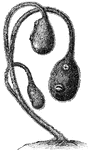
Social Tunicates
Ascidia Podunculata (Milne-Edwards) "The Tunicata would not probably by taken for animals at first sight.…
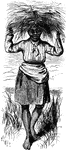
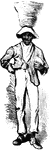
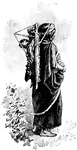
Field Hand
An illustration of an African American slave who is employed as a field hand, notice the basket balancing…
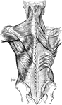
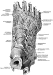
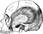
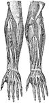
Back Muscles
Muscles of the back. On the left side is exposed the first layer; on the right side, the second layer…
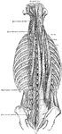
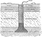
Diagram of Nitrate Bed
The nitrate is found on the east side of a low range of hills from fifteen to ninety miles back from…
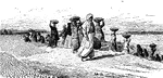
Mother Carrying Child
Almost as soon as one of these Indian babies is born it is wrapped in a skin or cloth and tied to a…
Hair of the Head
A hair of the head still in the course of growth, with hair bulb in longitudinal section.
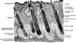
Hair of the Head in Longitudinal Section
Vertical section through the skin of head. The hair of the head in longitudinal section.
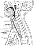
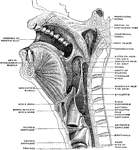
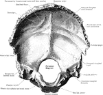
Sagittal Section of the Head and Neck
Sagittal median section of the head and neck. The head is thrown backward into complete extension which…
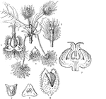
Medusa's Head Spurge
Euphorbia caputmedusae or the Medusa's Head is a spurge from the diverse genus, Euphorbia.
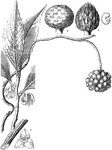
Common Beech
"Fagus sylvatica; 1. male catkins; 2. female do.; 3. the latter, with the scales of the involucre stripped…
Kadsura
"Kadsura japonica. 1. a calyx; 2. a head of stamens; 3. a pistil; 4. a section of a seed." -Lindley,…
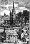
Hatfield, Herts
Dating back to Saxon times, the village of Hatfield was first known as "Hetfelle" and then became known…
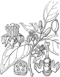
Micromelum
"Micromelum monophyllum. 1. a flower; 2. the pistil when the calyx is rolled back; 3. a cross section…
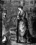
Woman Watching Fire Place
An illustration of a woman resting upon the back of a chair gazing into a fire.
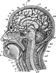
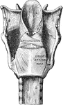
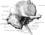
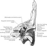
Section of the Head and Neck
Section of head and neck from front to back. Labels: 1, windpipe; 2, larynx; 3, spinal marrow; 4, pharynx;…
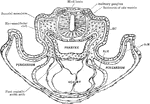
Transverse Section of a Rat Embryo
Transverse section of a rat embryo. Showing relation of the paraxial mesoderm of the head to the lateral…
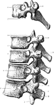
Thoracic Vertebrae
First, ninth, tenth, eleventh, and twelfth thoracic vertebrae from the left side. 1, Inferior articular…
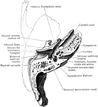
Anterior Half of Section Through Temporal Bone
The anterior half of a vertical transverse section through the left temporal bone.
Posterior Half of Section Through Temporal Bone
The posterior half of a vertical transverse section through the left temporal bone.
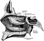
Horizontal Section of Temporal Bone
Horizontal section through left temporal bone showing lower half of section.
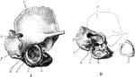
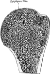
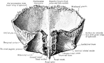
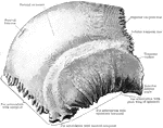
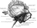
Temporal Bone at Birth
A, The outer surface of the right temporal bone at birth. B, The same with squamozygomatic portion removed.…
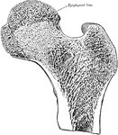
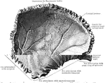
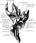
Temporal Bone at Birth
Inner surface of right temporal bone at birth. am squamozygomatic; b, petrosquamosal suture and foramen…