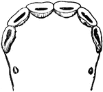
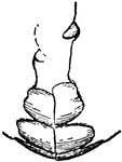
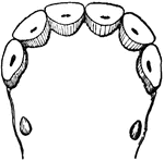
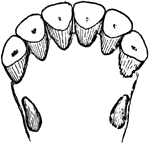
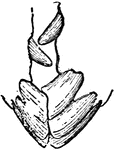
The lower nippers of a colt at three years of age.

Lower nippers at three years

The lower nippers of a colt at three years of age.

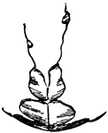
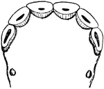
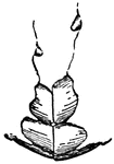
The lower nippers of a colt at four years.

Lower nippers at four years

The lower nippers of a colt at four years.

Lower nippers of a colt at five years of age.

Lower nippers at five years

Lower nippers of a colt at five years of age.

A colt's nippers at five years of age (side view)

Nippers at five years

A colt's nippers at five years of age (side view)

Lower nippers of a colt at six years of age.

Lower nippers at six years

Lower nippers of a colt at six years of age.

Side view of a colt's nippers at six years of age.

Nippers at six years

Side view of a colt's nippers at six years of age.

Side view of a colt's nippers at seven years of age.

Nippers at seven years

Side view of a colt's nippers at seven years of age.

A colt's lower nippers at eight years.

Lower nippers at eight years

A colt's lower nippers at eight years.

Lower nippers of an old horse.

Lower Nippers of an Old Horse

Lower nippers of an old horse.

Side view of the nippers of an old horse.

Nippers of an Old Horse

Side view of the nippers of an old horse.

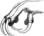
Foot of the eagle owl, with sharp talons for grasping prey.

Foot of the Eagle Owl

Foot of the eagle owl, with sharp talons for grasping prey.

"In human anatomy, certain regions into which the abdomen is arbitrarily divided for the purpose of mapping its surface with reference to the viscera which lie beneath these reions respectively."-Wright, 1902

Abdominal

"In human anatomy, certain regions into which the abdomen is arbitrarily divided for the purpose of…
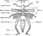
"Anatomy of the external skeleton of an insect" — Goodrich, 1859

External Anatomy of an Insect Skeleton

"Anatomy of the external skeleton of an insect" — Goodrich, 1859

"The structure of the mouth in insects exhibits very remarkable modifications, and these are of the utmost importance in the classification of these creatures. In some insects the mouth is formed exclusively for biting; in others, as exclusively for suction; while in others again it is fitted for the performance of both these actions; and the form of its constituent parts of course undergoes corresponding changes; but the same organs really exist in all, modified in apearance, indeed, so as sometimes to be scarecely recognizable." — Goodrich, 1859

Mouth and Tongue of a Bee

"The structure of the mouth in insects exhibits very remarkable modifications, and these are of the…

"a, head, antennae, &c; b, pharynx; c, crop; d, gizzard; e, chyle-forming stomach; f, biliary vessels; g, small intestine; h, secreting organs" — Goodrich, 1859

Digestive Apparatus of an Insect

"a, head, antennae, &c; b, pharynx; c, crop; d, gizzard; e, chyle-forming stomach; f, biliary vessels;…
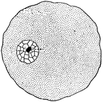
"In a general what we may describe a cell as a tiny mass of jelly in which floats another still smaller mass of slightly different composition, called the <em>nucleus</em> of the larger one."

Cell

"In a general what we may describe a cell as a tiny mass of jelly in which floats another still smaller…

"A, columnar cells found lining various parts of the intestines (called <em>columnar epthelium</em>); B, cells of a fusiform or spindle shape found in the loose tissue under the skin and in other parts (called <em>connective-tissue cells</em>); C, cell having many processes or projections - such are found in some kinds of connective tissue; D, primitive cells composed of protoplasm with nucleus, and having no cell wall." &mdash; Blaisedell, 1904

Various forms of cells

"A, columnar cells found lining various parts of the intestines (called columnar epthelium);…
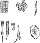
"A, columnar cells of intestine; B, polyhedral cells of the conjuctiva; C, ciliated conical cells of the trachea; D, ciliated cell of frog's mothl E, inverted conical cell of the trachea; F, squamous cell of the cavity of the mouth, seen on its broad surface; G, squamous cell, seen edgeways." &mdash; Blaisedell, 1904

Various kinds of epithelial cells

"A, columnar cells of intestine; B, polyhedral cells of the conjuctiva; C, ciliated conical cells of…

"One of the simplest of the tissues in the body is called the <em>epithelium</em>, and its cells are called epithelial cells. It serves as a lining of various cavities, a covering for the surface of the body, and the essential part of various organs called glands." — Blaisedell, 1904

Cross-Section of the Epithelium

"One of the simplest of the tissues in the body is called the epithelium, and its cells are…

"The connective tissue with white fibers sometimes forms a very thin sheet, as in the delicate covering of a bone known as the periosteum, or it may be made up into ropelike bands, as in the ligaments of joints and the tendons of muscles." &mdash; Blaisedell, 1904

White fibrous tissue

"The connective tissue with white fibers sometimes forms a very thin sheet, as in the delicate covering…
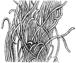
"Along with white fibrous tissue, the yellow fibrous tissue... makes the coats of the arteries, and certain ligaments, elastic." &mdash; Blaisedell, 1904

Yellow elastic tissue

"Along with white fibrous tissue, the yellow fibrous tissue... makes the coats of the arteries, and…

"Consisting of a very fine network of fibrils, around which are cells of various sizes." — Blaisedell, 1904

Connective Tissue from a Lymphatic Gland

"Consisting of a very fine network of fibrils, around which are cells of various sizes." — Blaisedell,…

"Showing (1) cartilage with martrix and cells; (2) cartilage with matrix containing cells and white fibers." &mdash; Blaisedell, 1904

Longitudinal section of cartilage

"Showing (1) cartilage with martrix and cells; (2) cartilage with matrix containing cells and white…

"There are in all two hundred and six seperate bones in the adult skelton. The teeth are not bones, but are a part of the skin." — Blaisedell, 1904

Human skeleton

"There are in all two hundred and six seperate bones in the adult skelton. The teeth are not bones,…

"The Right Femur sawed in Two Lengthwise." — Blaisedell, 1904

Cross Section of a Femur

"The Right Femur sawed in Two Lengthwise." — Blaisedell, 1904

"Little openings (Haversian canals) are seen, and around them are arranged rings of bone with little dark spaces (<em>lacunae</em>), from which branch out fine dark lines (<em>analiculi</em>)." &mdash; Blaisedell, 1904

Cross-section from a shaft of a long bone

"Little openings (Haversian canals) are seen, and around them are arranged rings of bone with little…
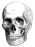
"A, frontal bone; B, parietal bone; C, temporal bone; D, sphenoid bone; E, malar bone; F, upper jawbone; G, lower jawbone" &mdash; Blaisedell, 1904

Human skull

"A, frontal bone; B, parietal bone; C, temporal bone; D, sphenoid bone; E, malar bone; F, upper jawbone;…
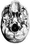
"A, Palate process of upper jawbone; B, zygoma, forming zygomatic arch; C, condyle, for forming articulation with atlas; D, foramen magnum; E, occipital bone" &mdash; Blaisedell, 1904

Base of skull

"A, Palate process of upper jawbone; B, zygoma, forming zygomatic arch; C, condyle, for forming articulation…

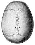
"The Top of the Skull, showing the Sutures." &mdash; Blaisedell, 1904

Top of skull

"The Top of the Skull, showing the Sutures." — Blaisedell, 1904

"The <em>spine</em> or backbone, serves as a support for the whole body. It is made up of a number of seperate bones called vertebrae, between which are placed elastic pads, or cushions, of cartilage." &mdash; Blaisedell, 1904

Spinal column

"The spine or backbone, serves as a support for the whole body. It is made up of a number of…
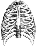
"The <em>ribs</em> are long, flat, and curved bones which bend round the chest somewhat like the hoops of a barrel. There are twenty-four ribs, twelve on each side." &mdash; Blaisedell, 1904

Thorax

"The ribs are long, flat, and curved bones which bend round the chest somewhat like the hoops…
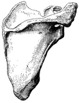
"The <em>shoulder-blade</em> is a large, flat, three-sided bone, which is placed on the upper and back part of the chest. On the outer side it has a saucerlike cavity on which the rounded head of the arm bone rests." &mdash; Blaisedell, 1904

Scapula

"The shoulder-blade is a large, flat, three-sided bone, which is placed on the upper and back…

"The <em>humerus</em>, a long, hollow bone, rests against a shallow socket on the shoulder blade. It is joined at the elbow to the bones of the forearm." &mdash; Blaisedell, 1904

Humerus

"The humerus, a long, hollow bone, rests against a shallow socket on the shoulder blade. It…

"The <em>ulna</em>, or elbow bone, is the larger of these two bones. It is joined to the humerus by a hinge joint at the elbow. It is on the same side as the little finger. The <em>radius</em>, queerly named because it is supposed to resemble one of the spokes of a wheel, is the long, slightly curved, outer bone fo the forearm. It is on the same side as the thumb. Its upper end is fastened both to the ulna and the humerus." — Blaisedell, 1904

Ulna and Radius

"The ulna, or elbow bone, is the larger of these two bones. It is joined to the humerus by…

"The <em>femur</em>, or thigh bone, the largest and heaviest bone in the body, reaches from the hip to the knee. It has a rounded head, which fits into the cuplike cavity in the hip bone which has already been mentioned." &mdash; Blaisedell, 1904

Femur

"The femur, or thigh bone, the largest and heaviest bone in the body, reaches from the hip…

"The <em>leg</em> consists, like the forearm, of two bones. The larger, a strong, three-sided bone with a sharp edge in front, is called the <em>tibia</em>. It is commonly known as the shin bone. The smaller bone, bound at both ends to the tibia, as a pin is to a brooch, is called the <em>fibula</em>, meaning a buckle or clasp. It is a long, slender bone on the outside of the leg, and is lower end forms the outer ankle." — Blaisedell, 1904

Tibia and Fibula

"The leg consists, like the forearm, of two bones. The larger, a strong, three-sided bone with…

"The foot is built in the form of a half-dome or half-arch. This is to afford a broad, strong support surface for the support of the weight of the body. The bones of the toes and the heel form the piers, while the little bones wedged in between the metatarsal bones and the heel make up the keystone of the arch. This arch gives a certain amount of spring and elasticity to the feet, and hence it is of the upmost importance in preventing jars and jolts." — Blaisedell, 1904

Bones of the Foot

"The foot is built in the form of a half-dome or half-arch. This is to afford a broad, strong support…

"Showing how the Ends of the Bones are shaped to form the Elbow Joint. The cut ends of a few ligaments are seen." — Blaisedell, 1904

Elbow Joint

"Showing how the Ends of the Bones are shaped to form the Elbow Joint. The cut ends of a few ligaments…

"The bones are fastened together, kept in place, and their movements limited, by tough and strong bands, or straps called <em>ligaments</em>, from a word meaning to bind." — Blaisedell, 1904

Powerful Ligament at the Hip Joint

"The bones are fastened together, kept in place, and their movements limited, by tough and strong bands,…

"The bones are fastened together, kept in place, and their movements limited, by tough and strong bands, or straps, called <em>ligaments</em>, from a word meaning to bind." — Blaisedell, 1904

Ligaments of the Foot and Ankle

"The bones are fastened together, kept in place, and their movements limited, by tough and strong bands,…

"When a bone is broken, blood trickles out between the injured parts, and afterwards gives place to a sticky, watery fluid, which gradually becomes thicker, like a syrup or jelly. This is slowly replaced by a new bone structure and forms a kind of cement to gold together the broken ends." — Blaisedell, 1904

Broken Radius

"When a bone is broken, blood trickles out between the injured parts, and afterwards gives place to…
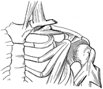
"When a bone is broken, blood trickles out between the incjured parts, and afterwards gives place to a sticky, watery fluid, which gradually becomes thicker, like a syrup or jelly. This is slowly replaced by a new bone structure and forms a kind of cement to gold together the broken ends." — Blaisedell, 1904

Broken Clavicle

"When a bone is broken, blood trickles out between the incjured parts, and afterwards gives place to…

"When a bone is broken, blood trickles out between the injured parts, and afterwards gives place to a sticky, watery fluid, which gradually becomes thicker, like a syrup or jelly. This is slowly replaced by a new bone structure and forms a kind of cement to gold together the broken ends." — Blaisedell, 1904

Broken Tibia

"When a bone is broken, blood trickles out between the injured parts, and afterwards gives place to…
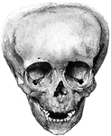
"Showing how the Bones of the Skull may be artificially deformed by "head-binding." From the photograph of a "triangular" skull found in an Indian grave in Ancon, Peru." &mdash; Blaisedell, 1904

Deformed skull

"Showing how the Bones of the Skull may be artificially deformed by "head-binding." From the photograph…

"A Portion of a Striped Muscular Fiber. Highly magnified. A, fiber separating into disks; B, fibrillae; C, cross-section of a disk." — Blaisedell, 1904

Striped Muscular Fiber

"A Portion of a Striped Muscular Fiber. Highly magnified. A, fiber separating into disks; B, fibrillae;…
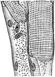
"A Portion of Striped Muscular Fiber, showing Stripes and Nuclei. Highly magnified." — Blaisedell, 1904

Striped Muscular Fiber

"A Portion of Striped Muscular Fiber, showing Stripes and Nuclei. Highly magnified." — Blaisedell,…

"<em>The involuntary muscles</em> consist of ribbon-shaped bands which surround hollow tubes or cavities in the body. They are never attached to bony levers nor are they furnished with tendons, as are the voluntary muscles." — Blaisedell, 1904

Spindle Cell of Involuntary Muscle

"The involuntary muscles consist of ribbon-shaped bands which surround hollow tubes or cavities…

"A single muscle rarely or never contracts alone, but always in harmony with a number of other muscles. Even the simplest movement we can make requres the combined action of several muscles to carry it out. If the movement is at all difficult, such as playing on the piano or riding on the bicycle, the different muscles must contract in a certain order and with a certain strength and rapidity." — Blaisedell, 1904

Superficial Muscles of the Body

"A single muscle rarely or never contracts alone, but always in harmony with a number of other muscles.…

"Tendons are white, glistening cords, or straps, which connect the muscles with the bones." &mdash; Blaisedell, 1904

Achillles tendon

"Tendons are white, glistening cords, or straps, which connect the muscles with the bones." —…

"Tendons on the Back of the Left Hand" &mdash; Blaisedell, 1904

Tendons of the hand

"Tendons on the Back of the Left Hand" — Blaisedell, 1904

"Tendons on the Top of the Right Foot." &mdash; Blaisedell, 1904

Tendons of the foot

"Tendons on the Top of the Right Foot." — Blaisedell, 1904

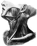
"Principal Muscles on the Left Side of the Neck." &mdash; Blaisedell, 1904

Muscles of the neck

"Principal Muscles on the Left Side of the Neck." — Blaisedell, 1904

"Some of the Larger Muscles on the Back of the Shoulder and the Arm." — Blaisedell, 1904

Muscles of the Back and Shoulder

"Some of the Larger Muscles on the Back of the Shoulder and the Arm." — Blaisedell, 1904

"Some of the Larger Muscles on the back of the Thigh. Powerful tendons at the hip and on the back of the knee are well shown." — Blaisedell, 1904

Thigh Muscles

"Some of the Larger Muscles on the back of the Thigh. Powerful tendons at the hip and on the back of…

"The head is represented as having been thrown back, and the tongue drawn forward. <em>A</em>, <em>B</em> and <em>C</em>, cainine; <em>D, E</em> bicuspids; <em>F, H, K,</em> molars; <em>M</em> anterior pillar of the fauces; <em>N</em>, tonsil; <em>L</em> uvula; <em>O</em>, upper part of the pharynx; <em>P</em>, tongue drawn forward; <em>R</em> linear ridge, or raphe</em>." &mdash; Blaisedell, 1904

Back view of the adult mouth

"The head is represented as having been thrown back, and the tongue drawn forward. A, B

"The principal muscles are seen on the left, and superficial veins on the right." — Blaisedell, 1904
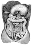
Principal Organs of the Thorax and Abdomen

"The principal muscles are seen on the left, and superficial veins on the right." — Blaisedell, 1904

"<em>A</em>, simple tubular gland; <em>B</em>, gland with mouth shut and sac formed; <em>C</em> gland with a coiled tube; <em>D</em>, plan of part of a racemose gland." &mdash; Blaisedell, 1904

Secreting glands

"A, simple tubular gland; B, gland with mouth shut and sac formed; C gland…

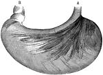
"<em>A</em>, cardiac end; <em>B</em>, pyloric end." &mdash; Blaisedell, 1904

Stomach

"A, cardiac end; B, pyloric end." — Blaisedell, 1904

"Showing the Relations of the Stomach, Liver, Intestines, Spleen, and other Organs of the Abdomen. <em>A</em>duodenum</em>; <em>B</em>, upper end of the small intestine; <em>C</em>, lower end of the small intestine; <em>D</em>, caecum; <em>E</em>, bladder. The liver and stomach are drawn up and portions of the lower intesting have been cut away." &mdash; Blaisedell, 1904

Digestive system

"Showing the Relations of the Stomach, Liver, Intestines, Spleen, and other Organs of the Abdomen. Aduodenum;…
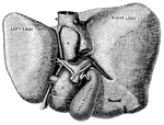
"The Liver seen from Below and Behind. <em>A</em> and </em>B</em>, smaller lobes of the liver; <em>C</em>, portal vein; <em>D</em> hepatic artery; <em>E</em>, inferior vena cava; <em>F</em>, trunk of the left hepatic vein; <em>G</em>, hepatic vein." &mdash; Blaisedell, 1904

Liver

"The Liver seen from Below and Behind. A and B, smaller lobes of the liver; C,…