The human nervous system includes the brain, spinal cord, and the nerves. Labels: A, cerebrum; B, cerebellum.
Nervous System

The human nervous system includes the brain, spinal cord, and the nerves. Labels: A, cerebrum; B, cerebellum.

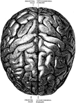
The surface of the cerebrum.

Surface of the Cerebrum

The surface of the cerebrum.

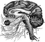
The brain and the origin of the twelve pairs of cranial nerves. Labels: F, E, the cerebrum; D, the cerebellum, showing the arbor vitae; G, the eye; H, the medulla oblongata; A, the spinal cord; C and B, the first two pairs of spinal nerves.

The Brain and the Cranial Nerves

The brain and the origin of the twelve pairs of cranial nerves. Labels: F, E, the cerebrum; D, the cerebellum,…

Next to the choroid and comprising about 1/4 the entire thickness of the retina is a multitude of transparent, colorless, microscopic rods (a) evenly arranged and packed side by side, like the seeds in the disk of a sunflower. Among them at regular intervals, are interspersed the cones (b). Delicate nerve fibers pass from the ends of the rods and cones, each expanding into a granular body (c) thence weaving a mesh (d) and again expanding into the granules (f). Last is a layer of fine nerve fibers (g) and gray, ganglionic cells (h) like the gray matter of the brain, whence filaments extend into the fibers of the optic nerve (i).

The Structure of the Retina

Next to the choroid and comprising about 1/4 the entire thickness of the retina is a multitude of transparent,…
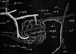
Diagrammatic representation of the submaxillary gland of the dog with its nerves and blood vessels. Labels: sm. gld., the submaxillary gland into the duct (sm. d.) of which a cannula has been tied. The sublingual gland and duct are not proceeding from the facial nerve, becoming conjoined with the lingual at n.l'., and afterwards diverging and passing to the gland along the duct; sm. gl., submaxillary ganglion with its roots; n. l., the lingual nerve proceeding to the tongue; a. car., the carotid artery. two branches of which, a. sm. a. and r. sm. p. pass to the anterior and posterior parts of the gland; v. sym., the conjoined vagus and sympathetic trunks; gl. cer. s., the superior cervical ganglion, two branches of which forming a plexus, a.f., over the facial artery are distributed (n.sym.sm.) along the two glandular arteries to the anterior and posterior portion of the gland. The arrows indicate the direction taken by the nervous impulses; during reflex stimulations of the gland they ascend to the brain by the lingual and descend by the chorda tympani.

Submaxillary Gland of Dog with Nerves and Blood Vessels

Diagrammatic representation of the submaxillary gland of the dog with its nerves and blood vessels.…

View of the cerebrospinal axis of the nervous system. The right half of the cranium and trunk of the body have been removed by a vertical section; the membranes of the brain and spinal cord have also been removed, and the roots and first part of the fifth and ninth cranial, and of all the spinal nerves of the right side, have been dissected out and laid separately on the wall of the skull and on the several vertebrae opposite to the place of their natural exit from the craniospinal cavity.

Central Nervous System

View of the cerebrospinal axis of the nervous system. The right half of the cranium and trunk of the…
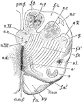
Anterior or dorsal section of the medulla oblongata in the region of the superior pyramidal decussation. a.m.f, anterior median fissure; f.a., superficial arciform fibers emerging from the fissure; py., pyramid, n.ar., nuclei of arciform fibers; f.a., deep arciform becoming superficial; o, lower end of olivary nucleus; n.l., nucleus lateralis; f.r., formatio reticularis; f.a. 2, arciform fibers proceeding from the formatio reicularis; g, substantia gelatinosa of Rolando; a.V., ascending root of fifth nerve; n.c., nucleus cuneatus; n.c.', external cuneate nucleus; n.g., nucleus gracilis; f.g., funiculus gracilis; p.m.f., posterior median fissure; c., central canal surrounded by gray matter, in which are n. XI., nucleus of the spinal accessory, and n. XII., nucleus of the hypoglossal; s.d., superior pyramidal decussation.

Medulla Oblongata

Anterior or dorsal section of the medulla oblongata in the region of the superior pyramidal decussation.…
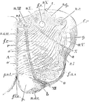
Section of the medulla oblongata at about the middle of the olivary body. f.l.a., anterior median fissure; n.ar., nucleus arciformis; p., pyramid; XII., bundle of hypoglossal nerve emerging from the surface; at b, it is seen coursing between the pyramid and the olivary nucleus, o.; f.a.e., external arciform fibers; n.l., nucleus lateralis; a., arciform fibers passing towards restiform body, partly through the substantia gelatinosa, g., partly superficial to the ascending root of the fifth nerve, a.V.; X., bundle of vagus root emerging; f.r., formatio reticularis; c.r., corpus restiform, beginning to be formed, chiefly by arciform fibers, superficial and deep; n.c., nucleus of the funiculus teres; n.am., nucleus ambiguus; r., raphe; A., continuation of the anterior column of cord; o', o", accessory olivary nucleus; p.o., pedunculus olivae.

Medulla Oblongata

Section of the medulla oblongata at about the middle of the olivary body. f.l.a., anterior median fissure;…
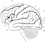
Lateral view of the brain. F, Frontal lobe; P, Parietal lobe; O, Occipital lobe; T, Temporal lobe; S, fissure of Sylvius; S', horizontal; S", ascending ramus of the same; c, sulcus centralis (fissure of Rolando); A, ascending frontal convolutions; fr, superior, f2, inferior frontal sulcus; f3, precentral sulcus; P1, superior parietal lobule; P2, inferior parietal lobule consisting of P2, supramarginal gyrus, and P2', angular gyrus; ip, interparietal sulcus; cm, termination of callosomarginal fissure; O1, first O2, second, 03, third occipitals inferior; T1, first T2, second, T3, third temporal convolutions; tr, first , t2, second temporal fissures.

Lateral View of the Brain

Lateral view of the brain. F, Frontal lobe; P, Parietal lobe; O, Occipital lobe; T, Temporal lobe; S,…
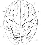
View of brain from above. F, Frontal lobe; P, Parietal lobe; O, Occipital lobe; T, Temporal lobe; S, fissure of Sylvius; S', horizontal; S", ascending ramus of the same; c, sulcus centralis (fissure of Rolando); A, ascending frontal convolutions; fr, superior, f2, inferior frontal sulcus; f3, precentral sulcus; P1, superior parietal lobule; P2, inferior parietal lobule consisting of P2, supramarginal gyrus, and P2', angular gyrus; ip, interparietal sulcus; cm, termination of callosomarginal fissure; O1, first O2, second, 03, third occipitals inferior; T1, first T2, second, T3, third temporal convolutions; tr, first , t2, second temporal fissures. Sr, end of horizontal ramus of fissure of Sylvius.

View of Brain from Above

View of brain from above. F, Frontal lobe; P, Parietal lobe; O, Occipital lobe; T, Temporal lobe; S,…

View of the right hemisphere in the median aspect. CC, corpus callosum longitudinally divided; Gf, gyrus fornicatus; H, gyrus hippocampi; h, sulcus hippocampi; U, uncinate gyrus; cm, calloso marginal fissure; F1, median aspect of first frontal convolution; c, terminal portion of sulcus centralis (fissure of Rolando); A, ascending frontal; B, ascending parietal convolution; P1', praecuneus; Oz, cuneus; po, parieto-occipital fissure; o, sulcus occipitalis transversus; oc, calcarine fissure; oc', superior; oc", inferior ramus of the same; D, gyrus descendens; T4, gyrus occipito-temporalis lateralis (lobulus fusiformis) ; T5, gyrus occipito-temporalis medialis (lobulus lingualis).

Right Hemisphere of the Brain

View of the right hemisphere in the median aspect. CC, corpus callosum longitudinally divided; Gf, gyrus…

Diagrammatic horizontal section of a vertebrate brain. Mb, midbrain: what lies in front of this is the fore-, and what lies behind, the hindbrain; Lt, lamina terminalis; Olf, olfactory lobes; Hmp, hemispheres; Th. E, thalamencephalon; Pn, pineal gland; Py, pituitary body; F. M., foramen of Munro; cs, corpus striatum; Th, optic thalamus; CC, crura cerebri; the mass lying above the canal represents the corpora quadrigemina; Cb, cerebellum; I-IX, the nine pairs of cranial nerves; 1, olfactory ventricle; 2, lateral ventricle; 3, third ventricle; 4, fourth ventricle; +, iter a tertio ad quartum ventriculum.

Horizontal Section of a Vertebrate Brain

Diagrammatic horizontal section of a vertebrate brain. Mb, midbrain: what lies in front of this is the…

Longitudinal and vertical diagrammatic section of a vertebrate brain. Mb, midbrain: what lies in front of this is the fore-, and what lies behind, the hindbrain; Lt, lamina terminalis; Olf, olfactory lobes; Hmp, hemispheres; Th. E, thalamencephalon; Pn, pineal gland; Py, pituitary body; F. M., foramen of Munro; cs, corpus striatum; Th, optic thalamus; CC, crura cerebri; the mass lying above the canal represents the corpora quadrigemina; Cb, cerebellum; I-IX, the nine pairs of cranial nerves; 1, olfactory ventricle; 2, lateral ventricle; 3, third ventricle; 4, fourth ventricle; +, iter a tertio ad quartum ventriculum. Lamina terminalis is represented by the strong black line joining Pn and Py.

Vertical Section of a Vertebrate Brain

Longitudinal and vertical diagrammatic section of a vertebrate brain. Mb, midbrain: what lies in front…
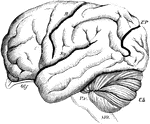
Brain of the Orangoutang, showing arrangement of the convolutions. Sy, fissure of Sylvius; R, fissure of Rolando; E P, external perpendicular fissure; Olf, olfactory lobe; Cb, cerebellum; PV, pons Varolii; MO, medulla oblongata. As contrasted with the human brain, the frontal lobe is short and small relatively, the fissure of Sylvius is oblique, the temporo-sphenoidal lobe very prominent, and the external perpendicular fissure very well marked.

Brain of the Orangutan

Brain of the Orangoutang, showing arrangement of the convolutions. Sy, fissure of Sylvius; R, fissure…

Brain of dog, viewed from above and in profile. F, frontal fissure sometimes termed crucial sulcus, corresponding to the fissure of Rolando in man. S, fissure of Sylvius, around which the four longitudinal convolutions are concentrically arranged; 1, flexion of head on the neck, in the median line; 2, flexion of head on the anterior limb; 5, 6, flexion and extension of posterior limb; 7, 8, 9, contraction of orbicularis oculi, and the facial muscles in general. The unshaded part in that exposed by the opening of the skull.

Brain of a Dog

Brain of dog, viewed from above and in profile. F, frontal fissure sometimes termed crucial sulcus,…

Diagrams of monkey's brain to show the effects of electric stimulation of certain spots. Labels: 1. causes movement of supination and flexion of forearm; 7, elevation of the upper lip; 8, conjoint action of elevation of upper lip and depression of lower; 9, opening mouth and protrusion of tongue; 10, retraction of tongue; 11, action of platysma; 12, elevation of eyebrows and eyelids, dilation of pupils, and turning head to opposite side and upwards, with usually contraction of the pupils; 13', similar action, but eyes usually directed downwards; 14, retraction of opposite ear, head turn to the opposite side, the eyes widely opened, and pupils dilated; 15, stimulation of this regions, which corresponds to the tip of the uncinate convolution, caused torsion of the lip and nostril of the same side.

Brain of a Monkey to Show effects of Electric Stimulation

Diagrams of monkey's brain to show the effects of electric stimulation of certain spots. Labels: 1.…

Diagram to indicate position of centers on the external surface of the brain.

Motor Centers of the Brain

Diagram to indicate position of centers on the external surface of the brain.
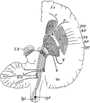
Diagram to show the connecting of the Frontal Occipital Lobes with the Cerebellum. The dotted lines passing in the crusta, outside the motor fibers, indicate the connection between the temporo-occipital lobe and the cerebellum. F.C., the fronto-cerebellar fibers, which pass internally to the motor tract in the crusta; I.F., fibers from the caudate nucleus to the pons. FR., frontal lobe; Oc, occipital lobe; AF. ascending frontal; AP., ascending frontal convolution; FR., fissure of Rolando; IPF., interparietal fissure, a section of crus is lettered on the left side. SN., substantia nigra; PY. pyramidal motor fiber, which on the right is shown as continuous lines converging to pass through the posterior limb of IC. internal capsule ( the knee or elbow of which is shown thus) upwards into the hemisphere and downwards through the pons to cross at the medulla in the anterior pyramids.

Brain Showing Connection of Frontal Occipital Lobe with Cerebellum

Diagram to show the connecting of the Frontal Occipital Lobes with the Cerebellum. The dotted lines…
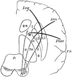
Diagram to show the relative position of several motor tracts in their course from the cortex to the crus. The section through the convolutions is vertical; that through the internal capsule, I, C, horizontal; that through the crus again vertical. C, N, caudate nucleus; O, H, optic thalamus; L2 and L3, middle and outer part of lenticular nucleus; f, a, l, face, arm, and leg fibers. The words in italic indicate corresponding cortical centers.

Motor Tracts of the Brain

Diagram to show the relative position of several motor tracts in their course from the cortex to the…
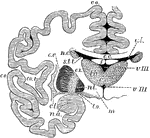
Vertical section through the cerebrum and basic ganglia to show the relation of the latter. Labels: co, cerebral convolutions; c, corpus callosum; v.l., lateral ventricle; f, fornix; vIII., third ventricle; n.c., caudate nucleus; th, optic thalamus; n.l., lenticular nucleus; c.i., internal capsule; c.l., claustrum; c.e., external capsule; m, corpus mammillare; t.o., optic tract; s.t.t., stria terminalis; n.a., nucleus amygdala; cm, soft commissure.

The Cerebrum and Basic Ganglia of the Brain

Vertical section through the cerebrum and basic ganglia to show the relation of the latter. Labels:…
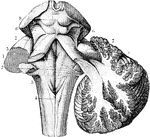
The cerebellum in section and fourth ventricle, with the neighboring parts. Labels: 1, median groove of fourth ventricle, ending below in the calamus scriptorius, with the longitudinal eminence formed by the fasciculi teretes, one on each side; 2, the same groove, at the place where the white streaks of th auditory nerve emerge from it to cross the floor of the ventricle; 3, in inferior crus or peduncle of the cerebellum, formed by the restiform body; 4, posterior pyramid; above this is the calamus scriptorius; 5, superior crus of cerebellum, or processus e cerebello ad cerebrum( or ad testes); 6, fillet to the side of the crura cerebri; 7, lateral grooves of the crura cerebri; 8, corpora quadrigemina.

The Cerebrum and Fourth Ventricle of the Brain

The cerebellum in section and fourth ventricle, with the neighboring parts. Labels: 1, median groove…

Outline sketch of a section of the cerebellum, showing the corpus dentatum. The section has been carried through the left lateral part of the pons, so as to divide the superior peduncle and pass nearly through the middle of the left cerebellar hemisphere. The olivary body has also been divided longitudinally so as to expose in section its corpus dentatum. c r, crus cerebri; f, fillet; q, corpora quadrigemina; s p, superior peduncle of the cerebellum divided; m p, middle peduncle or lateral part of the pons Varolii, with fibers passing from it into the white stem; a v, continuation of the white stem radiating towards the arbor vitae of the folia; c d, corpus dentatum; o, olivary body with its corpus dentatum; p, anterior pyramid.

The Cerebellum of the Brain

Outline sketch of a section of the cerebellum, showing the corpus dentatum. The section has been carried…

Vertical section of dog's cerebellum. Labels: p m, pia mater; p, corpuscles of Purkinje, which are branched nerve cells lying in a single layer and sending single processed downwards and more numerous ones upwards, which branch continuously and extend through the deep "molecular layer" towards the free surface; g, dense layer of ganglionic corpuscles., closely resembling nuclear layers of retina; f, layer of nerve fibers, with a few scattered ganglionic corpuscles. This last layer (f) constitutes part of the white matter of the cerebellum, while the layer between it and the free surface are gray matter.

Cerebellum of Dog's Brain

Vertical section of dog's cerebellum. Labels: p m, pia mater; p, corpuscles of Purkinje, which are branched…
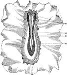
Portion of the germinal membrane, with rudiments of the embryo, from the ovum of a dog. The primitive groove, A, is not yet closed, and at its upper or cephalic end presents three dilatations, B, which correspond to the three divisions or vesicles of the brain. At its lower extremity the groove presents a lancet-shaped dilation (sinus rhomboidalis) C. The margins of the groove consist of clear pellucid nerve substance. Along the bottom of the groove is observed a faint streak, which is probably the chorda dorsalis. D. Vertebral plates.

Germinal Membrane of a Dog

Portion of the germinal membrane, with rudiments of the embryo, from the ovum of a dog. The primitive…
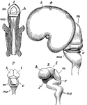
Early stages in development of human brain. 1, 2, 3, are from an embryo about seven weeks old; 4, about three months old. m, middle cerebral vesicle (mesencephalon); c, cerebellum; m o, medulla oblongata; i, thalamencephalon; h, hemispheres; i', infundibulum; Fig. 3 shows the several curves which occur in the course of development; Fig. 4 is a lateral view, showing the great enlargement of the cerebral hemispheres which have covered in the thalami, leaving the optic lobes, m, uncovered.

Development of the Brain

Early stages in development of human brain. 1, 2, 3, are from an embryo about seven weeks old; 4, about…

Side view of fetal brain at six months, showing commencement of formation of the principal fissures and convolutions. F, frontal lobe; P, parietal; O, occipital; T, temporal; a, commencing frontal convolutions; s, Sylvian fissure; s', its anterior division; c, within it the central lobe or island of Reil; r, fissure of Rolando; p, perpendicular fissure.

Fetal Brain at Six Months

Side view of fetal brain at six months, showing commencement of formation of the principal fissures…
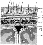
Diagram showing the layers of the scalp and membranes of the brain in section. Labels: a, skin; b, subcutaneous tissue with hair roots and vessels; c, epicranius; d, subepicranial layer; e, pericranium; fm parietal bone; g, dura mater; k, arachnoid; l, pia mater; m, cortex; n, in subdural space near a Pacchionian body projecting within the superior longitudinal sinus.

Layers of the Scalp and Membrane of the Brain

Diagram showing the layers of the scalp and membranes of the brain in section. Labels: a, skin; b, subcutaneous…
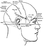
Showing the lines which indicate the position of the principal fissures of the brain.

Principle Fissures of the Brain

Showing the lines which indicate the position of the principal fissures of the brain.
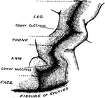
The convolutionary projections of the precentral gyrus, and their relationship to motor areas.

Precentral Gyrus in the Brain

The convolutionary projections of the precentral gyrus, and their relationship to motor areas.
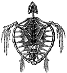
"The Tortoise will continue to live on for six months after it is deprived of its brain."

Skeleton of a Turtle

"The Tortoise will continue to live on for six months after it is deprived of its brain."
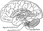
Scheme showing relations of the ventricles to the surface of the brain.

Relations of the Ventricles to the Surface of the Brain

Scheme showing relations of the ventricles to the surface of the brain.
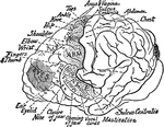
Semidiagrammatic view of the cerebrospinal axis.

Cerebrospinal Axis

Semidiagrammatic view of the cerebrospinal axis.

The lobes of the convex surface of the hemisphere.

Lobes of the Brain

The lobes of the convex surface of the hemisphere.

Location of the motor areas in the brain of a chimpanzee. The extent of the motor area is indicated by stippling; it lies entirely in from of the fissure of Rolando (sulcus centralis). Much of the motor area is hidden in the sulci. The regions marked eyes indicated the area whose stimulation gives conjugate movements of the eyeballs. It is doubtful however, whether these represent motor areas proper.

Motor Area of the Brain of a Chimpanzee

Location of the motor areas in the brain of a chimpanzee. The extent of the motor area is indicated…
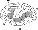
Lateral view of a brain hemisphere; cortical area V, damage to which produces "mind blindness" (word blindness); cortical area H, damage to which produced "mind deafness" (word deafness); cortical area S, damage to which caused the loss of audible speech; cortical area W, damage to which abolished the power of writing.

Association Area of the Brain

Lateral view of a brain hemisphere; cortical area V, damage to which produces "mind blindness" (word…
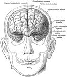
The brain in relation to the skull and face, anterior view.

Brain in Relation to Skull and Face

The brain in relation to the skull and face, anterior view.

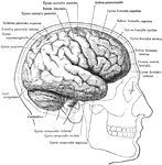
The brain in relation to the skull and face, lateral view.

Brain in Relation to Skull and Face

The brain in relation to the skull and face, lateral view.

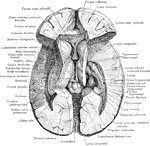
Dissection of the brain showing basal ganglia, third ventricle and adjacent structures viewed from above.
Dissection of the Brain

Dissection of the brain showing basal ganglia, third ventricle and adjacent structures viewed from above.

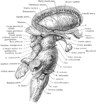
The right lateral aspect of the brain stem after the cerebral hemisphere (except the Corpus striatum) and the cerebellum (except the Nucleus dentatus) have been removed.

Brain Stem and Adjacent Structures

The right lateral aspect of the brain stem after the cerebral hemisphere (except the Corpus striatum)…
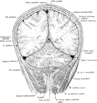
Frontal section of the head passing through the parietal and occipital cerebral lobes and he cerebellar hemispheres, viewed from behind.

Frontal Section of the Head

Frontal section of the head passing through the parietal and occipital cerebral lobes and he cerebellar…
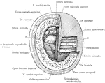
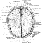
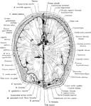
Section two inches above supraorbital border. Upper surface. The (*) on right indicates subaponeurotic tissue. (*) on left indicates pia mater.

Cross Section of Head

Section two inches above supraorbital border. Upper surface. The (*) on right indicates subaponeurotic…
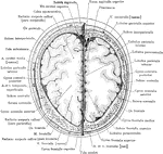
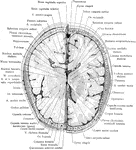
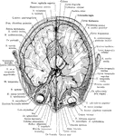
Section of head 4 cm above supraorbital border.

Cross Section of Head 4 cm above Supraorbital Border

Section of head 4 cm above supraorbital border.
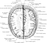
Section of head 3 cm above supraorbital border.

Cross Section of Head 3 cm above Supraorbital Border

Section of head 3 cm above supraorbital border.
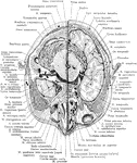
Section of head 2 cm above orbit.

Cross Section of Head 2 cm above Supraorbital Orbit

Section of head 2 cm above orbit.

Section of head 1 cm above orbit.

Cross Section of Head 1 cm above Orbit

Section of head 1 cm above orbit.

Section of the head at supraorbital margin.

Cross Section of Head at Supraorbital Margin

Section of the head at supraorbital margin.

Section of the head through the eyeballs.

Cross Section of Head Through Eyeballs

Section of the head through the eyeballs.

Section of the head through lower portion of orbit.

Cross Section of Head Through Lower Portion of Orbit

Section of the head through lower portion of orbit.
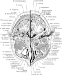
Section of the head immediately below the orbits, at the level of Reid's base line exposing the maxillary sinus.

Cross Section of Head Exposing Maxillary Sinus

Section of the head immediately below the orbits, at the level of Reid's base line exposing the maxillary…
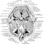
Section of the head through the nasopharynx.

Cross Section of Head Through the Nasopharynx

Section of the head through the nasopharynx.

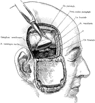
Exposure of the Gasserian ganglion and middle meningeal artery though a flap incision of the scalp and skull. The dura mater and brain are retracted upwards. The (*) points to the Foramen spinosum through which the middle meningeal artery passes as it enters the cranial cavity.

Incision of the Head Showing Gasserian Ganglion

Exposure of the Gasserian ganglion and middle meningeal artery though a flap incision of the scalp and…
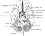
Simplified drawing of brain as seen from below, showing relations of brain stem to spinal cord and cerebrum.

Relation of Brain Stem to Spinal Cord

Simplified drawing of brain as seen from below, showing relations of brain stem to spinal cord and cerebrum.

Simplified drawing of brain as seen in mesial section, showing relation of brain stem, cerebrum and cerebellum, and ventricular spaces.

Brain in Mesial Section

Simplified drawing of brain as seen in mesial section, showing relation of brain stem, cerebrum and…
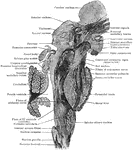
Sagittal section of brain stem; plane of section is somewhat lateral to midline.

Sagittal Section of Brain Stem

Sagittal section of brain stem; plane of section is somewhat lateral to midline.
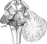
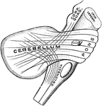
The peduncles of the cerebellum. On the left the three peduncles have been cut at their entrance into the cerebellum; on the right they are shown penetrating he cerebellar hemisphere.

Peduncles of the Cerebellum

The peduncles of the cerebellum. On the left the three peduncles have been cut at their entrance into…
Semidiagram of the three cerebellar peduncles.

Peduncles of the Cerebellum

Semidiagram of the three cerebellar peduncles.

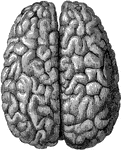
The cerebral hemispheres viewed from above.

Brain Viewed From Above

The cerebral hemispheres viewed from above.

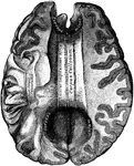
Shown is the corpus callosum. The corpus callosum is a thick stratum of transversely directed nerve fibers, by which almost every part of one cerebral hemisphere is connected with the corresponding part of the other cerebral hemisphere.

Brain Showing Corpus Callosum

Shown is the corpus callosum. The corpus callosum is a thick stratum of transversely directed nerve…

The fornix is a paired structure consisting of bilaterally symmetrical halves composed of longitudinally directed fibers which arch on each side from the region of the uncus to the corpus albicans. Shown is a diagram of the fornix.

Fornix

The fornix is a paired structure consisting of bilaterally symmetrical halves composed of longitudinally…
Typical arrangement of the cell layers of the cerebral cortex.

Cell Layers of the Cerebral Cortex

Typical arrangement of the cell layers of the cerebral cortex.