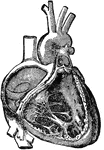
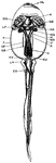
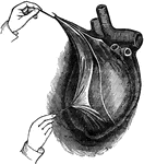
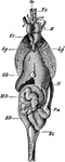
Bronchia and Veins of the Lungs
View of the bronchia and veins of the lungs, exposed by dissection, as well as the relative position…
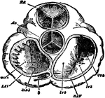
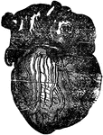
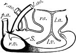
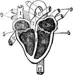
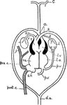
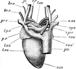
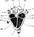
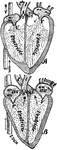
Heart
"Heart and blood vessels: A, B, Superior and inferior venae cavae; C, right auricle; D, right ventricle;…

Edgar Allan Poe
(1809-1849) Famous poet and story writer best known for The Raven, Annabel Lee, The Fall of the House…
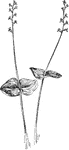
Heart-Leaved Twayblade
Of the orchid family (Orchidaceae), heart-leaved twayblade or Listera cordata.
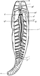
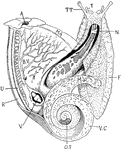
Lob Worm
"Dissection of lob-worm from dorsal surface. m., Opening of retracted buccal cavity; i., gullet; gl.,…
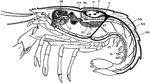
Lobster Organs
"Longitudinal section of lobster, showing some of the organs. H., Heart; AO., ophthalmic artery; SA.,…
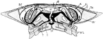
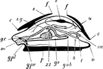
Crab Cephalothorax
"Section through cephalothorax of a crab. H., Heart; Te., extension of the tergum; ST., sternum; PL.,…
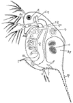
Daphnia
"Daphnia. E., Eye; A.2, second antenna; A.1, first antenna; dg., digestive caeca; s.g., shell gland;…
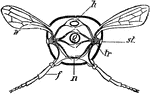
Insect Section
"Transverse section of insect. h., Heart; g., gut; n., nerve-cord; st., stigma; tr., trachea; w., wing;…
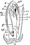
Mollusc Parts
"Ideal mollusc. m., Mouth; g.c., cerebral ganglia; c., edges of mantle skirt; z.g., duct of right lobe…
Dissection
"Dissection of snail. T., Short horn; TT., long horn with eye; N., cerebral ganglia; S.G., salivary…
Cuttlefish Structure
"Diagram of the structure of Sepia. a., Eight short arms around mouth; l.a., one of the two long arms;…
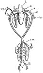
Cuttlefish Circulatory and Excretory Systems
"Diagram of circulatory and excretory systems in a Decapod-like Sepia. 1, Gill; 2, renal sac; 3, afferent…
Doliolum Mulleri
"'Nurse' of Doliolum mulleri. I., Inhalant, E., exhalant aperture; C., ciliated band round pharynx (P.);…
Doliolum Mulleri
"Sexual individual of Doliolum mulleri. G., gonads; B., gill-slits; I., Inhalant, E., exhalant aperture;…
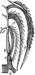
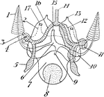
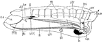
Respiration in Fishes
This figure shows the mode of respiration in fishes. The gills are seen bent over in the form of a feather.…
Salpa Africana
"Diagram of Salpa africana. o.a., Oral aperture; d.t., dorsal tubercle; te., tentacle; g., ganglion;…
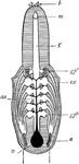
Hagfish Respiratory System
"Respiratory system of hag, from ventral surface. b., Barbules; m., mouth opening on ventral surface;…
Skate Heart
"Heart and adjacent vessels of skate. v., Ventricle; c.a., conus arteriosus; p.i., posterior innominate;…
Frog Arterial System
"Arterial system of frog. l., Lingual; c., carotid; s., systemic; cu., cutaneous; p., pulmonary; v.,…
Tadpole Dissection
"Dissection of tadpole. DL., Lower lip; H., ventricle of heart; DE., oesophagus; NA., head kidney; A.,…
Turtle Heart
"Dissection of Chelonian heart. r.v., Right half of ventricle; S., septum; l.v., left half of ventricle;…
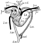
Tortoise Heart
"Heart and associated vessels of tortoise. r.a., Right auricle; superior venae cavae (s.v.c.) and inferior…

Pigeon Arterial System
"Heart and arterial system of pigeon. R.A., right auricle; R.V., right ventricle; L.V., left ventricle;…
Pigeon Venous System
"Heart and venous system of pigeon. R.A., Right auricle; R.V., right ventricle; L.V., left ventricle;…
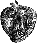
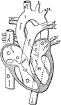
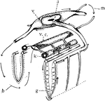
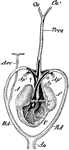
Heart in the Pericardium
View of the heart enclosed in its bag, or pericardium, which is a serious membrane. It is here laid…
Heart and its Chambers
View of the heart with its several chambers exposed and the vessels in connection with them. Labels:…
Bivalve Circulation
"Diagram showing the heart and general course of the circulation in the lamellibranchs. Only a short…
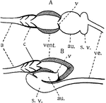
Vertebrate Heart
"Diagrams of the structure of the heart in the lower Vertebrates. A, primitive condition; B, the position…
Teleost Heart
"Diagram of the heart, the branchial arches, and the principal veins in the Teleosts. Ventral view.…
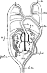
Queensland Lungfish Heart
"Diagram of heart and branchial arches in Ceratodus (one of the Dipnoi). a.b., air bladder (lung); p.a.,…
African Lungfish Heart
"Diagram of the heart and branchial arches in Protopterus (one of the Dipno). pre.c., precaval vein,…
Frog Heart
"Diagram of the heart and branchial arches in the Frog. c.g., carotid gland; l., lungs; l.a., left auricle;…
Reptile Heart
"Diagram of the heart and branchial arches in a Reptile...a, aorta; au., auricle; c, carotid; c.v.,…
Mammal Heart
"Diagram of the heart and the branchial arches in Mammals. A dotted outline of the arches of the Fish…
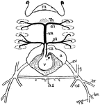
Crayfish Thorax
"Transverse section of thorax of crayfish, diagrammatic. abm, ventral abdominal muscles; bf, leg; bm,…
Ascidian Stages
"Diagram of the metamorphosis of the free, tailed larva into the fixed Ascidian. A, stage of free-swimming…
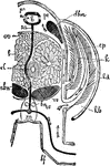
Sand Lizard Viscera
"Lacerta agilis. General view of the viscera in their naturaal relations. Bl, urinary bladder; Ci, post-caval…
Monitor Lizard Heart
"Heart of monitor (Varanus) dissected to show the cavity of the ventricle and the vessels leading out…
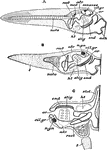
Pigeon Heart
"A, heart of the pigeon, dorsal aspect. a. ao, arch of aorta; br. a, brachial artery; br. v, bachial…
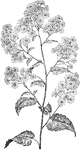
Heart-Leaved Aster
Of the Composite family (Compositae), the heart-leaved aster (Aster cordifolius).
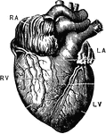
The Heart
The heart. Labels: RA, right auricle, RV; right ventricle; LA, left auricle; LV, left ventricle.
Interior of the Heart
Diagram of the interior of the heart. Labels: A, aorta; PA, pulmonary artery; VCI and VCS, vena cava…
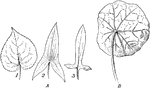
Leaf Bases
"A, Shapes of bases of leaves; B, Peltate leaf of Tropaeolum. 1, heart-shaped; 2, arrow-shaped; 3, halberd-shaped."…
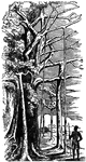
Banyan Tree
"This tree, a native of India, is remarkable for its vast branches. It is a species of fig; has ovate,…
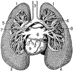
Lungs
"The Lungs. 1, Summit of lungs. 2, Base of lungs. 3, Trachea. 4, Right bronchus. 5, Left bronchus. 6,…

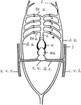
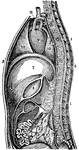
Thorax and Abdomen
"Thorax and abdomen. 1, 1, 1, 1. Muscles of the chest. 2, 2, 2, 2. Ribs. 3, 3, 3. Upper, middle and…

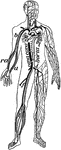
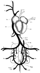
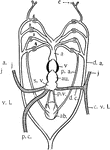
Veins and Arteries of the Body
Chief veins and arteries of the body. Labels: a, place of the heart; the veins are in the back. On the…
The Course of Blood in the Heart
Diagram of the rush of blood when the heart beats. The valves (v) open above are closed below while…
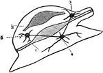
Diagram of a Mollusca
A diagram of a mollusca. Mollusca are soft-bodied animals that are usually protected by an external…
A Side View of the Lacteals and Thoracic Duct
A side view of the lacteal and thoracic duct. Labels: 1, Small intestine. 2, Lacteals. 3, Thoracic duct.…
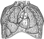
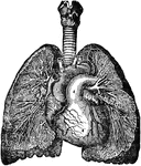
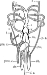
The Lungs
The lungs. Labels: 3, The lobes of the right lung. 4, The lobes of the left lung. 5, 6, 7, The heart.…
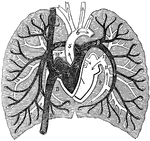
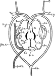
A Diagram of Pulmonary Circulation
A diagram of pulmonary circulation. Labels: 1, Descending vena cava. 2, Ascending cava vein. 3, Chamber…