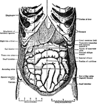
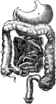
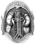
Abdomen
"The Abdominal Viscera in situ, as seen when the abdomin is laid open and the great omentum…
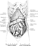
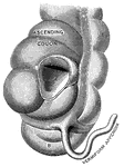
Abdomen Showing Displacement Caused by Corset
Abdomen of female showing displacement resulting from tight lacing. The liver is much enlarged, and…
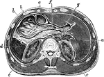
Horizontal Section Through Abdomen
Horizontal section through upper part of abdomen. Labels: a, liver; b, stomach; c, transverse colon;…
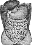
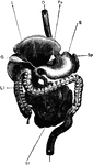
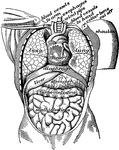
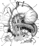
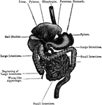
Abdominal Organs
Abdominal organs. Labels: 1, liver turned up; 2, gall bladder; 3, stomach; 4, large intestine; 5, small…
Ailmentary Canal
"ailmentary canal of a honey bee. at, honey stomach; s, true stomach; nt, intestine; o, esophagus; sg,…
Alimentary Canal
Diagram of the abdominal part of the alimentary canal (digestive system). Labels: C, the cardiac, and…
General View of the Alimentary Canal
Labels: O, esophagus; S, stomach; SI, small intestine; LI, large intestine, Sp spleen; L, liver (raised…
Vermiform appendix
"A, a portion of the colon laid open to show the valve between the large and small intestine;…
Vertical Section of the Back
"The spinal column below the twelfth dorsal vertebra at A has been removed, as well as the…
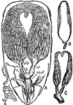
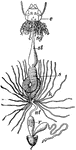
Bee Ovaries
"Ovaries of Queen and Workers (Apis). A, Abdomen of queen, under side. P, Petiole. o, o, Ovaries. hs,…
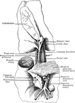
Biliary Apparatus
Portions of liver, duodenum, and pancreas, showing biliary and pancreatic ducts, head of pancreas turned…
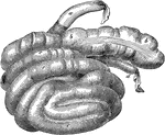
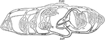
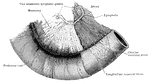
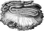
Caecum and Colon of a Dog
Caecum and colon of a dog-inflated. Labels: a, ileum; b, caecum; c, colon.
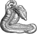
Caecum and Colon of a Hog
Caecum and colon of a hog-inflated. Labels: a, ileum; b, caecum; c, colon; d, rectum.
Caecum and Colon of Horse
Caecum and great colon of a horse. Labels: a, caecum; b, c, its muscular bands; d, termination of the…
Caecum of an Ox
Caecum and origin of colon of an ox- inflated. Labels: a, terminal portion of the ileum; b, caecum;…
Intestinal Tract from Canis Vulpes
Intestinal tract of Canis vulpes. S, cut end of duodenum; C, caecum; R, cut end of rectum.
Intestinal Tract of Chauna Chavaria
cc. Colic caeca, d. Duodenum. g. Glandular patch, l.l. Meckel's tract, l.i. Hind-gut, p.v. Cut root…
Columnar Epithelium
"In the stomach, intestines and elsewhere the epithelial cell is obling in profile and is called columnar…
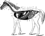
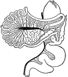
Digestive Apparatus of the Horse
The digestive apparatus of the horse. Labels: a, mouth; 2, pharynx; 3, esophagus; 4, diaphragm; 5, spleen;…
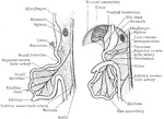
Digestive Organs
The stomach, pancreas, liver, and duodenum, with part of the rest of the small intestine and the mesentery;…
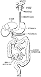
Digestive System
A diagram of the organs of digestion. Labels:1, The upper jaw. 2, The lower jaw. 3, The tongue. 4, The…
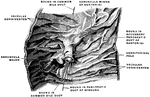
Meckel's Diverticulum
To show Meckel's diverticulum, the remains of he vitello-intestinal duct, and of the artery to the yolk…
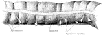
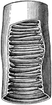
Longitudinal Section of Duodenum
Longitudinal section of duodenum; valvulae conniventes cut across, showing relation of these folds to…

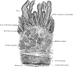
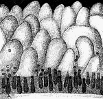
A Magnified Section of the Duodenum
A vertical section of the duodenum, highly magnified. Labels: 1, a fold-like villus; 2, epithelium,…
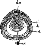
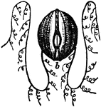
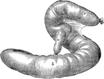
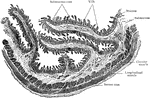
Earthworm
"Transverse section of the intestine of the Earthworm. ty, typhlosole, an infolded longitudinal ridge…
Various kinds of epithelial cells
"A, columnar cells of intestine; B, polyhedral cells of the conjuctiva; C, ciliated conical cells of…
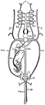
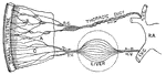
Fish Circulation
"Diagram of the principal vessels in the circulation of a fish, ventral view. a, aorta; au., auricle;…
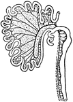
Fowl Digestive System
"Digestive system of the common Fowl. o, Gullet; c, Crop; p, Proventriculus; g, Gizzard; sm, Small intestine;…
Intestinal Tract of Giraffe
S, cut end of duodenum; R, cut end of rectum; C, caecum; P.C.L., post-caecal loop; S.P., spiral loop;…
Heart
"The heart and blood-vessels diagrammatically represented. L, lung; M, intestine; P, liver; dotted lines…
Mucous Membrane of the Ileum
The mucous membrane of the ileum. Labels: 1, cellular structure of the epithelium, or outer layer; 2,…
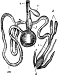
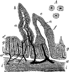
Digestive Apparatus of an Insect
"a, head, antennae, &c; b, pharynx; c, crop; d, gizzard; e, chyle-forming stomach; f, biliary vessels;…
Intestinal absorption
"A, a fold of peritoneum; B, lacteals and lymphatic glands; C, veins of intestines;…
Development of the Intestinal Canal
Two diagrams to illustrate the development of the intestinal canal. The figure to the right shows the…
Intestine of a Chick
Rudiments of the liver on the intestines of a chick at the fifth day of incubation. Labels: 1, heart;…
Section of Intestine wall
"A tiny block cut from the wall of the intestine showing villi and the mouths of glands at a; b, villus…
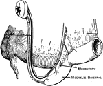
Small Intestine with Mesentery and Vessels
A portion of the intestine, with mesentery and vessels. The peritoneal coat has been removed from the…
Large Intestine
A piece of transverse colon from a child two years old. The three chief characteristics of the large…
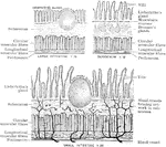
Structure of the Intestine
Diagram to show the structure of the small and large intestine and duodenum.
Transverse Section of Small Intestine
Transverse section of small intestine (lower part of duodenum), showing general arrangement of coats.
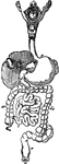
Intestines of an Ox
Mesentery and intestines of an ox. Labels: 1, duodenum; 2, small intestines; 3, caecum; 4, colon; 5,…
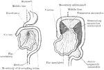
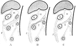
Development of the Mesenteries
Two diagrams to illustrate the development of the mesenteries. In the first figure the rotation of the…
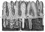
A Portion of the Mucous Membrane from the Small Intestine
Portion of the mucous membrane from the small intestine, magnified, showing the villi on its free surface,…
Mucous Membrane of the Small Intestine
Free surface of the mucous membrane of the small intestine, showing villi, solitary glands, and opening…
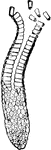
Muscular System
"Diagram showing the muscular system. M, ventral, N, dorsal valve; l, loop; V, mouth; Z, extremity of…
Development of the Great Omentum
Diagram to illustrate the development of the great omentum. A, shows the beginning of the great omentum…
Rabbit's Intestinal Mucous Membrane
Vertical section of the intestinal mucous membrane of the rabbit. Two villi are represented, in one…
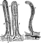
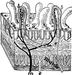
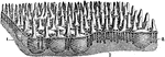
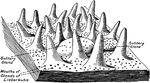
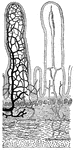
Small Intestine Villi
Villi of the small intestine, magnified about 80 diameters. In the left-hand figure the lacteals, a,b,c,…
Glands and villi of the small intestine
"A, B, glands seen in vertical section with their orifices at C opening upon the membrane…
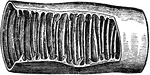
Piece of small intestine
"Piece of small intestine cut open to show wrinkling of inner coat bearing villi." —Davison, 1910
Transverse section of the small intestine
"In the figure on the left are seen the artery and vein of a villus. In the right figure are represented…
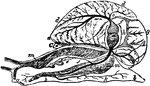
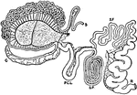
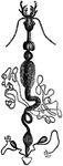
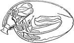
Snail Anatomy
"Anatomy of the Snail: a, the mouth; bb, foot; c, anus; dd, lung; e, stomach, covered above by the salivary…

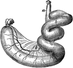
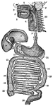
The Stomach and Intestines
The stomach and intestines. Labels: 1, stomach; 2, duodenum; 3, small intestine; 4, termination of the…