Diagram of circulation. Labels: L, left side of heart; R, right side of heart; a,a,a arterial system; b,b, capillaries; c,c,c veins; Alim., alimentary canal; Liv., liver; p, portal vein; H,hepatic vein; Lymph., lymphatic duct and tributaries; Pulm., lungs; Pa, pulmonary artery; Pu, pulmonary vein.

Diagram of Circulation

Diagram of circulation. Labels: L, left side of heart; R, right side of heart; a,a,a arterial system;…

"Among the cells of the body there is, besides the blood capillaries, a system of fine, thin-walled lymphatic capillaries." — Ritchie, 1918

Lymphatic Vessels in the Fingers

"Among the cells of the body there is, besides the blood capillaries, a system of fine, thin-walled…
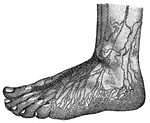
"In nearly every tissue of the body there is a marvelous network of vessels, precisely like the lacteals, known as the <em>lymphatics</em>. They seem to start out of the part in which they are found, like the rootlets of a plant in the soil. The tiny roots join together and make larger roots. They carry a fluid called <em>lymph</em>, very much like blood without the red corpuscles. It is to be remembered that the lacteals are really the lymphatics which begin in the villi of the small intestine." — Blaisedell, 1904

Superficial Lymphatics of the foot

"In nearly every tissue of the body there is a marvelous network of vessels, precisely like the lacteals,…

Superficial lymphatics of the digits and of the dorsal aspect of the hand.

Lymphatics of the Hand

Superficial lymphatics of the digits and of the dorsal aspect of the hand.

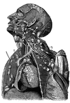
Lymphatics and lymphatic glands of the head and neck.

Lymphatics of the Head and Neck

Lymphatics and lymphatic glands of the head and neck.

"The heart and blood-vessels diagrammatically represented. L, lung; M, intestine; P, liver; dotted lines represent lymphatic vessels." &mdash;Martin, 1917

Heart

"The heart and blood-vessels diagrammatically represented. L, lung; M, intestine; P, liver; dotted lines…

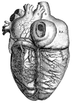
"<em>L.A.</em>, left auricle; <em>R.A.</em>, right auricle; <em>R.V.</em>, right ventricle; <em>A</em>, opening of the inferior vena cava; <em>B</em>, superior vena cava; <em>C</em>, right pulmonary veins; <em>D</em>, left pulmonary veins; <em>E</em>aorta; <em>F</em>, left branch of pulmonary artery; <em>G</em>, great cardiac vein; <em>H</em>middle cardiac vein. the cardiac lymphatics which follow the course of the cardiac veins are also shown." — Blaisedell, 1904

Posterior View of the Heart

"L.A., left auricle; R.A., right auricle; R.V., right ventricle; A,…

"Consisting of a very fine network of fibrils, around which are cells of various sizes." — Blaisedell, 1904

Connective Tissue from a Lymphatic Gland

"Consisting of a very fine network of fibrils, around which are cells of various sizes." — Blaisedell,…

Diagrammatic section of lymphatic gland. Labels: a.l., afferent lymphatic; e.l., efferent lymphatic; c, capsule, or envelope; tr, trabeculae; l.s., lymph-sinus; l.h., pulpy substance of gland.

Diagrammatic Section of a Lymphatic Gland (Lymph Node)

Diagrammatic section of lymphatic gland. Labels: a.l., afferent lymphatic; e.l., efferent lymphatic;…
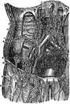
Diagram of the principle groups of the Lymphatic vessels.

Lymphatic Vessels

Diagram of the principle groups of the Lymphatic vessels.

Femoral iliac and aortic lymphatic vessels and glands. Labels: 1, saphena magna vein; 2, external iliac artery and vein; 3, primitive iliac artery and vein; 4, aorta; 9, superior set of inguinal lymphatic glands; 10, chain of lymphatics; 11, lymphatics which accompany the circumflex iliac vessels; 12, lumbar an aortic lymphatics; 13, origin of thoracic duct; 14, thoracic duct at its commencement.

Lymphatic Vessels and Glands

Femoral iliac and aortic lymphatic vessels and glands. Labels: 1, saphena magna vein; 2, external iliac…

The lymphatic vessels and glands. Labels: 1, 2, 3, 4, 5, 6, Lymphatic vessels and glands of the lower limb. 7, Lymphatic glands. 8, The receptacle of the thoracic duct. 9, The lymphatics of the kidney. 10, The stomach. 11, Of the liver. 12, Of the lungs. 13, 14, 15, The lymphatics and glands of the arm. 16, 17, 18, Of the face and neck. 19, 20, Large veins. 21, The thoracic duct.

The Lymphatic Vessels and Glands

The lymphatic vessels and glands. Labels: 1, 2, 3, 4, 5, 6, Lymphatic vessels and glands of the lower…
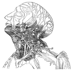
This diagram shows the lumphatics of the head and neck. It shows the glands, and B, the thoracic duct as it empties into the left innominate vein at the junction of the left jugular and subclavian veins.

Lymphatics

This diagram shows the lumphatics of the head and neck. It shows the glands, and B, the thoracic duct…
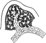
Origin of lymphatics. Labels: S, lymph spaces communicating with lympathic vessel; A, origin of lymphatic by union of lymp spaces; E, E, endothelial cells forming wall of lymph vessel.

Lymphatics

Origin of lymphatics. Labels: S, lymph spaces communicating with lympathic vessel; A, origin of lymphatic…

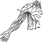
Superficial lymphatics and glands. Labels: G. Axillary glands.

Lymphatics

Superficial lymphatics and glands. Labels: G. Axillary glands.

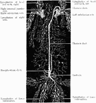
The lymphatic vessels. The thoracic duct occupies the middle of the figure. it lies upon the spinal column, at the sides of which are seen portions of the ribs (1). Labels: a, the receptacle of the chyle; b, the trunk of the thoracic duct, opening at c, into the junction of the left jugular (f) and subclavian (g) veins as they unite into the left innominate vein, which has been cut across to show the thoracic duct running behind it; d, lymphatic glands placed in the lumbar regions; h, the superior vena cava formed by the junction of the right and left innominate veins.

Lymphatics

The lymphatic vessels. The thoracic duct occupies the middle of the figure. it lies upon the spinal…
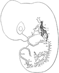
Developing lymphatics in rabbit embryo of 14 days. Lymphatic vessels are heavily shaded; veins are light. Labels: In.J., Ex.J., internal and external jugular veins; Pr.U., primitive ulnar; Ex.M., external mammary; Az., azygos; VCI., inferior vena cava; G., gastric; S.M., superior mesenteric; V., vitelline; Sc., subcardinal; R.A., renal anastomosis of subcardinals; Pr.Fi.,primitive fibular; c.b., connecting branch; An.T., anterior tibial; c., caudal; 3,4,5,6, position of corresponding cervical nerves.

Lymphatics in a Rabbit Embryo

Developing lymphatics in rabbit embryo of 14 days. Lymphatic vessels are heavily shaded; veins are light.…

Superficial lymphatics of right groin and upper part of thigh. Labels: 1, Upper inguinal glands. 2, 2'. Lower or inguinal or femoral glands. 3, 3'. Plexus of lymphatics in the course of the long saphenous vein.

Lymphatics of Groin and Thigh

Superficial lymphatics of right groin and upper part of thigh. Labels: 1, Upper inguinal glands. 2,…
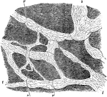
Lymphatics of central tendon of rabbit's diaphragm, stained with silver nitrate. The ground substance has been shaded diagrammatically to bring out the lymphatic clearly. Lymphatics, l, lined by long narrow endothelial cells, and showing valves, v, at frequent intervals.

Lymphatics of Rabbit's Diaphragm

Lymphatics of central tendon of rabbit's diaphragm, stained with silver nitrate. The ground substance…
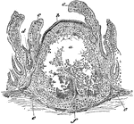
Lymphatic vessels near the surface of the skin.

Superficial Lympatics

Lymphatic vessels near the surface of the skin.

Section through the lymphoid tissue of a solitary gland. Labels: a, center of the gland, with the lymphoid tissue fallen away; b, epithelium of mucous membrane; c, c, villi, with epithelium partly broken away; d, crypts, or glands, of Lieberkuhn.

Lymphoid Tissue

Section through the lymphoid tissue of a solitary gland. Labels: a, center of the gland, with the lymphoid…

Vertical section of a portion of a Peyer's Patch, with lacteal vessels injected. Labels: a, villi, with their lacteals colored black; d, surface of rounded follicle, or solitary gland; e, central part; f,g,h,i, and k, lymph-channels, or lacteal vessels, colored black. "Closely connected with the lymphatic vessels in the intestines are small, rounded bodies of the size of a small pin's head, called solitary glands or follicles. These bodies consist of a rounded mass of fine lymphoid tissue, the meshes of which are crowded with leucocytes. A Peyer's patch, or agminated gland, as it is often called, is simply a collection of these follicles."&mdash; Kimber, 1907

Peyer's Patch

Vertical section of a portion of a Peyer's Patch, with lacteal vessels injected. Labels: a, villi, with…

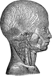
Lymphatic vessels are used to transfer lymph throughout the body.

Lymphatic Vessels

Lymphatic vessels are used to transfer lymph throughout the body.