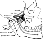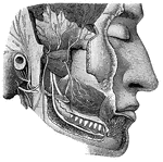
"Dental Branch of One of the Divisions of the Fifth Pair of Cranial Nerves, supplying the Lower Teeth. Branches from the motor root, distributed to various muscles, are also shown." — Blaisedell, 1904

Cranial Nerves

"Dental Branch of One of the Divisions of the Fifth Pair of Cranial Nerves, supplying the Lower Teeth.…
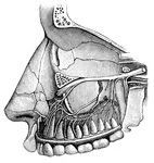
"Dental Branches of One of the Divisions of the Fifth Pair of Cranial Nerves, supply the Upper Teeth." — Blaisedell, 1904

Cranial Nerves

"Dental Branches of One of the Divisions of the Fifth Pair of Cranial Nerves, supply the Upper Teeth."…
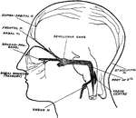
Surface markings for the second and third division of the fifth nerve.

Facial Nerves

Surface markings for the second and third division of the fifth nerve.

Diagram to show the proximity of the sensory nuclei of the fifth and tenth cranial and first and second cervical nerves.

Facial Nerves

Diagram to show the proximity of the sensory nuclei of the fifth and tenth cranial and first and second…

"Part of the nervous system viewed in front." —Davison, 1910

Nervous System

"Part of the nervous system viewed in front." —Davison, 1910

"Diagram illustrating the General Arrangement of the Nervous System. (posterior view.)" — Blaisedell, 1904

Nervous System

"Diagram illustrating the General Arrangement of the Nervous System. (posterior view.)" — Blaisedell,…
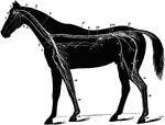
The nervous system of the horse. Labels: 1, brain; 2, optic nerve; 3, superior maxillary nerve (5th); 4, inferior maxillary nerve (5th); 5, pneumogastric nerve; 6, medulla oblongata; 6', spinal cord; 7, right brachial plexus; 8, musculo-cutaneous nerve; 9, median nerve; 10, radial nerve; 11, cubital nerve; 12, pneumogastric nerve; 13, solar plexus; 14, semilunar ganglion; 15m lumbo-sacral plexus; 16, anterior crural nerve; 17, great sciatic nerve; 18, external popiteal nerve; 19, external saphenic nerve; 20, tibial nerve; 21, metatarsal nerve; 22, radial portion of median nerve; 23, metacarpal nerves; 24, digital branches; s, sympathetic chain; c, inferior cervical plexus; g, gutteral ganglion; Sp, great splanchnic nerve; pm; posterior mesenteric plexus; p, pelvic plexus.

Nervous System of the Horse

The nervous system of the horse. Labels: 1, brain; 2, optic nerve; 3, superior maxillary nerve (5th);…

"Part of the sympathetic nervous system seen from in front, n, one of the two chief cords, t, i, and p, branches to organs." —Davison, 1910

Sympathetic Nervous System

"Part of the sympathetic nervous system seen from in front, n, one of the two chief cords, t, i, and…

"Showing a motor cell with its long, unbranched process (with two little lateral offshoots), with motor endings in striated, voluntary muscular tissue." — Blaisedell, 1904

Neuron

"Showing a motor cell with its long, unbranched process (with two little lateral offshoots), with motor…
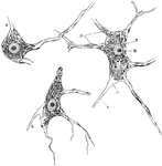
"A nerve cell from the brain." — Ritchie, 1918

Neuron

"A nerve cell from the brain." — Ritchie, 1918

Diagram of typical neuron.

Neuron

Diagram of typical neuron.

Diagram of nerve cell of type II, in which axone is not prolonged as nerve fiber.

Neuron

Diagram of nerve cell of type II, in which axone is not prolonged as nerve fiber.

Nerve cells of human spinal cord stained to show Nissl bodies. Labels: D, dendrites; A, axons; C, implantation cone; N, nucleus; M, nucleolus.

Neuron of Spinal Cord

Nerve cells of human spinal cord stained to show Nissl bodies. Labels: D, dendrites; A, axons; C, implantation…

Semidiagrammatic representation of structure of a neuron; a, axon.

Structure of a Neuron

Semidiagrammatic representation of structure of a neuron; a, axon.

Multipolar nerve cells of various forms. Labels: A, from spinal cord; B, from cerebral cortex; C, from cerebellar cortex (Purkinje cell); a, axon; c, implantation cone.

Various Forms of Neurons

Multipolar nerve cells of various forms. Labels: A, from spinal cord; B, from cerebral cortex; C, from…
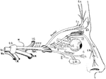
Scheme of the distribution of the ophthalmic nerve. Labels: Vs, trigeminal nerve, afferent root; Mo, efferent root; G.G, gasserian ganglion; M, meningeal branch; I.C, branch to internal carotid artery; Oph, opthalmic nerve; S.M, superior maxillary nerve; I.M, inferior maxillary nerve; III, communication to oculomotor nerve; IV, to trochlear nerve; L, branches to upper eyelid; L.G, long root to lenticular ganglion; Sy, root from sympathetic (on carotid artery); III, short root from motor oculi nerve; C, short ciliary branches; L.C, long ciliary nerves; I.T, infra-trochlear nerve; E.N, external nasal nerve; I.N, internal nasal nerve; O, orbital branch of superior maxillary nerve; L.Gl, lachrymal gland; C, conjuctival branch; L, branch to eyelids and face.

Ophthalmic Nerve

Scheme of the distribution of the ophthalmic nerve. Labels: Vs, trigeminal nerve, afferent root; Mo,…
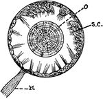
"Otocyst in a mollusk. n, nerve; ;o, otolith; s.c., sensory cells in wall of otocyst." — Galloway

Otocyst

"Otocyst in a mollusk. n, nerve; ;o, otolith; s.c., sensory cells in wall of otocyst." — Galloway
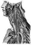
"Showing its distribution by its branches and ganglia to the larynx, pharynx, heart, lungs, and other parts." — Blaisedell, 1904

Trunk of the Pneumogastric Nerve

"Showing its distribution by its branches and ganglia to the larynx, pharynx, heart, lungs, and other…

"Illustrating the path of a simple nervous reflex action." — Blaisedell, 1904

Reflex Action

"Illustrating the path of a simple nervous reflex action." — Blaisedell, 1904

"Development of the lateral eyes of a scorpion. h, Epidermic cell-layer; mes, mesoblastic connective tissue; n, nerves; II, III, IV, V, depressions of the epidermis in each of which a cuticular lens will be formed." — The Encyclopedia Britannica, 1910

Scorpion Eyes

"Development of the lateral eyes of a scorpion. h, Epidermic cell-layer; mes, mesoblastic connective…
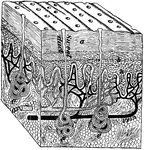
"A block out of the skin. a, dead part and d live part of the epidermis; e, sweat glands; n, nerve endings." —Davison, 1910

Section of skin

"A block out of the skin. a, dead part and d live part of the epidermis; e, sweat glands; n, nerve endings."…

Diagram to show the structure of the Skin. Labels: E.c, epidermis, corneous part; E.m, epidermis, Malpighian part; D.c, connective tissue of dermis; p, papilla; gl, sweat gland, the coils of the tube cut across or lengthwise; d, its duct; f, fat; v, blood vessels; n, nerve; t.c, tactile corpuscle.

Structure of the Skin

Diagram to show the structure of the Skin. Labels: E.c, epidermis, corneous part; E.m, epidermis, Malpighian…

Scheme of the origin, connection, and distribution of the spinal accessory nerve. Labels: Sp.Acc, spinal accessory nerve; C.1-4, first four cervical nerves (dorsal roots); Va, pneumogastric nerve; R, ganglion of the root; t, ganglion of the trunk; G.Ph, glossopharyngeal nerve; S.M, nerves to sternocleidomastoid; Tr, nerves to trapezius; F.M, foramen magnum; J.F, jugular foramen.

Spinal Accessory Nerve

Scheme of the origin, connection, and distribution of the spinal accessory nerve. Labels: Sp.Acc, spinal…

The skull and spinal canal of a child from behind with the Dura Mater slit open and ribs with the transverse processes of vertebrae dissected out.

The Position of the Spinal Cord and Spinal Nerves in the Spinal Canal

The skull and spinal canal of a child from behind with the Dura Mater slit open and ribs with the transverse…

"Diagram of a slice across the spinal cord, showing the roots of a spinal nerve to the arm on the left. The arrows show which way the messages travel. The little circles are the cut ends of fibers extending up and down the cord. Only a few of the thousands of fibers really present are here shown." —Davison, 1910

Section of Spinal Cord

"Diagram of a slice across the spinal cord, showing the roots of a spinal nerve to the arm on the left.…

"The spine, sawn in two lengthwise, showing the spinal canal and the holes between the vertebrae, where nerves and blood-vessels pass out." — Tracy, 1888

Spine

"The spine, sawn in two lengthwise, showing the spinal canal and the holes between the vertebrae, where…

The sympathetic ganglions and their connection with other nerves. Labels: A, The semilunar ganglion and solar plexus. D, The thoracic (chest) ganglions. E, The external and internal branched of the thoracic ganglions. G, H, The right and left coronary (heart) plexus. I, N, Q , The inferior, middle and superior cervical (neck) ganglions. 1, The renal plexus of nerves. 2, The lumbar (loin) ganglions. 3, Their internal branches. 4, Their external branches. 5, The aortic plexus of nerves.

The Sympathetic Ganglions and their Connection to other Nerves

The sympathetic ganglions and their connection with other nerves. Labels: A, The semilunar ganglion…
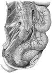
"Showing the distribution of some of the great plexuses of the sympathetic nerve in the lumbar and sacral regions." — Blaisedell, 1904

Plexuses of the Sympathetic Nerves

"Showing the distribution of some of the great plexuses of the sympathetic nerve in the lumbar and sacral…

Diagrammatic view of the Sympathetic cord of the right side, showing its connections with the principal cerebrospinal nerves and the main prae-aortic plexuses. Cerebrospinal nerves. VI, a portion of the sixth cranial as it passes through the cavernous sinus, receiving two twigs from the carotid plexus of the sympathetic nerve; O, opthalmic ganglion connected by a twig with the carotid plexus; M, connection of the spheno-palatine ganglion by the Vidian nerve with the carotid plexus; C, cervical plexus; Br, brachial plexus; D 6, sixth intercostal nerve; D 12, twelfth; L 3, third lumbar nerve; S 5, fifth; Cr, anterior crural nerve winding round the subclavian artery. Sympathetic Cord. c, superior cervical ganglion; c', second or middle; c", inferior; from each of these ganglia cardiac nerves (all deep on this side) are seen descending to the cardiac plexus; d 1, placed immediately below the first dorsal sympathetic ganglion; d 6, is opposite he sixth; l 1, first lumbar ganglion. Preaortic and Visceral Plexuses. p, pharyngeal, and, lower down, laryngeal plexus; pl, post-pulmonary plexus spreading from the vagus on the back of the right bronchus; c a,on the aorta , the cardiac plexus, towards which , in addition to the cardiac nerves from the three cervical sympathetic ganglia, other branches are seen descending from the vagus and recurrent nerves; co, right or posterior and co1, left or an. coronary plexus; o, esophageal plexus in long meshes on the gullet; sp, great splanchnic nerve formed by branched from the fifth, sixth, seventh, eight, and ninth dorsal ganglia; +, small splanchnic from the ninth and tenth; ++, smallest or third splanchnic from the eleventh: the first and second of theses are shown joining the solar plexus, s o; the third descending to the renal plexus, r e; connecting branches between the solar plexus and the vagi are also represented; pn', above the place where the right vagus passes to the lower or posterior surface of the stomach; pn", the left distribution on the anterior side or upper surface of the cardiac portion of the organ: from the solar plexus large branches are seen surrounding the arteries of the coeliac axis, and descending from the solar and sup. mesenteric plexuses and from the lumbar ganglia; mi, the inf. mesenteric plexus surrounding the corresponding artery; hy, hypogastric plexus; pl, the right pelvic or inf. hypogastric plexuses; pl, the right pelvic plexus; from this the nerves descending are joined by those from the plexus on the sup. hemorrhoidal vessels, mi', by nerves from the third and fourth sacral spinal nerves, and there are thus formed the rectal, vesical, and other plexuses, which ramify upon the viscera, ,as towards ir, and v, the rectum and bladder.

Sympathetic System

Diagrammatic view of the Sympathetic cord of the right side, showing its connections with the principal…

Nerves of the thigh. Labels: 1, gangliated cord of sympathetic; 2, third lumbar nerve; 3, branches to iliacus internus; 4, fourth lumbar nerve; 5, anterior crural nerve; 6, lumbosacral nerve; 7, branch to psoas; 8, obturator nerve; 9, external cutaneous nerve (cut); 10, nerve to pectineus; 11, superficial division of obturator nerve; 13, 13, sartorius muscle; 14, 14, adductor longus; 15, branch to rectus; 16, deep division of obturator nerve; 17, branches to vastus externus and crureus; 18, adductor brevis; 19, branch to vastus internus; 20, adductor magnus; 21, vastus externus; 22, 22, internal saphenous nerve; 23, rectus femoris; 24, pattelar branch of saphenous nerve; 25, vastus internus; 26, gracilis.

Nerves of the Thigh

Nerves of the thigh. Labels: 1, gangliated cord of sympathetic; 2, third lumbar nerve; 3, branches to…