Anterior view of the brain and spinal marrow. Labels: 1, 1, hemispheres of the cerebrum; 2, great middle fissure; 3, cerebellum; 4, olfactory nerves; 5, optic nerves; 6, corpora albicantia; 7, motor oculi nerves; 8, pons Varolii; 9, fourth pair of nerves; 10, lower portion of the medulla oblongata; 11, 11, medulla spinalis in its whole length; 12, 1, spinal nerves; 13, cauda equina.

Brain and Spinal Cord

Anterior view of the brain and spinal marrow. Labels: 1, 1, hemispheres of the cerebrum; 2, great middle…

The brain and spinal cord. Labels: 1, 1, hemispheres of cerebrum; 2, great middle fissure; 3, cerebellum; 4, olfactory nerves; 5, optic nerves; 6, corpora albicanlia; 7, motor oculi; 8, pons Varolii; 9, fourth nerve; 10, medulla oblongata; 11, 11, medulla spinalis; 12, 12, spinal nerves; 18, cauda equina.

Brain and Spinal Cord

The brain and spinal cord. Labels: 1, 1, hemispheres of cerebrum; 2, great middle fissure; 3, cerebellum;…

Human fetus in the third month of development, with the brain and spinal cord exposed from behind.

Brain and Spinal Cord of Fetus

Human fetus in the third month of development, with the brain and spinal cord exposed from behind.

Simplified drawing of brain as seen in mesial section, showing relation of brain stem, cerebrum and cerebellum, and ventricular spaces.

Brain in Mesial Section

Simplified drawing of brain as seen in mesial section, showing relation of brain stem, cerebrum and…
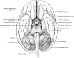
Simplified drawing of brain as seen from below, showing relations of brain stem to spinal cord and cerebrum.
Relation of Brain Stem to Spinal Cord

Simplified drawing of brain as seen from below, showing relations of brain stem to spinal cord and cerebrum.

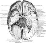
The base of the brain. The under part of the left temporal and occipital lobes has been sliced off so as to open into the lateral ventricle. The left hemisphere of the cerebellum has also been removed.

Base of the Brain

The base of the brain. The under part of the left temporal and occipital lobes has been sliced off so…

View of the cerebrospinal axis of the nervous system. The right half of the cranium and trunk of the body have been removed by a vertical section; the membranes of the brain and spinal cord have also been removed, and the roots and first part of the fifth and ninth cranial, and of all the spinal nerves of the right side, have been dissected out and laid separately on the wall of the skull and on the several vertebrae opposite to the place of their natural exit from the craniospinal cavity.

Central Nervous System

View of the cerebrospinal axis of the nervous system. The right half of the cranium and trunk of the…
Semidiagrammatic view of the cerebrospinal axis.

Cerebrospinal Axis

Semidiagrammatic view of the cerebrospinal axis.

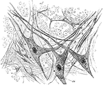
Section of gray matter of anterior cornu of a calf's spinal cord; a, nerve fibers of white matter in transverse section, showing axis cylinder in the center of each; r, large stellate nerve cells with nuclei and three prolongations.

Gray Matter of Spinal Cord

Section of gray matter of anterior cornu of a calf's spinal cord; a, nerve fibers of white matter in…
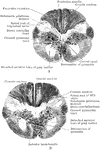
Two section through the junction between the cord and medulla of the Orang. A is at a slightly lower level than B.

Section Through the Junction of the Medulla and Cord of the Orang

Two section through the junction between the cord and medulla of the Orang. A is at a slightly lower…
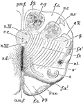
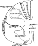
Anterior or dorsal section of the medulla oblongata in the region of the superior pyramidal decussation. a.m.f, anterior median fissure; f.a., superficial arciform fibers emerging from the fissure; py., pyramid, n.ar., nuclei of arciform fibers; f.a., deep arciform becoming superficial; o, lower end of olivary nucleus; n.l., nucleus lateralis; f.r., formatio reticularis; f.a. 2, arciform fibers proceeding from the formatio reicularis; g, substantia gelatinosa of Rolando; a.V., ascending root of fifth nerve; n.c., nucleus cuneatus; n.c.', external cuneate nucleus; n.g., nucleus gracilis; f.g., funiculus gracilis; p.m.f., posterior median fissure; c., central canal surrounded by gray matter, in which are n. XI., nucleus of the spinal accessory, and n. XII., nucleus of the hypoglossal; s.d., superior pyramidal decussation.

Medulla Oblongata

Anterior or dorsal section of the medulla oblongata in the region of the superior pyramidal decussation.…
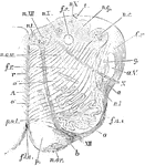
Section of the medulla oblongata at about the middle of the olivary body. f.l.a., anterior median fissure; n.ar., nucleus arciformis; p., pyramid; XII., bundle of hypoglossal nerve emerging from the surface; at b, it is seen coursing between the pyramid and the olivary nucleus, o.; f.a.e., external arciform fibers; n.l., nucleus lateralis; a., arciform fibers passing towards restiform body, partly through the substantia gelatinosa, g., partly superficial to the ascending root of the fifth nerve, a.V.; X., bundle of vagus root emerging; f.r., formatio reticularis; c.r., corpus restiform, beginning to be formed, chiefly by arciform fibers, superficial and deep; n.c., nucleus of the funiculus teres; n.am., nucleus ambiguus; r., raphe; A., continuation of the anterior column of cord; o', o", accessory olivary nucleus; p.o., pedunculus olivae.

Medulla Oblongata

Section of the medulla oblongata at about the middle of the olivary body. f.l.a., anterior median fissure;…
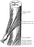
Throughout the dorsal region, the spinal cord presents a uniform girth and a very nearly circular outline when seen in transverse section. Shown is the roots and origin of the seventh dorsal nerve.

Seventh Dorsal Nerve

Throughout the dorsal region, the spinal cord presents a uniform girth and a very nearly circular outline…

Diagram of the human nervous system showing the brain, spinal cord, and nerves.

Nervous System

Diagram of the human nervous system showing the brain, spinal cord, and nerves.

Diagram if the spinal origin of the spinal accessory nerve.

Spinal Accessory Nerve

Diagram if the spinal origin of the spinal accessory nerve.

The conus and medullaris and the filum terminale exposed within the spinal canal.

Sacral Region of Spinal Canal

The conus and medullaris and the filum terminale exposed within the spinal canal.

Side view of the spinal column. Labels: C 1-7, cervical; D 1-12, dorsal; L 1-15 lumbar; S 1, sacrum; Co 1-4, coccygeal.

Side View of the Spinal Column

Side view of the spinal column. Labels: C 1-7, cervical; D 1-12, dorsal; L 1-15 lumbar; S 1, sacrum;…

Diagrammatic view from before of the spinal cord and medulla oblongata, including the roots of the spinal and some of the cranial nerves, and on one side of the gangliated chain of the sympathetic. The spinal nerves are enumerated in order on the right side of the figure. Labels: Br, brachial plexus; Cr, anterior crural, O, obturator, and Ss, great sciatic nerves, coming off from lumbo-sacral plexus; x, x, filum terminale; a,b,c, superior, middle, and inferior cervical ganglia of the sympathetic, the last united with the first thoracic, d; d', the eleventh thoracic ganglion; l, the twelfth thoracic (or first lumbar); below ss, the chain of sacral ganglia.

Spinal Cord

Diagrammatic view from before of the spinal cord and medulla oblongata, including the roots of the spinal…

The spinal cord (medulla spinalis).

The Spinal Cord

The spinal cord (medulla spinalis).

The spinal cord is a double cord, the two halves being joined by a narrow portion, but the outside sheath of connective tissue gives it the appearance of being single.

Spinal Cord

The spinal cord is a double cord, the two halves being joined by a narrow portion, but the outside sheath…
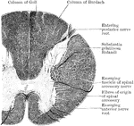
Section through the upper part of the cervical region of the cord of an orangoutang. Showing the origin of the spinal part of the spinal accessory nerve.

Spinal Cord

Section through the upper part of the cervical region of the cord of an orangoutang. Showing the origin…

The spinal cord and medulla oblongata. Labels: A, from the ventral, and B, from the dorsal aspect; C to H, cross-sections at different levels.

The Spinal Cord and Medulla Oblongata

The spinal cord and medulla oblongata. Labels: A, from the ventral, and B, from the dorsal aspect; C…
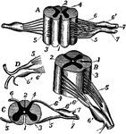
Diagrams of spinal cord and nerve roots. Labels: A, a small portion of the cord seen from the ventral side; B, the same seen laterally; C, a cross-section of the cord; D, the two roots of a spinal nerve; 1, anterior (ventral) fissure; 2, posterior (dorsal) fissure; 3, surface groove along the line of attachment of the anterior nerve roots; 4, line of origin of the posterior roots; 5, anterior root filaments of a spinal nerve; 6, posterior root filaments; 6', ganglion of the posterior root; 7, 7', the first two divisions of the nerve trunk after its formation by the union of the two roots.

Spinal Cord and Nerve Roots

Diagrams of spinal cord and nerve roots. Labels: A, a small portion of the cord seen from the ventral…
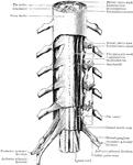
Scheme of the arrangement of the membranes of the spinal cord and the roots of the spinal nerves.

Spinal Cord and Spinal Nerves

Scheme of the arrangement of the membranes of the spinal cord and the roots of the spinal nerves.
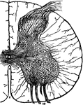
A thin transverse section of half the spinal cord magnified about 10 diameters. Labels: 1, anterior fissure; 2, posterior fissure; 3, central canal ;8, pia mater enveloping the cord; 6,7, bands of pia mater penetrating the cord and supporting its nerve elements; 9, a posterior root; 10, bundles of an anterior root; a,b,c,d,e, groups of nerve cells in the gray matter.

Spinal Cord Section

A thin transverse section of half the spinal cord magnified about 10 diameters. Labels: 1, anterior…
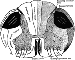
Diagrammatic transverse section of the spinal cord showing the conduction paths.

Section of the Spinal Cord

Diagrammatic transverse section of the spinal cord showing the conduction paths.
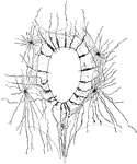
Diagram of spinal cord as seen from behind.

Spinal Cord Seen from Behind

Diagram of spinal cord as seen from behind.

Section through the central canals of the spinal cord of a human embryo, showing ependymal (A) and neuroglial cells (B).

Section Through Spinal Cord Showing Neuroglial Cell

Section through the central canals of the spinal cord of a human embryo, showing ependymal (A) and neuroglial…

Three stages of development of the spinal cord. Labels: AC., anterior column; AH., anterior horn of gray matter; AL., alar lamina; AMF., antero-median fissure; AR., anterior nerve root; BC., column of Burdach; BL., basal lamina; E., ependyma; GC., column of Goll; LC., lateral column; MDL., mid-dorsal lamina; MVL., mid-ventral lamina, PC., early posterior column; PH., posterior horn of gray matter; PMF., postero-median fissure; PR., posterior nerve root.

Development of Spinal Cord

Three stages of development of the spinal cord. Labels: AC., anterior column; AH., anterior horn of…

Diagram of development of spinal cord. Labels: c, central canal; af, anterior fissure; pf, posterior fissure; g, gray matter; w, white matter.

Development of the Spinal Cord

Diagram of development of spinal cord. Labels: c, central canal; af, anterior fissure; pf, posterior…
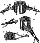
Different views of a portion of the spinal cord from the cervical region, with roots of the nerves slightly enlarged. Labels: In A, the anterior surface of the specimen is shown, the anterior nerve root of its right side being divided: in B, a view of the right side is given; in C, the upper surface is shown; in E, the nerve roots and ganglion are shown from below; 1, the anterior median fissure; 2, posterior median fissure; 3, anterior lateral depression, over which the anterior nerve roots are seen to spread; 4, posterior lateral groove, into which the posterior roots are seen to sink; 5, anterior roots passing the ganglion; 5', in A, the anterior root divided; 6, the posterior roots, the fibers of which pass into the ganglion 6'; 7, the united or compound nerve; 7', the posterior primary branch, seen in A and D to be derived in part from the anterior and in part from the posterior root.

Different View of the Spinal Cord

Different views of a portion of the spinal cord from the cervical region, with roots of the nerves slightly…

Lowered end of the spinal cord and the cauda equina, dorsal aspect. The dorsal roots of the right side have been removed.

Lower End of the Spinal Cord

Lowered end of the spinal cord and the cauda equina, dorsal aspect. The dorsal roots of the right side…
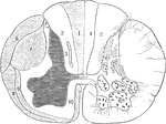
Section of a spinal cord, one half of which shows the tracts of the white matter, and the other half (right) shows the position of the nerve cells in the gray matter. 7, 10, 9, and 3 are tracts of descending degeneration, 1, 4, 6, and 8, of the ascending degeneration.

Section of the Spinal Cord

Section of a spinal cord, one half of which shows the tracts of the white matter, and the other half…
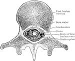
Section through the conus medullaris and the cauda equina as they lie in the spinal canal.

Section Through Spinal Cord

Section through the conus medullaris and the cauda equina as they lie in the spinal canal.

Transverse section through upper part of the cervical region of the cord.

Section Through Upper Part of Spinal Cord

Transverse section through upper part of the cervical region of the cord.
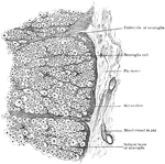
Peripheral part of transverse section of spinal cord, showing nerve fibers subdivided into groups by ingrowth of subpial layer of neuroglia.

Transverse Section of Spinal Cord

Peripheral part of transverse section of spinal cord, showing nerve fibers subdivided into groups by…
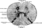
Transverse section of the spinal cord at the middle of the thoracic region. The neuroglia septum has been removed from between the dorsal columns.

Transverse Section of the Spinal Cord

Transverse section of the spinal cord at the middle of the thoracic region. The neuroglia septum has…
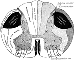
Diagrammatic representation of a transverse section through the spinal cord. The nerve tracts in the white matter and the clusters of the nerve cells in the gray matter are shown.

Transverse Section Through Spinal Cord

Diagrammatic representation of a transverse section through the spinal cord. The nerve tracts in the…
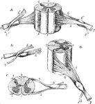
Different views of a portion of the spinal cord from the cervical region, with the roots of the nerves. A, the anterior surface of the specimen is shown; the anterior nerve root of its right side being divided. B, a view of the right side. C, the upper surface. D, the nerve roots and ganglion are shown from below. Labels: 1, The anterior median fissure; 2, posterior median fissure; 3, anterior lateral depression, over which the anterior nerve roots are seen to spread; 4, posterior lateral groove, into which the posterior roots are seen to sink; 5, anterior roots passing the ganglion; 5', the anterior root divided; 6, the posterior roots, the fibers of which pass into the ganglion 6'; 7, the united or compound nerve; 7', the posterior primary branch, seen is A and D to be derived in part from the anterior and in part from the posterior root.

Views of a Spinal Cord

Different views of a portion of the spinal cord from the cervical region, with the roots of the nerves.…

Transverse section through the white matter of the cord, as seen through the microscope.

White Matter of Spinal Cord

Transverse section through the white matter of the cord, as seen through the microscope.

Diagrammatic representation of the roots and ganglia of the spinal nerves, showing their position in relation to the spinal column. The nerves are shown as thick black lines on the left side.

Spinal Nerves

Diagrammatic representation of the roots and ganglia of the spinal nerves, showing their position in…

Diagram on frozen section, showing relations of bodies and spines of vertebrae to levels at which spinal nerves escape from vertebral canal.

Spine

Diagram on frozen section, showing relations of bodies and spines of vertebrae to levels at which spinal…