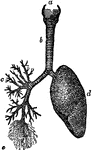
Branchi and Blood Vessels
Branchi of the lungs, the heart, and blood vessels. Labels: 1, left auricle; 2, right auricle; 3, left…
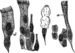
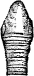
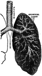
A Bronchial Tube
A small bronchial tube. Labels: a, dividing into its terminal branches, c; these have pouched or sacculated…
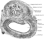
Lateral section of the chest
"A, a muscle which aids in pushing the food down the esophagus; B, esophagus; C,…
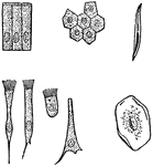
Various kinds of epithelial cells
"A, columnar cells of intestine; B, polyhedral cells of the conjuctiva; C, ciliated conical cells of…
Ciliated Epithelium Cells
Ciliated epithelium from the human trachea, highly magnified. Labels: a, large ciliated cell; d, cell,…
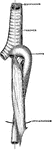
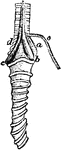
Esophagus, Trachea, and Aorta in an Infant
The relationship of the esophagus, trachea and aorta in an infant.
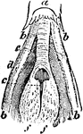
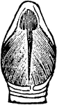
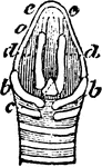
The Glottis of a Rook
"Glottis, or opening of trachea in the mouth; a, base of tongue; b, b, horns of hyoid bone; c, rima…
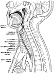
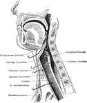
Sagittal Section of the Head and Neck
Sagittal median section of the head and neck. The head is thrown backward into complete extension which…
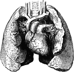
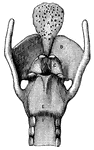
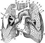
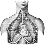
Heart and Lungs
The heart and lungs. 1, right ventricle; 3, right auricle (atrium); 6, 7, pulmonary artery; 9, aorta;…
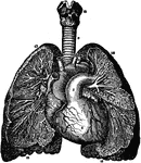
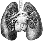
Heart and Lungs
The heart, showing its relative position to the lungs. The heart is almost wholly covered up by the…
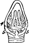
The Laryngeal Muscles of a Rook
"a, b, c, d, inferior laryngeal or syringeal muscles, not well made out in this figure; But typical…
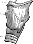
Larynx
External view of the left side of Larynx. 1: Front portion of hyoid bone; 2: Upper edge of larynx; 3:…
Larynx
Cross section of the larynx above the vocal cords. 1: Right vocal cord. 2: Left vocal cord. 3: Cartilages…
Vertical Section of Larynx
This illustration shows a vertical section of the larynx and its many parts (A. Thyroid Cartilage; B.…
The Larynx Muscles of a Rook
"Muscles of the larynx. thyro-arytenoids, or openers of the glottis" Elliot Coues, 1884
The Larynx Muscles of a Rook
"Muscles of the larynx. Thyro-cricoids, posterior thyro-cricoids." Elliot Coues, 1884
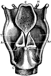
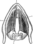
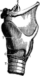
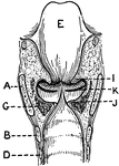
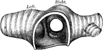
Back View of the Larynx
Labels: T, thyroid cartilage: C, cricoid cartilage; Tr, trachea; H, hyoid bone; E, epiglottis; I, joint…

Lateral Aspect of Larynx
This illustration shows a lateral aspect of the larynx and its multiple parts (A. Thyroid Cartilage;…
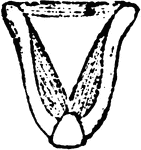
Posterior view of the larynx
"Cartilages and Ligaments of the Larynx. (Front view.) A, epiglottis; B, thyroid cartilage;…
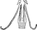
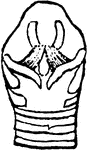
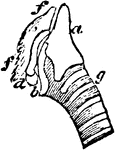
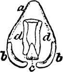
The Larynx of a Rook
"Larynx viewed from before (below); a, thyroid bone or cartilage." Elliot Coues, 1884
The Larynx of a Rook
"Larynx viewed from behind (above); a, thyroid bone; b, b, its appendages; c, cricoid; d, d, arytenoids;…
The Larynx of a Rook
"Larynx viewed from the right side; a, thyroid; b, appendage; c, cricoid; d, arytenoid; f, f, cartilage…
The Larynx of a Rook
"Larynx viewed from behind; a, thyroid; b, b, its appendages; c, cricoid; d, d, arytenoid." Elliot Coues,…
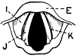
Side View of the Larynx
Labels: T, thyroid cartilage: C, cricoid cartilage; Tr, trachea; H, hyoid bone; E, epiglottis; I, joint…
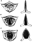
The Larynx in Different Conditions of the Glottis
The larynx as seen by means of the laryngoscope in different conditions of the glottis. Labels: A, while…
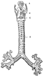
Front View of the Cartilages of the Larynx, Trachea and Bronchi.
Front view of cartilages of larynx, trachea and Bronchi.
Longitudinal Section of Larynx Seen from Behind
This illustration shows a longitudinal section of the larynx as seen from behind (A. Thyroid Cartilage;…
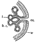
Lungs
This is a diagram illustrating lungs or tracheae. b.c., the cavity in which the body fluids circulate;…
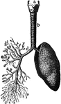
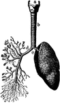
Lungs
"The Lungs. 1, Summit of lungs. 2, Base of lungs. 3, Trachea. 4, Right bronchus. 5, Left bronchus. 6,…
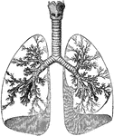
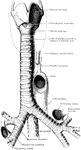
Lungs and Air Passages
The lungs and air passages seen from the front. On the left of the figure the pulmonary tissue has been…
The Lungs and Air Passages
The lungs and air passages seen from the front. On the left of the figure the pulmonary tissue has been…
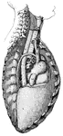
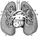
Anterior View of the Lungs and Heart
Anterior view of the lungs and heart. Labels: 1, heart; 2, inferior vena cava; 3, superior vena cava;…
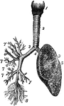
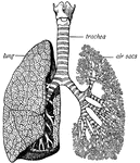
Lungs and Trachea
The lungs and windpipe (trachea). Labels: 1, larynx; 2, windpipe (trachea); 3, right lung, showing bronchi…
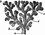
Structure of the Lungs
Diagram showing the structure of the lungs. At d is the left lung, and at c are represented…
The lungs
"The lungs fill up most of the cavity of the chest. One lies on either side of the heart which is in…
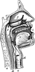
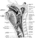
The Mouth, Nose, and Pharynx
The mouth, nose, and pharynx, with the commencement of gullet (esophagus) and larynx, as exposed by…
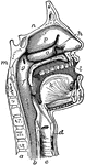
The Mouth, Nose, and Pharynx
The mouth, nose, and pharynx, with the larynx and commencement of gullet (esophagus), seen in section.…
Respiratory Mechanism
"The respiratory mechanism consists of the lungs, a series of minute air chambers with a network of…
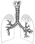
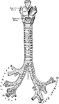
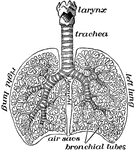
Respiratory system
"Larynx, trachea, and bronchi, showing the manner of division, and the rings of cartilage." —…
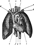
Respiratory System
The respiratory system. Labels: 1. the larynx; 2. the trachea; 3. right bronchia; 4. left bronchia;…
Respiratory System
The respiratory system. Labels: 1, larynx; 2, trachea; 3, right lung; 4, left lung; 5, heart.
Upper Respiratory Tract
Operative approaches through the front of the neck to the larynx, pharynx, and trachea. a: Approach…
Transverse Section of Trachea and Esophagus
Transverse section of trachea and esophagus of child, seen from below.
Trachea and lungs
"The trachea has in its walls stiff rings of cartilage that hold it open so that the air can…
Transverse Section of Trachea Showing Arrangement of Walls
Transverse section of trachea, showing general arrangement of its wall.
The Trachea of a Rook
"Last entire tracheal ring, viewed from below, crossed by the pessulus." Elliot Coues, 1884
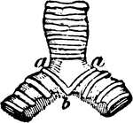
The Trachea of a Rook
"Bifurcation of trachea, and bronchi, viewed from below; a, pessulus, the bolt-bar, or "bone of divarication";…
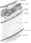
Section of the Trachea
Section of the trachea. Labels: a, columnar ciliated epithelium; b and c, proper structure of the mucous…
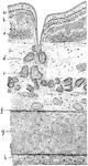
Sections Through Trachea
Transverse section through the trachea and its immediate surroundings at the level of each of the upper…
Transverse Section of the Trachea
Transverse section of the trachea, just above its bifurcation, showing the carina tracheae.
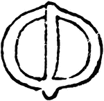
The Tracheal Rings of a Bird
"1, 2, left, two tracheal rings, separate. b; 1, 2, right hand, the same put together." Elliot Coues,…