The Mammal Anatomy: Skeleton ClipArt gallery provides 277 views of bones, teeth, and skeletal system of various mammals.

"Pectoral girdle of Echidna. sc., Scapula; cl., clavicle; i.cl., prosternum or "interclavicle"; co., coracoid or metacoracoid; e.co., procoracoid or precoracoid; st., mesosternum." -Thomson, 1916

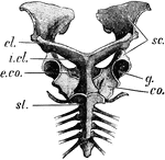
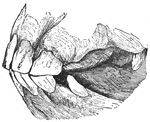
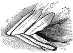
Echidna Pectoral Girdle

"Pectoral girdle of Echidna. sc., Scapula; cl., clavicle; i.cl., prosternum or "interclavicle"; co.,…
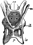
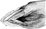
"The pelvis of the Echidna; sa, sacrum; il, illum; is, ischium; p, pubis; m, marsupial bone." — Chambers, 1881

Echidna Pelvis

"The pelvis of the Echidna; sa, sacrum; il, illum; is, ischium; p, pubis; m, marsupial bone." —…
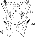
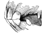
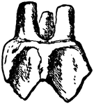
"Pelvis of Echidna. S., Sacrum; Ep., epipubic bones; Ac., acetabulum; o.f., obturator foramen between ischium and pubis (p.)." -Thomson, 1916

Echidna Pelvis

"Pelvis of Echidna. S., Sacrum; Ep., epipubic bones; Ac., acetabulum; o.f., obturator foramen between…
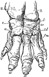
"Right fore foot of Indian Elephant. U, ulna; R, radius; c, cunelform; l, lunar; sc, scaphold; u, unciform; m, magnum; td, trapezoid; tm, trapezium; I to V, first to fifth digit." —The Encyclopedia Britannica, 1903

Elephant Foot

"Right fore foot of Indian Elephant. U, ulna; R, radius; c, cunelform; l, lunar; sc, scaphold; u, unciform;…

"Skeleton and Outline of African Elephant (Elephas or Loxodon africanus). fr, frontal; ma, mandible; ma', malar; fi, "finger" at end of trunk; C, cervical vertebrae; D, dorsal vertebrae; D, dorsal vertebrae; pe, pelvis; sc, scapula; st, sternum; hu, humerus; ul, ulna; ra, radius; mc, metacarpus; fe, femur; pat, patella; tib, tibia; fib, fibula; met, metatarsus." -Whitney, 1911

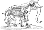
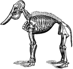
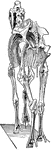
African Elephant Skeleton

"Skeleton and Outline of African Elephant (Elephas or Loxodon africanus). fr, frontal; ma, mandible;…

"Shows how the bones of the arm (q), forearm (q'x), and foot (o), are twisted to form an osseous screw."—Pettigrew, 1875

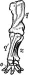
Anterior Extremity of Elephant

"Shows how the bones of the arm (q), forearm (q'x), and foot (o), are twisted to form an osseous screw."—Pettigrew,…
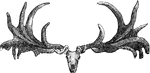
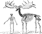
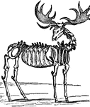
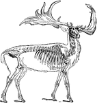
The fossilized skeleton of an elephant.

Fossil elephant

The fossilized skeleton of an elephant.

"Skull and antlers of the Irish elk." -Taylor, 1904

Irish Elk Skull

"Skull and antlers of the Irish elk." -Taylor, 1904

The giant elk ranged from Ireland to the borders of Italy. The Irish Elk or Giant Deer was a species of Megaloceros and one of the largest deer that ever lived. The name "Giant Deer" is preferred as it is not related to either of the living species of elk. The Skeleton of Extinct Giant Elk (Megacerous hibernicus) of Ireland, compared with the skeleton of man.

Extinct Giant Elk

The giant elk ranged from Ireland to the borders of Italy. The Irish Elk or Giant Deer was a species…
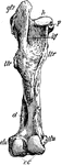
The fossilized skeleton of an elk.

Fossilized elk

The fossilized skeleton of an elk.

"Irish elk (Cervus megaceros)." -Taylor, 1904

Irish Elk

"Irish elk (Cervus megaceros)." -Taylor, 1904

"Skull of Eupleres goudoti." —The Encyclopedia Britannica, 1903

Eupleres Goudoti

"Skull of Eupleres goudoti." —The Encyclopedia Britannica, 1903

Posterior view of a left femur of a horse. Labels: 1, head; 2, trochanter major; 3, trochanter minor; 4, trochanter internus; 5, notch for the round ligament; 6, trochanteric fossa; 7, 8, places for attachment for the laterla ligaments; 11, the condlyes, separated by the intercondyloid groove; 10, supracondyloid fossae.

Femur of a Horse

Posterior view of a left femur of a horse. Labels: 1, head; 2, trochanter major; 3, trochanter minor;…
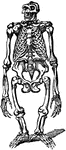
"Skull of Fiber zibethica." —The Encyclopedia Britannica, 1903

Fiber Zibethica

"Skull of Fiber zibethica." —The Encyclopedia Britannica, 1903

Foot of bear: plantigrade

Carnivore Foot

Foot of bear: plantigrade

Hind feet of Seal; Pinnigrada

Carnivore foot

Hind feet of Seal; Pinnigrada

An illustration of the skeleton of a kangaroo's right hind foot.

Kangaroo Foot

An illustration of the skeleton of a kangaroo's right hind foot.

Skeleton of the fore limb of the horse. <em>sc</em>, shoulder blade; <em>h</em>, humerus; <em>r</em>, radius; <em>ua</em>, ulna; <em>m</em>, splint bone or fourth metacarpal; <em>p</em>, phalanges.

Fore Limb

Skeleton of the fore limb of the horse. sc, shoulder blade; h, humerus; r,…

A small fruit eating flying bat.

Flying Fox

A small fruit eating flying bat.

The skeletal structure of a gorilla.

Gorilla Skeleton

The skeletal structure of a gorilla.

"Skeleton of male gorilla. cl., Clavicle; sc., tip of scapula; S., praesternum; H., humerus; r., radius; u., ulna; Il., ilium; C., coccyx; P., pubis; Is., ischium; F., femur; t., tibia; f., fibula." -Thomson, 1916

Gorilla Skeleton

"Skeleton of male gorilla. cl., Clavicle; sc., tip of scapula; S., praesternum; H., humerus; r., radius;…
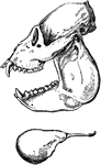
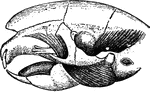
"Skull and teeth of Gray's Rat Kangaroo (Bellongia grayii). c, upper canine tooth. i1, i2, i3, first, second, and third upper incisors; pm, second or posterior premolar (the first having been already shed); m1, m2, m3, m4, the four true molars." &mdash;The Encyclopedia Britannica, 1910

Gray's Rat Kangaroo

"Skull and teeth of Gray's Rat Kangaroo (Bellongia grayii). c, upper canine tooth. i1, i2, i3, first,…

A comparison of vertebrate hands. A, hand or anterior foot of the dog; B, that of the hog; C, that of the elk; D, that of the ox. Digits: I, pollex; II, index; III, medius; IV, annukus; V, minimus.

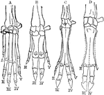
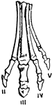
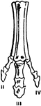
Hands of Vertebrates

A comparison of vertebrate hands. A, hand or anterior foot of the dog; B, that of the hog; C, that of…
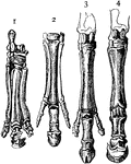
An illustration of a cows horns.

Cow Horns

An illustration of a cows horns.

"Feet of horse and its predecessors. 1, Palaeotherium; 2, Anchitherium; 3, Hippotherium; 4, Equus." -Thomson, 1916

Horse Feet

"Feet of horse and its predecessors. 1, Palaeotherium; 2, Anchitherium; 3, Hippotherium; 4, Equus."…

"Posterior View of Left Femur of a Horse. h, head; gtr, great trochanter; ttr, third trochanter; ltr, lesser trochanter; p, pit for round ligament; itf, intertrochanteric fossa; d, a depression or fossa; etu, itu, external and internal tuberosities; cc, the two condyles." -Whitney, 1911

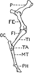
Posterior View of Left Femur of Horse

"Posterior View of Left Femur of a Horse. h, head; gtr, great trochanter; ttr, third trochanter; ltr,…

"Successive stages of modification of the feet of extinct forms of horse-like animals, showin gradual reduction of the outer and enlargement of the middle toe (III). Orohippus (Eocene)." &mdash; Encyclopedia Britannica, 1893

Horse Foot

"Successive stages of modification of the feet of extinct forms of horse-like animals, showin gradual…

"Successive stages of modification of the feet of extinct forms of horse-like animals, showin gradual reduction of the outer and enlargement of the middle toe (III). Meschippus (Early Miocene)." &mdash; Encyclopedia Britannica, 1893

Horse Foot

"Successive stages of modification of the feet of extinct forms of horse-like animals, showin gradual…

"Successive stages of modification of the feet of extinct forms of horse-like animals, showin gradual reduction of the outer and enlargement of the middle toe (III). Miohippus and Anchitherium (Late Miocene)." &mdash; Encyclopedia Britannica, 1893

Horse Foot

"Successive stages of modification of the feet of extinct forms of horse-like animals, showin gradual…

"Successive stages of modification of the feet of extinct forms of horse-like animals, showin gradual reduction of the outer and enlargement of the middle toe (III). Hipparion and Pliohippus (Pliocene)." &mdash; Encyclopedia Britannica, 1893

Horse Foot

"Successive stages of modification of the feet of extinct forms of horse-like animals, showin gradual…

"Successive stages of modification of the feet of extinct forms of horse-like animals, showin gradual reduction of the outer and enlargement of the middle toe (III). Equus (Pleistocene)." &mdash; Encyclopedia Britannica, 1893

Horse Foot

"Successive stages of modification of the feet of extinct forms of horse-like animals, showin gradual…

The colt's mouth at two years of age.

Horse Jaw

The colt's mouth at two years of age.

The mouth of a colt at two and a half years old.

Horse jaw

The mouth of a colt at two and a half years old.

The mouth of a colt at three years old.

Horse jaw

The mouth of a colt at three years old.

The side view of the jaw of a three year old colt.

Horse jaw

The side view of the jaw of a three year old colt.

The side view of the jaw of a four year old colt.

Horse jaw

The side view of the jaw of a four year old colt.

The mouth of a colt at four years old.

Horse jaw

The mouth of a colt at four years old.

The mouth of a colt at four and a half years old.

Horse jaw

The mouth of a colt at four and a half years old.

The mouth of a horse at five years of age.

Horse jaw

The mouth of a horse at five years of age.

The mouth of a horse at six years old.

Horse jaw

The mouth of a horse at six years old.

The mouth of a horse at seven years old.

Horse jaw

The mouth of a horse at seven years old.

The mouth of a horse at eight years old.

Horse jaw

The mouth of a horse at eight years old.

The mouth of a horse at twenty years old.

Horse jaw

The mouth of a horse at twenty years old.

The mouth of a horse at thirty years old.

Horse jaw

The mouth of a horse at thirty years old.

"Side view of second upper molar tooth of Anchitherium (brachyodont form)." &mdash; Encyclopedia Britannica, 1893

Horse Molar

"Side view of second upper molar tooth of Anchitherium (brachyodont form)." — Encyclopedia Britannica,…

"Corresponding tooth of horse (hypsidont form)." &mdash; Encyclopedia Britannica, 1893

Horse Molar

"Corresponding tooth of horse (hypsidont form)." — Encyclopedia Britannica, 1893

Skeleton of a horse that lived in Colorado more than a million years ago. From Guide Leaflet of the American Museum by Matthews.

Horse Skeleton

Skeleton of a horse that lived in Colorado more than a million years ago. From Guide Leaflet of the…
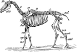
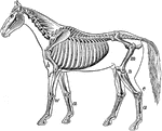
"Skeleton of Horse (Equus caballus). fr, frontal bone; C, cervical vertebrae; D, dorsal vertebrae; L, lumbar vertebrae; cd, caudal vertebrae; sc, scapula; pe, pelvis; ma, mandible; hu, humerus; ra, radius; cp, carpus; mc, metacarpus; fe, femur; tib, tibia; ca, calcaneum; tar, tarsus; mt, metatarsus; p, phalanges." -Whitney, 1911

Horse Skeleton

"Skeleton of Horse (Equus caballus). fr, frontal bone; C, cervical vertebrae; D, dorsal vertebrae; L,…
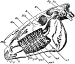
"Side view of skull of horse, with the bone removed so as to expose the whole of the teeth. PMx, premaxilla; Mx, maxilla; Na, nasal bone; Ma, malar bone; L, lacrymal bone; Fr, frontal bone; Sq, squamosal bone; Pa, parietal bone; oc, occipital condyle; pp, paroccipital process; i, the three incisor teeth; c, the canine tooth; pm1, the situation of the rudimentary first premolar, which has been lost in the lower, but is present in the upper jaw; pm2, pm3, and pm4, the three fully-developed premolar teeth; m, the three true molar teeth." &mdash; Encyclopedia Britannica, 1893

Horse Skull

"Side view of skull of horse, with the bone removed so as to expose the whole of the teeth. PMx, premaxilla;…
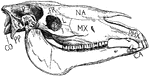
"Side view of horse's skull. P., Parietal; FR., frontal; NA., nasal; PMX., premaxilla; MX., maxilla; J., jugal; L., lachrymal; SQ., squamosal; PP., paroccipital process; CO., condyle CA., canine." -Thomson, 1916

Horse Skull

"Side view of horse's skull. P., Parietal; FR., frontal; NA., nasal; PMX., premaxilla; MX., maxilla;…

This illustration shows the leg of a horse.

Leg of Horse

This illustration shows the leg of a horse.

"Skeleton of the horse." &mdash; Davison, 1906

Skeleton of a Horse

"Skeleton of the horse." — Davison, 1906

"Side views of skull and hyoid bone of Howling Money." &mdash; Encyclopedia Britanica, 1893

Howling Monkey Skull

"Side views of skull and hyoid bone of Howling Money." — Encyclopedia Britanica, 1893

"Dentition of Hyaenodon leptorhynchus. The posterior molar is concealed behind the penultimate tooth." &mdash;The Encyclopedia Britannica, 1903

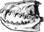
Hyaenodon Leptorhynchus

"Dentition of Hyaenodon leptorhynchus. The posterior molar is concealed behind the penultimate tooth."…
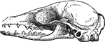
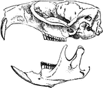
"Skull of Hydrochaerus Capybara." &mdash;The Encyclopedia Britannica, 1903

Hydrochaerus Capybara

"Skull of Hydrochaerus Capybara." —The Encyclopedia Britannica, 1903

"Skull of Hydropotes inermis (adult male), a deer without antlers, but with largely-developed upper canine teeth." &mdash;The Encyclopedia Britannica, 1903

Hydropotes Inermis

"Skull of Hydropotes inermis (adult male), a deer without antlers, but with largely-developed upper…

The hyoid series is composed of five distinct pieces- a body, or hyoid bone proper, two cornua or horns and two cornicula or lesser horns. Shown us the left aspect of the hyoid series of bones in a horse. Labels: a, proximal, end of left cornu; b, distal end of right cornu; c, proximal end of corniculum; d, hyoid bone. The left heel process points downwards and backwards, the spur process in the opposite direction.

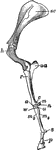
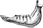
Hyoid Series of Bones in a Horse

The hyoid series is composed of five distinct pieces- a body, or hyoid bone proper, two cornua or horns…
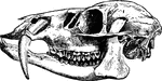
"Skull of Hystrix Eristata. t, temporal muscle; m, masseter. m', portion of masseter transmitted through the infra-orbital foramen, the superior maxillary nerve passing outwards between it and the maxillary bone." &mdash;The Encyclopedia Britannica, 1903

Hystrix Eristata

"Skull of Hystrix Eristata. t, temporal muscle; m, masseter. m', portion of masseter transmitted through…

Inferior maxilla of a horse-anterolateral view. Labels: a, body; b, b', rami; c, neck; d, mental foramen; e, buccinator; e', masseter surface; f, f', inner surface of ramus; g, molor alveoli; g', anterior border; g', bar, bounding diastema; h, h', h', posterior border. The angle lies between h' and h'; i, condyles; km coronoid processes; l, maxillary space; m, inferior dental foramen; n, sigmoid notch.

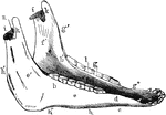
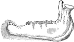
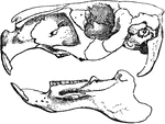
Inferior Maxilla of a Horse

Inferior maxilla of a horse-anterolateral view. Labels: a, body; b, b', rami; c, neck; d, mental foramen;…
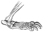
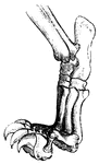
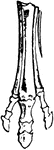
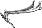
"Foot of young kangaroo. 2, 3, Small syndactylous toes; 4, large fourth toe; 5, fifth toe." -Thomson, 1916

Kangaroo Foot

"Foot of young kangaroo. 2, 3, Small syndactylous toes; 4, large fourth toe; 5, fifth toe." -Thomson,…