The Cellular Biology ClipArt gallery offers 242 illustrations of biology at the cellular level for many species and parts of the body, including blood cells and bone tissue, and also contains general images of cell, cell structure, and cell reproduction.
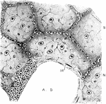
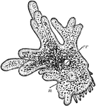
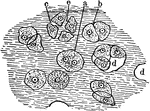
Air Cells from a Cat's Lung
From a section of the lung of a cat, stained with silver nitrate. Labels: A. D., alveolar duct or intercellular…
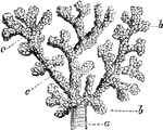
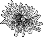
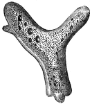
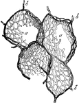
Air Cells of a Monkey
Terminal branch of a bronchial tube, with its infundibula and air cells from the margin of the lung…
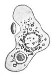
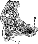
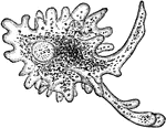
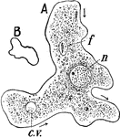
Amoeba
The streaming of Protoplasm in the Amoeba. The forward motion of the granules takes place more rapidly…
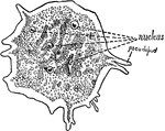
Amoeba
Amoung the simplest one-celled animals living in the ooze at the bottom of nearly every freshwater stream…
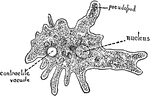
Amoeba
Difflugia one of several genera of amoebozoa that produce shells or tests from granules of sand. These…
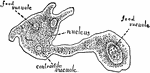
Amoeba
Gromia is a widespread genus of marine and freshwater amoeboids, closely resembling some foraminiferans.
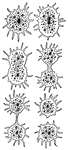
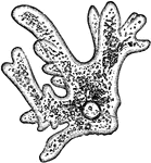
Amoeba Cell Division
"Direct cell division (Amoeba). A, active specimen with pseudopodia; B, becoming spherical preliminary…
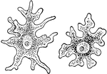
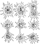
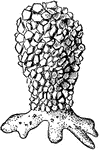
Amoeba Princeps (Ehrenberg)
"These little creatures frequently have the appearance of small, rounded masses, like drops of water.…
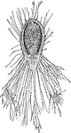
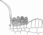
Section through Antheridium of Liverwort
The Antheridium is the male organ of plants. Within it are produced the sperms or their equivalents,…
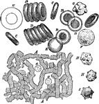
Subcutaneous Areolar Tissue from a Young Rabbit
Subcutaneous areolar tissue from a young rabbit, highly magnified. The white fibers are in wavy bundles,…
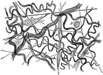
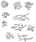
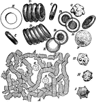
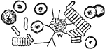
Bacilli
"Bacilli, or Rod-Shaped Bacteria. From a culture obtained in antharax, or malignant pustule, of the…
Bacillis Anthracis
"A. Bacilli mingled with blood corpuscules from the blood of a guinea-pig; some of the bacilli dividing.…
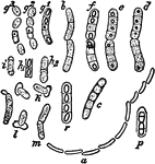
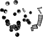
Bacillus Megaterium
"a, a chain of motile rodlets still growing and dividing (bacilli). b, a pair of bacilli actively growing…
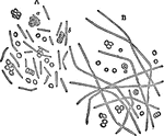
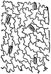
Milk bacteria
"Different Kinds of Milk Bacteria. It is not uncommon for a large number of person to be poisoned from…
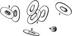
Balsam
"Epidermis of the garden Balsam, showing stomata st, of an elliptical form." — Encyclopedia Britanica,…
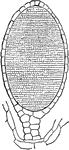
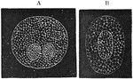
Blastoderm of an Egg
Vertical section of area pellucida and area opaca (left extremity of figure) of blastoderm of a fresh…
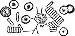
Red and White Blood Cells
Red and white corpuscles (cells) of the blood, magnified. Labels: A, moderately magnified, the red corpuscles…
Blood Cells
Blood corpuscles. Labels: A, magnified about 400 diameters. The red corpuscles have arranged themselves…
Blood Cells
Red corpuscles (blood cells) of the frog. The red blood cells of birds, reptiles, amphibians and fishes…
Blood Cells
White blood corpuscle (cell), sketched at successive intervals of a few seconds to illustrate the changes…

Blood Corpuscles
"Blood corpuscles (human). c, colored; l, leucocytes. The red cells tend to collect in rows with the…
Blood Corpuscles
Blood corpuscles (cells). Labels: A, magnified about 400 diameters. The red corpuscles have arranged…
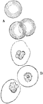
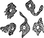
Colorless and Colored Blood Corpuscles
A, Three colored blood corpuscles. B, Three colorless blood corpuscles acted on by acetic acid; the…
Intracellular Network of Blood Corpuscles
The intracellular network of colorless and colored blood corpuscles. Labels: A, The colorless blood…
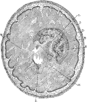
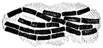
Formation of Compact Bone in a Kitten
Transverse section through the tibia of a fetal kitten. Labels: P, Periosteum. O, Osteogenetic layer…
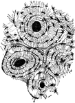
Bone Tissue of Humerus
Transverse section of compact tissue of humerus, magnified about 150 diameters. Three of the Haversian…
V. Sepium Bract
"Portion of a cross section through a nectariferous bract of Vicia sepium; n, nectar-secreting cells."…
Capillaries Showing Nucleated Endothelial Membrane
The walls of capillaries are composed of a single layer of elongated or radiate, flattened and nucleated…
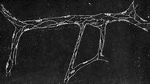
Capillary Vessels of Air Cells
The form of the capillary network presents considerable variety in the different textures of the body:…
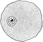
Cartilage Tissue Cells
A thin slice of cartilage, magnified, to how the cells embedded in the homogenous matrix. Labels: a,…
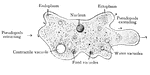
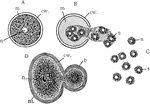
Cell
"In a general what we may describe a cell as a tiny mass of jelly in which floats another still smaller…
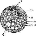
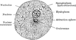
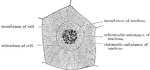
Cell
Diagram showing the principal parts of the cell and something of the protoplasmic architecture as it…
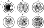
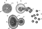
Cell Development
Every human body begin as a single nucleated cell. This cell, known as the ovum, divides or segments…
Cell Division
Diagram showing the change which occur in the centrosomes and nucleus of a cell n the process of mitotic…
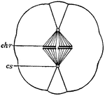
Cell Division
"Diagram of cell division. chr., Chromosomes forming an equatorial plate; cs., centrosome." -Thomson,…
Process of Cell Division
Indirect division of a cell (karyokinesis). Labels: 1, nesting cell; 2, cell preparing to divide-two…
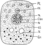
Cell Parts
"Diagram showing the principal parts of the cell as it appeaers when killed and stained. The protoplasm…
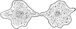
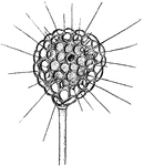
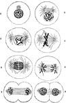
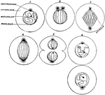
Cell Reproduction
Modes of cell reproduction. A, B, and C, stages in the reproduction of the Infusorian, Colpoda, by the…
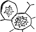
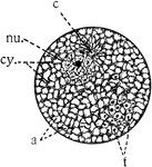
Cell Structure
"Diagram of cell structure. Pl. Plastids in cytoplasm. cc. Centrosome. n. Nucleolus. Chr. Chromosomes.…
Cell with its Reticulum Disposed
A cell with its reticulum disposed radically; from the intestinal epithelium in a worm.