Clipart tagged: ‘auricle’

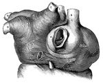
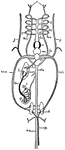
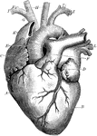
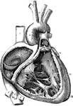
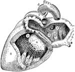
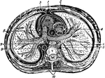
Right Atrium and Ventricle of the Heart
The right auricle (atrium) and ventricle of the heart opened, and a part of their right and anterior…
Muscular fibers of the auricle
L.A., left auricle; R.A., right auricle; A, opening of the inferior vena…
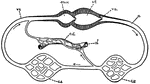
Blood Circulation
This illustration shows a representation of the circulation of the blood, in its essential features.…
Branchi and Blood Vessels
Branchi of the lungs, the heart, and blood vessels. Labels: 1, left auricle; 2, right auricle; 3, left…
Fish Circulation
"Diagram of the principal vessels in the circulation of a fish, ventral view. a, aorta; au., auricle;…
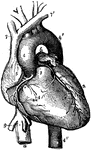
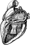
Heart
Front view of the heart and great vessels. The pulmonary artery has been cut short close to its origin.…
The Heart
A diagram of the heart. Labels: 1. Left auricle. 2, Right auricle. 3, Left ventricle. 4, Right ventricle.…
The Heart
A diagram of the heart. Labels: 1. Right auricle. 2, Left auricle. 3, Right ventricle. 4, Left ventricle.…
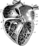
Heart
The heart. Labels: A, the right ventricle; B, the left ventricle; C, the right auricle; D, the left…
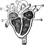
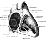
Heart and its Chambers
View of the heart with its several chambers exposed and the vessels in connection with them. Labels:…
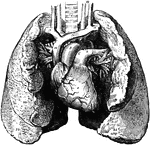
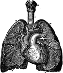
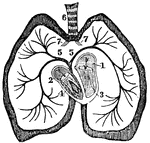
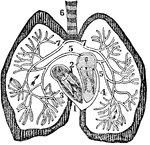
Heart and Lungs
The heart and lungs. 1, right ventricle; 3, right auricle (atrium); 6, 7, pulmonary artery; 9, aorta;…
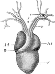
Heart of a Frog
The heart of a frog (Rana esculenta) from the front. Labels: V, ventricle, Ad, right auricle; As, left…
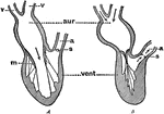
Diagram Showing the Pumping of the Heart
Diagram to illustrate the action (pumping) of the heart. Labels: aur., auricle; vent., ventricle; v,…
Heart with Left Auricle and Ventricle Laid Open
The left auricle and ventricle laid open, the posterior walls of both being removed.
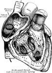
Heart with Right Auricle and Ventricle Laid Open
The right auricle and ventricle laid open, the anterior walls of both being removed.
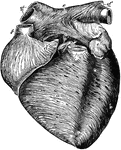
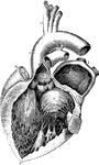
Anterior View of the Heart
Anterior view of the heart, dissected, after long boiling to show the superficial muscular fibers. The…

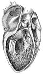
Auricle and Ventricle of the Heart
The cavities of the right auricle and right ventricle of the heart.
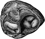
Cavities of the heart
"A, B, right pulmonary veins, S, openings of the left pulmonary veins; E, D, C,…
Chambers of the Heart
The chambers of the heart. Labels: A, right ventricle; B, left ventricle; C, right auricle; D, left…
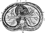
A Diagram of the Heart
The right auricle and ventricle opened, and a part of their right and anterior walls removed, so as…
A Diagram of the Heart
The left auricle and ventricle opened and a part of their anterior and left walls removed. The pulmonary…
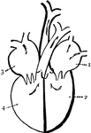
Diagram of the heart
"Diagram of the passages of the heart. 1. left auricle. 2. left ventricle, 3. Right auricle. 4. Right…
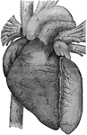
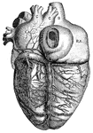
Heart, Front View of
A representation of the heart as it really appears showing the front view. At a is the right…
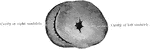
Left Side of Heart
Left side of heart. Labels: 1, cavity of left auricle (atrium); 3, opening of right pulmonary veins;…
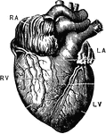
The Heart
The heart. Labels: RA, right auricle, RV; right ventricle; LA, left auricle; LV, left ventricle.
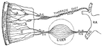
Intestinal absorption
"A, a fold of peritoneum; B, lacteals and lymphatic glands; C, veins of intestines;…
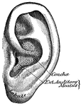
Pinna
"The outer ear consists of a plate of gristle, shaped somewhat like a shell, known as the pinna, or…
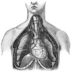
Thorax
"The transverse section of the thorax. 1, anterior mediastinum; 2, internal mammary vessels; 3, triangularis…
Transverse Section of the Thorax
Transverse section of the thorax. Labels: 1, anterior mediastinum; 2, internal mammary vessels; 3, triangularis…
Ventricle
The right ventricle is in the shape of a crescent moon and the left ventricle is in the shape of a circle.