The Bird Anatomy ClipArt gallery offers 411 illustrations of skeleton diagrams, arteries, digestive system, eggs, feathers, and both internal and external diagrams.
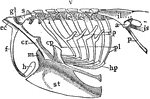
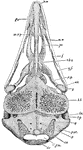
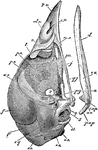
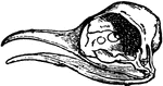
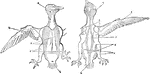
Bobolink Epipleurae
"Epipleurae.-- Thorax, scapular arch, and part of pelvic arch of a bobolink (Dolichonyx oryzivorus).…
Brain of a Bird
In birds the hemispheres are not united as in humans; the cerebellum is proportionately larger than…
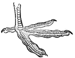
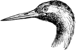
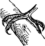
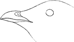
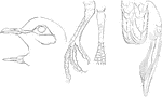
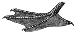
The Foot of a Bridled Foot
"Sterna anaisthetikos. Bridled Tern. The foot of a Bridled Tern; Tarsus .85; middle toe the same, with…
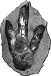
Footprint of brontozoum giganteum
A footprint of brontozoum giganeum, a now extinct relative of the cassowary. This example is eighteen…

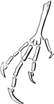
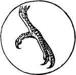
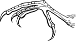
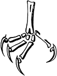
Foot of Honey-Buzzard
"In birds of prey the claws are powerful and hooked; in others the foot is flat, claws straight, and…
Phalanges of Caprimulgine
"Fig. 41 shows phalanges of caprimulgines foot, where the ratio is 2, 3, 4, 4." Elliot Coues, 1884
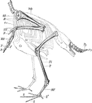
Skeleton of the Limbs and Tail of a Carinate Bird
"Skeleton of the Limbs and Tail of a Carinate Bird. (The skeleton of the body is indicated by dotted…
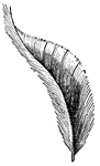
Cassowary Feather
"Casuarius (Cassowary). Feather, showing after-shaft and disconnected barbs." -Parker, 1900
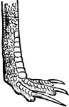
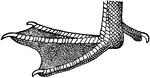
Foot of Cassowary
"In birds of prey the claws are powerful and hooked; in others the foot is flat, claws straight, and…
Scutellate Laminiplanter Tarsus of a Cat-bird
"Figure shows Scutellate laminiplanter tarsus of a cat-bird. A tarsus so disposed as to its podotheca…
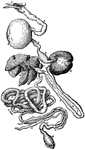
Intestinal Tract of Chauna Chavaria
cc. Colic caeca, d. Duodenum. g. Glandular patch, l.l. Meckel's tract, l.i. Hind-gut, p.v. Cut root…
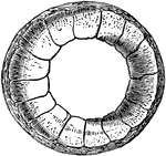
Chick Development
"Origin of lungs, liver, and pancreas in the chick. The mesoderm is shaded; the endoderm dark. lg.,…
Chick Head
"Fig 66 - Head of a chick, second stage, after five days of incubation, section in profile; x6 diameters.…
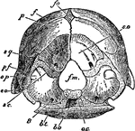
The Skull of a Chick Stage Three
"Skull of chick, third stage, viewed from below, x6 & 2/3 diameters. pn, prenasal cartilage, running…
The Skull of a Chick Stage Two
"Skull of chick, second stage, in profile, brain and membranes removed to show cartilaginous formations,…
Ripe Chick's Skull Profile
"Ripe chick's skull, longitudinal section, vied inside, x 3 diameters; after Parker. px, premaxillary;…
Ripe Chick's Skull
"Ripe chick's skull, longitudinal section, vied inside, x 3 diameters; after parker. In the mandible…
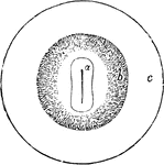
Vascular Area in the Chick
Diagram showing vascular area in the chick. A, area pellucida; b, area vasculosa; c, area vitellina.

Barn Swallow Claw
Side view of the Barn Swallow's claw. "Hirundo horreorum. Barn Swallow. Tarsi shorter than middle toe…
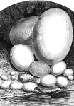
Comparative view of the size of the eggs of different animals
1, Epyornis; 2, Ostrich; 3, Cassoway; 4, Wild Goose; 5, hen; 6, Pigeon; 7, Humming-bird; 8, Eagle; 9,…
Condor Skeleton
"Entire skeleton of condor, showing the relative positions of the chief bones." -Thomson, 1916
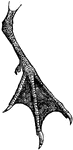
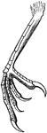
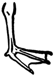
Foot of Coot
"The great toe is generally the strongest, but this is not an absolute law; a projection which is found…
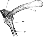
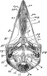
The Knee-joint of a Cormorant
"Phalacrocorax bicristatus. Cormorant. The knee-joint of a Cormorants. F, femur; P, patella; T, tibia;…
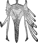
Cormorant Skull
"Phalacrocorax bicristatus. Red-Faced Cormorant. Skull showing sto, occipital style or nuchal bone;…
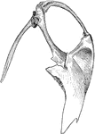
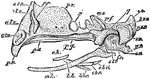
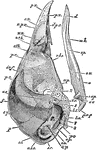
Cormorant Sternum and Shoulder
"Phalacrocorax bicristatus. Red-Faced Cormorant. Sternum and the shoulder from the skeleton of a Cormorant."…
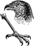
Bill of Cormorant
"The beak or bill of birds is composed of two bony pieces, called mandibles, surrounded by a horny substance,…
Neck-Covert
"From the head, backward to the tail, the body feathers increase in strength and size, also alter in…
Tail-Covert
"From the head, backward to the tail, the body feathers increase in strength and size, also alter in…
Sandhill Crane Windpipe
"Coiling of the windpipe in the sternum of Grus canadensis. Sandhill Crane." Elliot Coues, 1884
Whooping Crane Windpipe
"Very generally, in cranes and swans, the trachea enters the keel of the sternum, which is excavated…
Bill of Crane
"The beak or bill of birds is composed of two bony pieces, called mandibles, surrounded by a horny substance,…
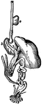
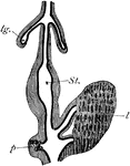
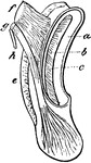
Crop and Digestive Organs
The swelling under the throat is called the crop, or first stomach. It is largely developed in some…
Bill of Cuckoo
"The beak or bill of birds is composed of two bony pieces, called mandibles, surrounded by a horny substance,…
Foot of a Cuckoo
The foot of a Cuckoo, a bird belonging to the Scansores order. Scansores is an order of birds, popularly…
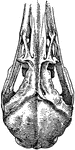
Curlew Skull
"Schizorhinal skull of curlew (top view), showing the long cleft, a, between upper and lower forks of…
Phalanges of Cypseline Foot
"Fig. 40 Phalanges of Cypseline foot, where the ratio is 2, 3, 3, 3 of Caprimulginae." Elliot Coues,…
Pterylosis of Cypselus Apus
"Fig. 24. - Pterylosis of Cyoselus apus, drawn by Coues after Nitzsch; right hand upper, left hand lower,…
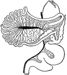
Fowl Digestion
The digestive system of a fowl. 1 is the tongue, 3 is the crop, 6 is the gizzard, 10 is the small intestine,…

Digestive Tract of a Chick
Diagram of part of digestive tract of a chick (4th day). The black line represents hypoblast , the outer…
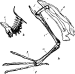
Diver Bones
"A. Pelvis and bones of the leg of the Leon or Diver; i, Innominate bone; f, Thighbone (femur); r, Tibia;…
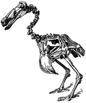
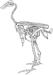
Dodo Skeleton
Illustration of a dodo bird skeleton. The dodo (Raphus cucullatus) was a flightless bird endemic to…
Sea Dove Bill
"Alle. Sea Dove. Size small. Bill very short, stout, and obtuse, as well as high at base, the sides…
White-fronted Dove Details
"Details of Engyptila albifrons (White-fronted Dove); head and foot natural size; wing and tail reduced.
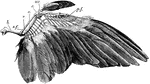
Dove Wing
"Wing of dove. h., Humerus; s.f., secondary feathers; r., radius; u., ulna; c., carpals; mc., carpo-metacarpus;…
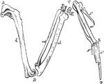
The Bones of the Right Wing of a Duck
"Fig 27. - Bones of the right wing of a duck, Clangula islandica, A, shoulder, omos; B, elbow, ancon;…
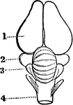
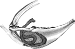
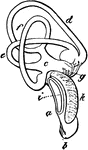
Eagle Cochlea
"Cochlea, X3. a, external, b, internal, cartilaginous prism; c, membranous zone; d, saccular extremity…
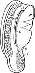
A Section of an Eagle's Cochlea
"Section of the cochlea, X3. a, vestibular surface of external cartilaginous prism, extending into d,…
Eagle Hind-Limb
"Bones of hind-limb of eagle. f., Femur; t.t., tibio-tarsus; fb., fibula; a., ankle-joint; m.t., tarso-metatarsus;…
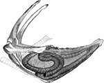
The Inner Ear of a White-tailed Eagle
"Membranous labyrinth of Haliaetus albicilla (White-tailed Eagle), X2. a,b, cochlea; b, its saccular…