The Bird Anatomy ClipArt gallery offers 411 illustrations of skeleton diagrams, arteries, digestive system, eggs, feathers, and both internal and external diagrams.
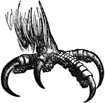
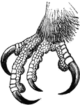
The talon of the Golden Eagle.

Eagle Talon

The talon of the Golden Eagle.

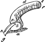
"Part of the superior vertical semicircular canal, showing its ampulla (which is the dilatation of the base of any semicircular canal), nerve of ampulla, artery connective tissue of the perilymph, X3, a, that part of the vestibule (alveus) next to the ampulla; b, the idlatation of the ampulla at its vestibular opening; c, where it passes into the canal proper; d, the canal, furnished with connective of the perilymph along its concave border and sides, as appears clearly at the sections e and f; g, nerve of the ampulla; h, artery of the connective tissue, running beneath it, remote from the wall of the duct." Elliot Coues, 1884

Eagle's Ampulal

"Part of the superior vertical semicircular canal, showing its ampulla (which is the dilatation of the…

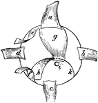
"Membranous labyrinth of Haliaetus albicilla (White-tailed Eagle), X2.  a,b, cochlea; b, its saccular extremity (or lagena); c, vestibule; g, its utricle; d, anterior of superior vertical semicircular canal; e, external or horizontal semicircular canal; f, posterior of inferior vertical semicircular canal; h, membranous canal leading into aqueduct of the vestibule; k, vascular membrane covering the scala vestibuli; opposite this, at i, are seen the edges of the cartilaginous prisms in the fenestra rotunda; from the edges of these cartilages proceeds the delicate membrane closing the opening of the cochlea (not shown in the fi.)" Elliot Coues, 1884

The Inner Ear of an Eagle

"Membranous labyrinth of Haliaetus albicilla (White-tailed Eagle), X2. a,b, cochlea; b, its saccular…

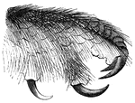
Foot of a bald eagle, well suited for grasping fish.

Foot of a Bald Eagle

Foot of a bald eagle, well suited for grasping fish.

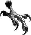
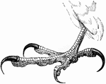
Foot of a bald eagle, showing claws for seizing its prey.

Claw of a Bald Eagle

Foot of a bald eagle, showing claws for seizing its prey.

"The beak or bill of birds is composed of two bony pieces, called mandibles, surrounded by a horny substance, differing both in form and thickness, according to the habits of the species."

Bill of Eagle

"The beak or bill of birds is composed of two bony pieces, called mandibles, surrounded by a horny substance,…
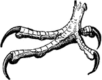
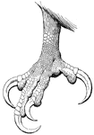
Foot of the royal eagle

Foot of the Royal Eagle

Foot of the royal eagle

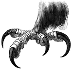
The claw of a golden eagle.

Golden Eagle Claw

The claw of a golden eagle.

"In birds of prey the claws are powerful and hooked; in others the foot is flat, claws straight, and adapted for walking."

Foot of White-Headed Eagle

"In birds of prey the claws are powerful and hooked; in others the foot is flat, claws straight, and…

"The more angular the wing of birds - that is to say, the longer the feathers on the edge of the wing - the more rapidly does it propel itself through the air."

Wing of an Eagle

"The more angular the wing of birds - that is to say, the longer the feathers on the edge of the wing…

A body formed in the females of birds, and some other animals, from which their young is produced.
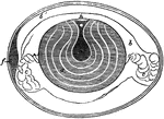
Egg

A body formed in the females of birds, and some other animals, from which their young is produced.

"Section of a Hen's Egg before Incubation. a, yolk, showing concentric layers; a', its semi-fluid center; b, inner dense part of the albumen; b', outer thinner part; c, twisted cords of albumen; h, the white spot, or germ cell." -Cooper, 1887

Egg Parts

"Section of a Hen's Egg before Incubation. a, yolk, showing concentric layers; a', its semi-fluid center;…

Cross-section of an egg, showing the different parts.

Cross-section of an egg

Cross-section of an egg, showing the different parts.

"Somateri mollissima. Somateri dresseri. Common Eider. Bill gibbous at base of upper mandible; outline of culmen variously curved; with long, acute or clubbed, tumid process extending in line with culmen variously curved; with long, acute or clubbed tumid process extending in line with culmen on each side of forehead, divided by extension of feathers on culmen. feathers of side of bill advancing to about under nostrils, far beyond those on culmen. No speculum. Male no black marks on chin. (mollissima - Frontal processes short, narrow, acute, parallel. Smaller.). (dresseri - Frontal processes long, broad, clubbed, divergent. Larger.)." Elliot Coues, 1884

The Bill of an Eider

"Somateri mollissima. Somateri dresseri. Common Eider. Bill gibbous at base of upper mandible; outline…
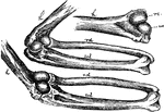
"Fig. 28. - Mechanism of elbow-joint. ..., where rc and uc show respectively the size, shape, and position of the radial condyle and ulnar condyle of the humerus. It is evident that in the flexed state of the elbow, as shown in the middle figure, the radius, rd, is do pushed upon that its end projects beyond ul, the ulna; while in the opposite condition of extension, shown in the lower figure, rd is pulled back to a corresponding extent." Elliot Coues, 1884

Mechanism of the Elbow-Joint

"Fig. 28. - Mechanism of elbow-joint. ..., where rc and uc show respectively the size, shape, and position…

Embryo chick (36 hours), viewed from beneath as a transparent object (magnified). Labels:pl, outline of pellucid area; FB, forebrain, or first cerebral vesicle: from its sides project op, the optic vesicles; SO, backward limit of somatopleure fold, "tucked in", under head; A, head-fold of true amnion; a', reflected layer of amnion, sometimes termed "false amnion;" sp, backward limit of splanchnopleure folds, along which run the omphalomesaraic veins uniting to form h, the heart, which is continued forwards into ba, the bulbus arteriosus; d, the foregut, lying behind the heart, and having a wide crescentic opening between the splanchnopleure folds; HB, hindbrain; MB, midbrain; pv, protovertebrae lying behind the foregut; mc, line of junction of medullary folds and of notochord; ch, front end of notochord; vpl, vertebral plated; pr, the primitive groove at its caudal end.

Embryo Chick

Embryo chick (36 hours), viewed from beneath as a transparent object (magnified). Labels:pl, outline…
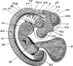
Embryo chick at fourth day, viewed as a transparent object, lying on its left side. CH, cerebral hemispheres; FB, forebrain or vesicle of third ventricle, with Pn, pineal gland projecting from its summit; MB, midbrain; Cb, cerebellum; IV V, fourth ventricle; L, lens; chs, choroidal slit; Cen V, auditory vesicle; s m, superior maxillary process; 1F, 2F, 3F,4F., first, second, third, and fourth visceral folds; V, fifth nerve, sending one branch (ophthalmic) to the eye, and another to the first pharyngeal nerve, passing to the third visceral arch; G.Pg, pneumogastric nerve, passing towards the fourth visceral arch; iv, investing mass; ch, notochord; its front end cannot be seen in the living embryo, and it does not end as shown in the figure, but takes a sudden bend downwards, and then terminated in a point; Ht, heart seen through the walls of the chest; MP, muscle-plates; W, wing, showing commencing differentiation of segments, corresponding to arm, forearm, and hand; H L, hind-limb, as yet a shapeless bud, showing no differentiation. Beneath it is seen the curved tail.

Embryo Chick at Fourth Day

Embryo chick at fourth day, viewed as a transparent object, lying on its left side. CH, cerebral hemispheres;…

A transverse section through an embryo chick (26 hours). Labels: a, epiblast; b, mesoblast; c, hypoblast; d, central portion of mesoblast, which is here fused with epiblast; e, primitive groove; f, dorsal ridge.

Cells of an Embryo Chick

A transverse section through an embryo chick (26 hours). Labels: a, epiblast; b, mesoblast; c, hypoblast;…

Transverse section of an embryo chick (third day). Labels: mr, rudimentary spinal cord; the primitive central canal has become constricted in the middle; ch, notochords; layer of mesoblast lining groove, which is not yet closed in to form the intestines; a o, one of the primitive aorta, u n, Wolffian body; ung; Wolffian duct; vc, vena cardinalis; h, epiblast; hp, somatopleure and its reflection to form af, amniotic fold; p, pleuroperitoneal cavity.

Transverse Section of an Embryo Chick

Transverse section of an embryo chick (third day). Labels: mr, rudimentary spinal cord; the primitive…
The eye of a common owl. The length from the anterior to posterior diameter lengthened.

Bird Eye

The eye of a common owl. The length from the anterior to posterior diameter lengthened.

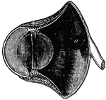
"Right Eyeball of Bird, seen from behind, showing the following muscles; a, rectus superior; b, rectus externus; c, rectus inferior; d, rectus internus; e, obliquus superior; f, obliquus inferior; g, quadratus; h, pyramidalis, with its tendon, k, passing through a pulley in the quadratus (as shown by dotted line) to keep it off the optic nerve, i, then passing around the edge of the ball to its insertion in the nictitating membrane." -Whitney, 1911

Right Eyeball of Bird

"Right Eyeball of Bird, seen from behind, showing the following muscles; a, rectus superior; b, rectus…

"A falcon. mn., Mandible; C., cere; N., nostril; E.C., ear covert; th.W., thumb wing; C., wing coverts; D., dorsal coverts; S., secondaries; P., primaries; R., rectrices; A., ankle; Mt., tarso-metatarsus; I., first toe." -Thomson, 1916

Falcon

"A falcon. mn., Mandible; C., cere; N., nostril; E.C., ear covert; th.W., thumb wing; C., wing coverts;…
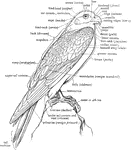
"The annexed figure explains the nomenclature of most of the outward of a Bird, but some further explanations may be given." A. H. Evans, 1900

A Labeled Diagram of a Falcon to Show the Nomenclature of the External Parts

"The annexed figure explains the nomenclature of most of the outward of a Bird, but some further explanations…

"Skeleton of the trunk of a Falcon. Ca, coracoid, which articulates with the sternum (St) at ; Cr, keel of sternum; Fu (Cl), furcula (clavicles); G, glenoid cavity for humerus; S, scapula; Un uncinate process; V, vertebral, and Sp, sternal, portion of rib. (From Wiedersheim.)" A. H. Evans, 1900

The Skeleton of the Trunk of a Falcon

"Skeleton of the trunk of a Falcon. Ca, coracoid, which articulates with the sternum (St) at ; Cr, keel…

The claw of a gerfalcon.

Gerfalcon Claw

The claw of a gerfalcon.

Foot of the Peregrine falcon, with talons for grasping prey.

Foot of a Peregrine Falcon

Foot of the Peregrine falcon, with talons for grasping prey.

One of the growths, generally formed each of a central quill and a vane on each side of it, which make up the covering of a bird.

Feather

One of the growths, generally formed each of a central quill and a vane on each side of it, which make…
"Parts of a Feather. a, quill; b, shaft; c, vane; d, down." -Cooper, 1887

Feather Parts

"Parts of a Feather. a, quill; b, shaft; c, vane; d, down." -Cooper, 1887

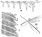
"Parts of a feather. I., Four barbs (B.) bearing anterior barbules (A.BB.) and posterior barbules (P.BB.); II., six barbs (B.) in section, showing interlocking of barbules; III., anterior barbule with barbicels (H.)." -Thomson, 1916

Feather Parts

"Parts of a feather. I., Four barbs (B.) bearing anterior barbules (A.BB.) and posterior barbules (P.BB.);…

"The feathers are horny productions, consisting of a hollow tube or barrel and a stem rising from it."

Feather Shaft

"The feathers are horny productions, consisting of a hollow tube or barrel and a stem rising from it."

"Fig. - 20 - Two barbs, a, a, of a vane, bearing anterior, b, b, and posterior, c, barbules; enlarged; after Nitzsch." Elliot Coues, 1884

Structure of a Feather

"Fig. - 20 - Two barbs, a, a, of a vane, bearing anterior, b, b, and posterior, c, barbules; enlarged;…

"The feathers are horny productions, consisting of a hollow tube or barrel and a stem rising from it."
Feather Tube

"The feathers are horny productions, consisting of a hollow tube or barrel and a stem rising from it."

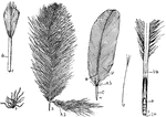
"Types of feathers. D., Down. 2, Developing feather in sheath (sh.). 3, Covert of heron showing aftershaft (A.S.). 4, Secondary feather of pigeon--C., calamus; A.S., aftershaft; R., rachis; V., vane. 5, Portion of quill showing inferior umbilicus (I.u.), superior umbilicus (S.u.), pith (P.); F., filoplume." -Thomson, 1916

Feather Types

"Types of feathers. D., Down. 2, Developing feather in sheath (sh.). 3, Covert of heron showing aftershaft…

"The feathers are horny productions, consisting of a hollow tube or barrel and a stem rising from it. The webs are convex above, and are thus enabled to resist flexion or fracture from beneath better than from any other direction."

Feather Web

"The feathers are horny productions, consisting of a hollow tube or barrel and a stem rising from it.…

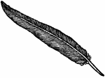
An illustration of a single black feather.

Black Feather

An illustration of a single black feather.

"A., Filoplume. B., very young feather within its sheath (sh.); c., the core of dermis; b., the barbs. C., the same, external view." -Thomson, 1916

Feathers

"A., Filoplume. B., very young feather within its sheath (sh.); c., the core of dermis; b., the barbs.…

"Filoplume. In ornithology, a thread-feather; a thread-like or hair-like feather, with a very slender stem, lacking webs in most or all of its length." -Whitney, 1911

Filoplume of a Goose

"Filoplume. In ornithology, a thread-feather; a thread-like or hair-like feather, with a very slender…

"Carpodacus. Purple Bullfinch. Bill smaller and less turgid than in Pinicol or Pyrrhula, more regularly conic and more acute; sides convex in all directions, but with distinct ridge prolonged in a point on forehead where not concealed by the antiae, its outline moderately curved; commissure decidedly angulated, about straight before and behind the bend; gonys quite straight. Nasal ruff little developed, barely cocealing the slight nasal fossae, thence falling over sides of bill, but discontinuous across culmen." Elliot Coues, 1884

The Bill of a Purple Finch

"Carpodacus. Purple Bullfinch. Bill smaller and less turgid than in Pinicol or Pyrrhula, more regularly…

"Sturnella magna. Field Lark. Old-field Lark. Meadow Lark. The colors, as above described, rich and pure, the prevailing aspect brown; black streaks prevailing on brown; yellow of chin usually confined between rami of under mandible; black bars on wings and tail usually confluent along the shaft of the feathers, leaving the gray in scallops. Sexes are similar: Female duller colored, the yellow paler. " Elliot Coues, 1884

Meadow Lark Foot and Bill

"Sturnella magna. Field Lark. Old-field Lark. Meadow Lark. The colors, as above described, rich and…
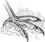
"Fig. 53 shows the lobate foot of a coot. In the lobate foot, a paddle results not from connecting webs, but from a series of lobes or flaps along the sides of the individual toes; as in the coots, grebes, phalaropes, and sun-birds. Lobation is usually associated with semipalmation, as is well seen in the grebes (Podicipedidae). In the snipe-like pharalopes (Phalaropodidae), lobation is present as a modification of a foot otherwise quite cursorial. The most emphatic cases of lobation are those in which each joint of the toes has its own flap, with a free convex border; the membranes as whole therefore present a scolloped outline." Elliot Coues, 1884

Coot Foot

"Fig. 53 shows the lobate foot of a coot. In the lobate foot, a paddle results not from connecting webs,…

"Quiscalus. Grackle. The feet are large and strong, and the birds spend much of their time on the ground, where they walk or run instead of advancing by leaps." Elliot Coues, 1884

Grackle Foot

"Quiscalus. Grackle. The feet are large and strong, and the birds spend much of their time on the ground,…
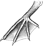
"Fig. 52 shows the totipalmate foot of a pelican. The totipalmate is a special case of palmation, in which all four toes are webbed; this characterizes the whole order Steganopodes." Elliot Coues, 1884

Pelican Foot

"Fig. 52 shows the totipalmate foot of a pelican. The totipalmate is a special case of palmation, in…

"Fig. 53 bis - shows the lobate foot of a phalarope. In the lobate foot, a paddle results not from connecting webs, but from a series of lobes or flaps along the sides of the individual toes; as in the coots, grebes, phalaropes, and sun-birds. Lobation is usually associated with semipalmation, as is well seen in the grebes (Podicipedidae). In the snipe-like pharalopes (Phalaropodidae), lobation is present as a modification of a foot otherwise quite cursorial. The most emphatic cases of lobation are those in which each joint of the toes has its own flap, with a free convex border; the membranes as whole therefore present a scolloped outline." Elliot Coues, 1884

Phalarope Foot

"Fig. 53 bis - shows the lobate foot of a phalarope. In the lobate foot, a paddle results not from connecting…

"In ornithology, pinnatiped; having pinnate feet, the toes being separately furnished with flaps, as in the grebes, coots, phalaropes, fin-foots, etc." -Whitney, 1911

Fin-Footed Coot Foot

"In ornithology, pinnatiped; having pinnate feet, the toes being separately furnished with flaps, as…
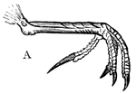
Foot of Peregrine Falcon.

Falcon Foot

Foot of Peregrine Falcon.

Foot of Tawny Owl.

Owl Foot

Foot of Tawny Owl.

Foot of Yellow Wagtail.

Wagtail Foot

Foot of Yellow Wagtail.

Foot of Water-ousel.

Water-ousel Foot

Foot of Water-ousel.

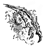
(Archeopteryx macrocrura), an ancient bird fossil showing claws.

Fossil

(Archeopteryx macrocrura), an ancient bird fossil showing claws.

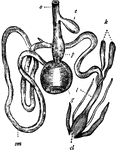
"Digestive system of the common Fowl. o, Gullet; c, Crop; p, Proventriculus; g, Gizzard; sm, Small intestine; k, Intestinal caeca; l, Large intestine; cl, Cloaca." — Winston's Encyclopedia, 1919

Fowl Digestive System

"Digestive system of the common Fowl. o, Gullet; c, Crop; p, Proventriculus; g, Gizzard; sm, Small intestine;…

"Mature stapes of fowl, about x4; after Parker. st, its foot, fitting fenestra ovalis; mst, main shaft, or medio-stapedial element; sst, supra-stapedial; est, extra-stapedial; ist, infra-stapedial, its end representing a rudimentary stylo-hyal; f, a fenestra in the extra-stapedial." Elliot Coues, 1884

The Ear Bone of Fowl

"Mature stapes of fowl, about x4; after Parker. st, its foot, fitting fenestra ovalis; mst, main shaft,…
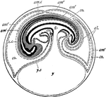
"Diagram of a longitudinal section through the embryo of a fowl, showing formation of amnion and allantois and the relation of these membranes to the embryo. am1, inner or true amnion; am2, outer or false amnion; am.c., amniotic cavity; al., allantois; c.c., central canal of the spinal cord; co., coelom; g, gut; ys., yolk sac." -Galloway, 1915

Fowl Embryo

"Diagram of a longitudinal section through the embryo of a fowl, showing formation of amnion and allantois…

The pelvis and caudal vertebrae of an adult fowl.

Fowl Pelvis

The pelvis and caudal vertebrae of an adult fowl.

The sacrum of a young fowl; natural size seen from below.

Fowl Sacrum

The sacrum of a young fowl; natural size seen from below.

The upper view of the skull of an old fowl.

Fowl Skull

The upper view of the skull of an old fowl.

The skull of an adult fowl.

Fowl Skull

The skull of an adult fowl.

The skull of an adult fowl. Here the temporal fossa is bridged over by the junction of the post-frontal and squamosal processes.

Fowl Skull

The skull of an adult fowl. Here the temporal fossa is bridged over by the junction of the post-frontal…

"Fig. 62 Skull of common fowl, enlarged. from nature by Dr. R.W. Shufeldt, U.S.A. The names of bones and some other parts are printed, requiring no explanation; but observe the following points: The distinction of none of the bones composing the brain-case (the upper back expanded part) can be found in a mature skull. The brain is contained between the occipital, sphenoidals, squamosals, parietals and part of frontal; the ethmoidals belong to the same group of cranial bones proper. All other bones, excepting the three otic ear-bones, are bones of the face and jaws. The lower jaw, of five bones, is drawn detached; it articulates by the black surface marked articular with the prominence just above- the quadratic bone. Observe that from this quadrate a series of bones quadrato-jugal, jugal, maxillary-makes a slender rod running to the premaxillary; this is the zygoma, or jugal bar. Observe from the quadrate also another series, composed of pterygoid and palatine bones, to the premaxillary; this is the pterygo-palatine bar; it slides along a median fixed axis of the skull, the rostrum, which bears the loose vomer at its end. The under mandible, quadrate, pterygoid, and vomer are the only movable bones of this skull. But when the quadrate rocks back and forth, as it does by its upper joint, its lower end pulls and pushes upon the upper mandible, by means of the jugal and pterygo-palatine bars, setting the whole scaffolding of the upper jaw in motion. This motion hinges upon the elasticity of the bones of the forehead, at the thin place just where the reference-lines from the words "lacrymal" and "mesethmoid" cross each other. The dark oval space behind the quadrate is the external orifice of the ear; the parts in it to which the three reference-lines go are diagrammatic, not actual representations; thus, the quadrate articulates with a large pro-otic as well as with the squamosal. The great excavation at the middle of the figure, containing the cirlet of the unshaded bones, is the left orbital cavity, orbit, or socket of the eye. The mesethmoid includes most of the background of this cavity, shaded diagonally. The upper one of the two processes of bone extending into it from behind is post-frontal or sphenotic process; the under one (just over the quadrate) is the squamosal process. A bone not shown, the presphenoid, lies just in front of the oval black space over the end of basisphenoid. This black oval is the optic foramen, through which the nerve of sight passes from the brain-cavity to the eye. The black dot a little behind the optic foramen is the orifice of exit of a part of the trifacial nerve. The black mark under the letters "on" of the word "frontal" is the olfactory foramen, where the nerve of smell emerges from the brain-box to go to the nose. The nasal cavity is the black space behind nasal and covered by that bone, and in the oval blank before it. The parts of the beak covered by horn are only premaxillary, nasal, and dentary. The condyle articulates with the first cervical vertebra; just above it, not shown, is the foramen magnum, or great hole through which the spinal medulla, or main nervous cord, passes from the spinal column. The basioccipital is hidden, excepting its condyle; so is much of the basisphenoid. The prolongation forward of the basisphenoid, marked "rostrum," and bearing the vomer at its end, is the parasphenoid, as far as its thickened under border is concerned. Between the fore end of the pterygoid and the basisphenoidal rostrum, is the site of the basipterygoid process, by which the bones concerned articulate by smooth facets; further forward, the palatines ride freely upon the parasphenoidal rostrum. In any passerine bird , the vomer would be thick in front, and forked behind, riding like the palatine upon the rostrum. The palatine seems to run into the maxillary in this view; but it continues on to premaxillary. The maxillo-palatine is an important bone which cannot be seen in the figure because it extends horizontally into the paper from the maxillary about where the reference life "maxillary" goes to that bone. The general line from the condyle to the end of the vomer is the cranial axis, basis cranii, or base of the cranium. This skull is widest across the post-frontal; next most so across the bulge of the jugal bar." Elliot Coues, 1884

Skull of a Common Fowl

"Fig. 62 Skull of common fowl, enlarged. from nature by Dr. R.W. Shufeldt, U.S.A. The names of bones…

"Schizognathous skull of common fowl, nat. size, from nature, by Dr. R.W. Shufeldt, U.S.A. Letters as before; Pa, palatine. Schizognathism is a kind of " cleft palate" shown by the columbine and gallinaceous birds, by the wader at large, and many of the swimmers. In this general case, the vomer, whether large or small, tapers to a point in front, while behind it embraces the basisphenoidal rostrum, between the palatines; these bones and the pterygoids are directly articulated with one another and with the basisphenoidal rostrum, not being borne upon the divergent posterior ends of the vomer; the maxillo-palatines, usually elongated and lamelar, pass inwards over (under, when the skull is viewed upside-down, as it usually is) the anterior part of the palatines, with which they unite and then bend backwards, along the inner edge of the palatines, leaving a broader or narrower fissure between themselves and the vomer, on each side, and do not unite with one another or with the vomer." Elliot Coues, 1884

Common Fowl Skull

"Schizognathous skull of common fowl, nat. size, from nature, by Dr. R.W. Shufeldt, U.S.A. Letters as…
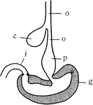
"Diagram of the stomach and esophagus of the fowl. o, esophagus; c, crop; p, proventriculus or glandular stomach; g, gizzard or grinding stomach; i, intestine." -Galloway, 1915

Fowl Stomach

"Diagram of the stomach and esophagus of the fowl. o, esophagus; c, crop; p, proventriculus or glandular…