The Cellular Botany ClipArt gallery offers 293 illustrations of plant tissue at the cellular level, illustrations of cell division, and examples of individual pollen grains.

Leaf Epidermis
"Lizard's tail (Saururus cernuus). Portions of leaf-epidermis; U, upper epidermis; L, lower epidermis;…

Leaf Resin Duct
"Resin duct in leaf of Pinus silvestris, in cross section at A, and in longitudinal section at B; h,…

Codonanthe Leaf Tissues
"Cross section of a portion of leaf of Codonanthe, showing the water-storage tissue at f, and the chlorophyll-bearing…

Blister Formed by Phytoptus Pyris by Leaf
In the middle of the blister, on its lower surface, is a small opening, which permits the gall mites…

Cross-section of a leaf
Cross section of a leaf, showing the breathing pores and intercellular spaces. The small dots are grains…
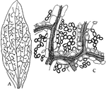
Dicotyledon Leaf
A: "Camera-lucida drawing of a bleached leaf of a Dicotyledon, showing the course of the vascular bundles,…

Diseased Leaf
"Transverse section of a diseased patch in the leaf showing the hyphae of the fungus pushing between…
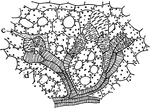
E. Splendens Leaf
"Water-storage tracheids in the leaf of Euphorbia splendens. b, b, water-storage tracheids; d, mesophyll…
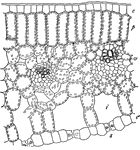
Lily Leaf
Cross section of leaf of lily, somewhat diagrammatic: e, upper epidermis,s, stomata in cross-section,…
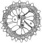
M. Forskalli Leaf
"Cross section of leaf of Mesembryanthemum Forskalii showing a large part of the leaf devoted to the…
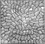
Microscopic view of a leaf
"The branch vascular bundles will be distinctly seen, resembling in some respects the arteries and veins…
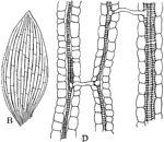
Monocotyledon Leaf
B: "Camera-lucida drawing of a bleached leaf of... a Monocotyledon, showing the anastomosis of the parallel…

P. Commune Leaf
"Cross section through a portion of leaf of Polytrichum commune. b, chains of chlorophyll-bearing cells;…

Pondweed Leaf
"Vertical section of the leaf of Potamogeton or Pondweed, showing air cavities or lecunae l, and parenehymatous…
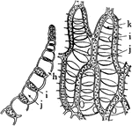
Sphagnum Leaf
"Portion of leaf of Sphagnum, in cross section on the left, and surface view on the right. h, hole through…
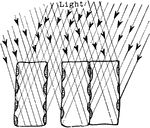
Plant Light Intake
"Diagram showing how the position of the chlorplasts against the vertical walls of the palisade cells…

Liverwort Life Cycle
"Life-history of Liverwort (Marchantia polymorpha): 1 and 2, developing thallus; 2 shows the cup with…
!["Longitudinal tangential section of [maple], showing the ends of the medullary rays." - Century, 1889](https://etc.usf.edu/clipart/73300/73379/73379_maple_mth.gif)
Longitudinal Tangential Section of Maple
"Longitudinal tangential section of [maple], showing the ends of the medullary rays." - Century, 1889

Longleaf Pine (Pinus palustris Mill.). Two-thirds natural size. cross sections (magnified) of leaves.
The longleaf pine commonly found in the South Atlantic and Gulf States. The leaves are 9 to 12 inches…

Longleaf Pine (Pinus palustris Mill.). Two-thirds natural size. cross sections (magnified) of leaves.
The longleaf pine commonly found in the South Atlantic and Gulf States. The leaves are 9 to 12 inches…
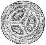
Longleaf Pine (Pinus palustris Mill.). Two-thirds natural size. cross sections (magnified) of leaves.
The longleaf pine commonly found in the South Atlantic and Gulf States. The leaves are 9 to 12 inches…
Longleaf Pine (Pinus palustris Mill.). Two-thirds natural size. epidermis of leaf (magnified)
The longleaf pine commonly found in the South Atlantic and Gulf States. The leaves are 9 to 12 inches…
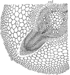
Lupine
This illustration shows a cross-section of a root of lupine showing the origin of the lateral rootlets.
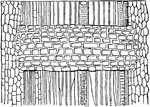
Maple
Surface of a young Maple wood from which the bark has been torn away, showing the bark (on the left)…

Bit of Young Maple Wood
Magnified view of surface of a bit of young Maple wood from which the bark has been torn away, showing…
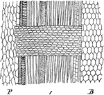
Medullary Rays of Maple
"Longitudinal radial section through the wood of a branch of maple one year old: P, pith; B, bark."…

Megaspore Formation Stage 1
"Stages in the formation of the megaspore, its germination, fertilization of the egg and endosperm cells.…

Megaspore Formation Stage 10
"Stages in the formation of the megaspore, its germination, fertilization of the egg and endosperm cells.…
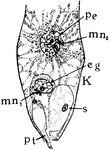
Megaspore Formation Stage 11
"Stages in the formation of the megaspore, its germination, fertilization of the egg and endosperm cells.…

Megaspore Formation Stage 12
"Stages in the formation of the megaspore, its germination, fertilization of the egg and endosperm cells.…

Megaspore Formation Stage 2
"Stages in the formation of the megaspore, its germination, fertilization of the egg and endosperm cells.…

Megaspore Formation Stage 3
"Stages in the formation of the megaspore, its germination, fertilization of the egg and endosperm cells.…
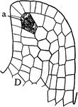
Megaspore Formation Stage 4
"Stages in the formation of the megaspore, its germination, fertilization of the egg and endosperm cells.…
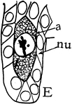
Megaspore Formation Stage 5
"Stages in the formation of the megaspore, its germination, fertilization of the egg and endosperm cells.…

Megaspore Formation Stage 6
"Stages in the formation of the megaspore, its germination, fertilization of the egg and endosperm cells.…

Megaspore Formation Stage 7
"Stages in the formation of the megaspore, its germination, fertilization of the egg and endosperm cells.…

Megaspore Formation Stage 8
"Stages in the formation of the megaspore, its germination, fertilization of the egg and endosperm cells.…

Megaspore Formation Stage 9
"Stages in the formation of the megaspore, its germination, fertilization of the egg and endosperm cells.…
Melon Fibers
"Spiral vessels of the Melon, showing the elastic fibers uncoiled, and the vessels overlapping at their…

Microspore Anther
The anther and archesporium in the stages of "formation of anthers and pollen grains or microspores…
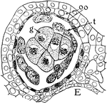
Microspore Anther Lobe
The cross section of a mature anther lobe in the stages of "formation of anthers and pollen grains or…
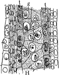
Microspore Anther Lobe
The cross section of a mature anther lobe in the stages of "formation of anthers and pollen grains or…
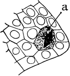
Microspore Archesporium
The archesporium in the stages of "formation of anthers and pollen grains or microspores of Silphium."…
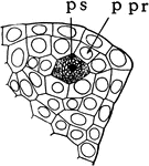
Microspore Cells
The primary sporogenous cell (ps) and the primary parietal layer (ppr) in the stages of "formation of…
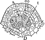
Microspore Cells
The sporogenous cells (s), tapetum (t), two parietal layers (oo) in the stages of "formation of anthers…

Microspore Cells
The sporogenous cells (s), tapetum (t), two parietal layers (oo) in the stages of "formation of anthers…
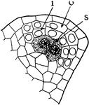
Microspore Layers
The inner layer (i), outer layer (o), sporogenous cells (s) in the stages of "formation of anthers and…

Microspore Pollen Grains
The pollen grains beginning to germinate in the stages of "formation of anthers and pollen grains or…
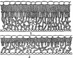
Oak Leaf Cross-Section
"Cross-sections of leaves of an oak (Quercus novimexicana), showing the effect of different light conditions…

Onion Cells
"A, embryonic cells from onion root tip; d, plasmatic membrane; c, cytoplasm; a, nuclear membrane enclosing…

Onion Cells
"B, older (onion) cells farther back from the root tip. The cytoplasm is becoming vacuolate; f, vacuole."…
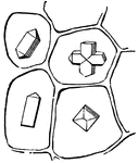
Onion Magnified
This is an illustration of crystals from the base of an onion, one of them a hemitrope or double.
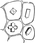
Onion-Peel
Four cells from dried Onion-peel, each holding a crystal of different shape, one of them twinned.

Orange Rind
"Vertical section of part of the rind of the Orange, showing glands containing volatile oil, R, R, R,…

Palisade Cell
"Diagrammatic representation of a single palisade cell, with chloroplasts lining the walls." -Stevens,…

Palisade Cell
"Diagram to show the activities going on in a palisade cell. The arrows from the chloroplasts into the…

