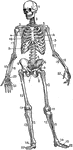
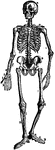
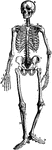
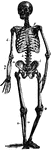
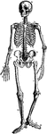
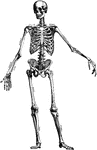
The Human Skeleton
The Human Skeleton. Labels: a, parietal bone; b, frontal; c, cervical vertebrae; d, sternum; e, lumbar…
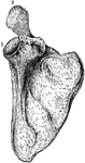
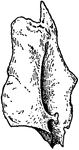
The Human Scapula
The human scapula bone (shoulder blade). Labels: 1, glenoid cavity; 2, end of the spine of scapula.
The Human Humerus
The human humerus bone, the longest and largest bone of the upper leg. Labels: a, rounded head; gt,…
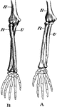
The Human Ulna and Radius
The Ulna and Radius. Labels: 1, radius; 2, ulna; o, olecranon process, on the anterior surface of which…
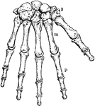
The Human Wrist and Hand Bones
Bones of the Wrist and Hand. Labels: m, metacarpal bones; p, phalanges; 3, bones of wrist.

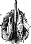
Part of the Human Pelvic Bone
The Os Innominatum, or nameless bone, so called from bearing no resemblance to any known object, is…
Human Femur Bone
The Femur (upper leg bone) is the longest, largest, and strongest bone in the skeleton. Labels: b, rounded…
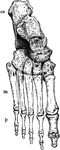
Bones of the Ankle and Foot
Bones of the Ankle and Foot. Labels: m, metatarsal bones; p, phalanges; ca, os calcis, or heel bone.
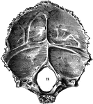
Occipital Bone of the Human Skull
Occipital bone of the human skull, inner surface. It is situated at the back and base of the skull.…
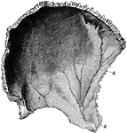
Parietal Bone of the Human Skull
Parietal bone of the human skull, inner surface. The parietal bones form the greater part of the sides…
Frontal Bone of the Human Skull
Frontal bone of the human skull, outer surface. The frontal bone forms the forehead, roof of the orbital…
Temporal Bone of the Human Skull
Temporal bone of the human skull. The temporal bones are situated at the sides and base of the skull.…
Sphenoid Bone of the Human Skull
Sphenoid bone, situated the anterior part of the base of the skull, articulating with all the other…
Ethmoid Bone of the Human Skull
Ethmoid bone, posterior surface. The ethmoid bone is an exceedingly light, spongy bone, placed between…
Human Lachrymal Facial Bone
Lachrymal Bone. The lachrymal are the smallest and most fragile bones fo the face. They are situated…
Human Vomer Nasal Bone
Vomer bone, a single bone placed at the back part of the nasal cavity, and forms part of the septum…
Human Malar (Cheek) Bone
Malar (cheek) bone. The malar bones form the prominence of the cheek, and part of the outer wall and…
Human Palate Bone
Palate bone. Palate bones form the back part of the roof of the mouth; part of the floor and outer wall…
Human Nostril Bone
Inferior turbinated bone, convex surface. The inferior turbinated bones are situated on the outer wall…
Human Maxillary (Upper Jaw) Bone
Superior maxillary bone. With it's fellow on the opposite side, it forms the whole of the upper jaw.…
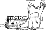
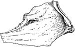
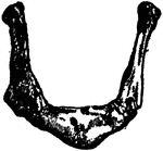
Human Maxillary (Upper Jaw) Bone
Inferior Maxillary Bone (lower jaw). It is the largest and strongest bone in the face and serves for…
Human Hyoid Bone
The hyoid, os hyoides, or tongue bone, is an isolated, U-shaped bone lying in front of the throat, just…
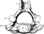
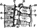
Human Cervical Vertebra Bone
A cervical vertebra of the spine, inferior surface. Labels: 1, spinous process, slightly bifid; 4, transverse…
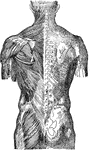
Human Spinal Column
Side view of spinal column, without sacrum and coccyx. Labels: 1 to 7, cervical vertebrae; 8 to 19,…
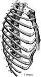
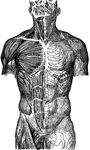
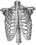
Human Thorax (Chest)
Thorax. The thorax, or chest, is an elongated conical-shaped cage, formed by the sternum and costal…
Human Sternum Bone
Sternum, front and side view. The sternum, or breast bone, is a flat narrow bone, situated in the median…
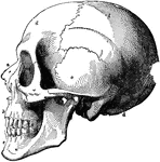
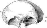
Human Skull
The skull. Labels: a, nasal bone; b, superior maxillary; c, inferior maxillary; d, occipital; e, temporal;…
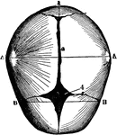
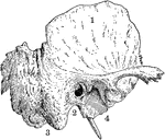
Human Skull at Birth
The skull at birth, superior suerface. The cranial bones of the infant at birth are not fullyformed…
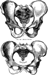
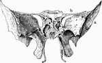
Human Pelvis, Male and Female
Male pelvis (top) and female pelvis (bottom). The pelvis is stronger and more massively constructed…
Human Joint, Mixed Articulation
A mixed articulation (slightly movable). In this form, the bony surfaces are usually joined together…
Human Joint, Dentated Suture
A toothed, or dentated suture. This is one type of immovable articulation. It is found in the union…
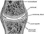
A Simple Complete Joint
A simple complete joint, one type of movable articulation. The synovial membrane is represented by dotted…
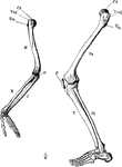
Arm and Leg Skeleton
The skeleton of the arm and leg. Labels: H, the humerus; Cd, its articular head which fits into the…
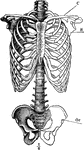
Skeleton of Trunk
The skeleton of the trunk and the limb arches seen from the front. Labels: c, clavicle; S, scapula;…
Arm Bones
Demonstration of the movement of a pivot joint. Labels: A, arm in supination (palm uppermost); B, arm…
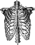
Thorax
The skeleton of the thorax. Labels: a, g, vertebral column; b, first rib; c, clavicle; d, third rib;…
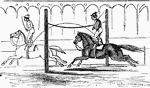
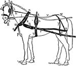
Inertia Demonstration
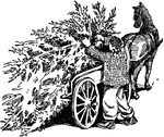
"While his horse is going at full speed, he jumps over a rope extended across the ring, and regains…
!["A horse without machinery can not lift a weight; but he does it readily with the aid of the simple apparatus shown [here].." —Quackenbos 1859](https://etc.usf.edu/clipart/36300/36331/pulley_sys_36331_mth.gif)
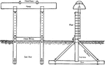
Pulley System
"A horse without machinery can not lift a weight; but he does it readily with the aid of the simple…
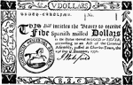
Paper Money, Five Dollars Bill, 1776
Five Dollars ($5) South Carolina currency from 1776. Image of a horse surrounded by the inscription…
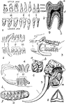
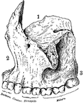
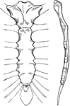
Teeth of Man and Several Animal Species
1. Dentition (teeth) of man. 2. Dentition of hyena. 3. Dentition of pig. 4. Dentition of Patagonian…
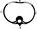
Segment of the Axial Skeleton
Diagrammatic representation of a segment of the axial skeleton V, a vertebra; C, Cv, ribs articulating…
Thorax Skeleton
The skeleton of the thorax. Labels: a, g, vertebral column; b, first rib; c, clavicle; e, seventh rib;…
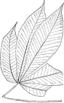
Genus Aesculus, L. (Buckeye, Horse Chestnut)
Leaves - compound (hand-shaped; leaflets, usually five, sometimes seven); opposite; edge toothed. Outline…
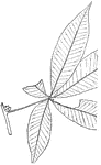
Genus Aesculus, L. (Buckeye, Horse Chestnut)
Leaves - compound (hand-shaped; leaflets, five); opposite; edge toothed. Outline - of leaflet, oval…