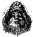
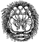
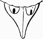
Plains Pocket Gopher
"Under Side of Head of Geomys bursarius, showing entrance of external cheek-pouches and sulcate superior…
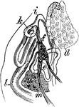
Reproductive Organs of Trematoid Worm
"Reproductive Organs of a Trematoid Worm (Aspidogaster conchicola). d, germarium; e, internal vas deferens;…

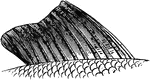
Parts of Fish Gills
"Gill of Fish. A, first branchial arch of left side of black-bass: 1, gill-rakers; 2, branchial lamellae.…
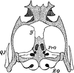
Frog Chondrocranium
"Chondrocranium of Frog (Rana esculenta). y, girdle-bone or os en ceinture; EO, exoccipital; PrO, proötic;…
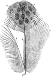
Feather from a Argus Pheasant
"Fig. 19 - A partly pennaceous, partly plumulaceous feather, from Argus pheasant; after Nitzsch. ad,…
Structure of a Feather
"Fig. - 20 - Two barbs, a, a, of a vane, bearing anterior, b, b, and posterior, c, barbules; enlarged;…
Single Barbule
"Fig. 21. -A single barbule, baring barbicels and hooklets; magnified; after Nitzsch. ...barbicels (another…
Barbs
"The arrangement shown in fig. 22, where a, a, a, a, are four barbs in transverse section, viewed from…
A Feather from the Tail of a Kingbird
"Fig. 23 - A feather from the tail of a kingbird, Tyrannus carolinensis, almost entirely pennaceous;…
Pterylosis of Cypselus Apus
"Fig. 24. - Pterylosis of Cyoselus apus, drawn by Coues after Nitzsch; right hand upper, left hand lower,…
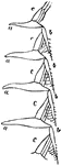
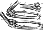
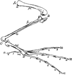
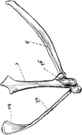
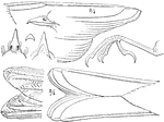
The Bones of the Right Wing of a Duck
"Fig 27. - Bones of the right wing of a duck, Clangula islandica, A, shoulder, omos; B, elbow, ancon;…
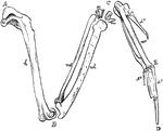
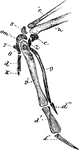
Mechanism of the Elbow-Joint
"Fig. 28. - Mechanism of elbow-joint. ..., where rc and uc show respectively the size, shape, and position…
The Wing Bones of a Young Grouse
"Fig. 29., from a young grouse (Centrocercus urophasianus, six months old), is designed to show the…
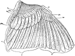
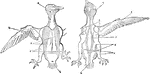
Feathers of a Sparrow's Wing
"Fig. 30., Feathers of a sparrow's wing. pc, covers of the primaries; msc, median upper secondary coverts;…
Red Shafted Woodpecker Ulna
"Fig. 31. - Ulna of Colaptes mexicanus, showing points of attachment of the secondaries. (Dr. R. W.…
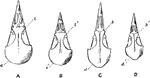
Bird Skulls
"Fig. 114. -Skulls of Turdidae and Sylvicolidae, nat. size; after Shufeldt. A, Oroscoptes montanus;…
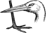
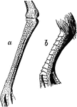
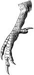
Stork Head and Leg
The head and leg of the stork, a bird in the Ciconiidae family of storks, herons, and egrets.
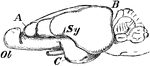
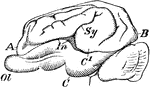
Rabbit Brain
"Brain of rabbit. Ol, olfactory lobe; A, B, C, frontal, occipital, and temporal lobes; Sy, Sylvian fissure."…
Pig Brain
"Brain of pig. Ol, olfactory lobe; A, B, C, frontal, occipital, and temporal lobes; C1, a portion of…
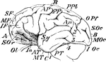
Chimpanzee Brain
"Brain of chimpanzee. Ol, olfactory lobe; A, B, C, frontal, occipital, and temporal lobes; C1, a portion…
Scutellate Laminiplanter Tarsus of a Cat-bird
"Figure shows Scutellate laminiplanter tarsus of a cat-bird. A tarsus so disposed as to its podotheca…
Reticulate Tarsus of a Plover
"Fig 38 a, Reticulate tarsus of a Plover. b, Scutellate and reticulate tarsus of a pigeon." Elliot Coues,…
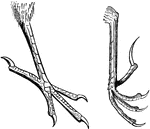
Tridactyle Foot of a Sanderling
"Fig. 39 shows a Tridactyle foot of a sanderling, Calidris arenaria." Elliot Coues, 1884
Phalanges of Cypseline Foot
"Fig. 40 Phalanges of Cypseline foot, where the ratio is 2, 3, 3, 3 of Caprimulginae." Elliot Coues,…
Phalanges of Caprimulgine
"Fig. 41 shows phalanges of caprimulgines foot, where the ratio is 2, 3, 4, 4." Elliot Coues, 1884
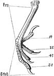
Bones of a Bird's Hind Limb
"Fig 34 - Bones of a bird's hind limb: from a duck, Clangula islandica. A, hip: B, knee: C, heel or…
Typical Passerine Bird Feet
"Fig. Typical passerine feet. The right hand fig. is plectrophanes lapponicus." Elliot Coues, 1884
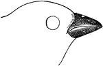
The foot of a Kingfisher
"Fig. 44- Syndactyle foot of a kingfisher. Thus a kingfisher shows what is called a syndactyle or syngnesious…
Zygodactyle Foot of a Woodpecker
"Fig. 45.- Zygodactyle foot of a woodpecker, Hylotomus pileatus. The zygodactyle or yoke-toed modification…
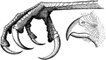
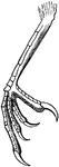
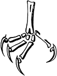
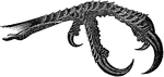
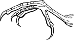
The Raptorial Foot of a Hawk
"Fig. 46. - Raptorial foot of a hawk, Accipiter cooperi. The raptorial is another modification of the…
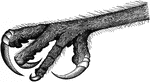
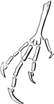
The Raptorial Foot of an Owl
"Fig. 47. - Raptorial foot of an owl, The raptorial is another modification of the insessorial foot.…
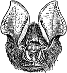
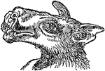
Bat Head
"Bat, one of the group of wing-handed, flying mammals, having the fore-limb peculiarly modified so as…
Chin Leafed Bat Head
The head of the chin leafed bat. "Bat, one of the group of wing-handed, flying mammals, having the fore-limb…
Flower-Nosed Bat Head
The head of the flower-nosed bat. "Bat, one of the group of wing-handed, flying mammals, having the…
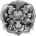
Hammer-Headed Bat Head
The head of the Hammer-Headed Bat (Hypsignathus monstrosus). Also known as the big-lipped bat, it is…
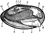
Mussel Anatomy
"Longitudinal Section through a Fresh-water Mussel. a, edge of mantle; b, foot, with position of ganglion…
Kangaroo Pelvis
An illustration of a kangaroo pelvis. "M, marsupial bones, borne upon P, pubis; Il, ilium; Is, ischium;…
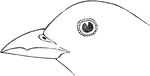
The Dentirostral Bill of a Tanager
"Fig 178 - the dentirostral bill of a Tanager (Pyranga hepatica)" Elliot Coues, 1884
Barn Swallow Details
"Fig 181 - Generic details of Hirundo horreorum(Barn Swallow) Deep lustrous steel-blue; forehead and…
Barn Swallow Tail
"Hirundo horreorum. Barn Swallow. Tail deeply fornicate, nearly or about as long as the wings; lateral…
Barn Swallow Bill
Aerial (top) view of Barn Swallow's bill "Hirundo horreorum. Barn Swallow. Bill of moderate size for…
Barn Swallow Claw
Side view of the Barn Swallow's claw. "Hirundo horreorum. Barn Swallow. Tarsi shorter than middle toe…
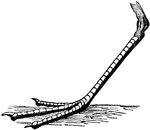
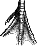
Half-Webbed Foot of a Sandpiper
"... basal webs generally run out to the end of the first, or along part of the second, phalanx of the…
Half-Webbed Foot of a Willet
The semipalmated (half-webbed) bases of toes on the foot of a Willet. "... basal webs generally run…
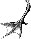
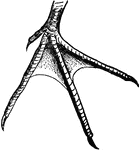
Webbed Foot of a Tern
"In the palmate or ordinary webbed foot, all the front toes are united by ample webs." Elliot Coues,…
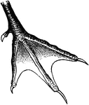
Incised Webbed Foot of a Tern
"...one or both webs may be so deeply incised, that is, cut away, that the palmation is practically…
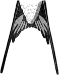
Sternum of a Robin
"Typical passerine sternum, pectoral arches, and sternal ends of ribs; from the robin, Turdus migratorius,…
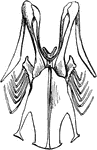
The Right Pectoral Arch of a Bird
"Right pectoral arch of a bird. s, scapula; c, coracoid; gl, glenoid, the cavity for head of humerus;…
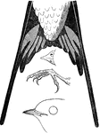
Generic Details of a Thrush
"Generic details of a Myiadestes townsendi (Townsend's Flycatching Thrush); bill and foot nat. size,…
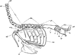
Axial Skeleton
"Fig 56 - Axial skeleton, minus the skull, of an owl, Asio wilsonianus, life size; from nature by Dr.…
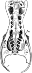
The Pelvis of a Heron
"Pelvis of a heron (ardea herodias), nat. size, viewed from below; from nature by Dr. R.W. Shufeldt,…
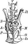
The Pelvis of a Young Grouse
"Pelvis of a young grouse, showing three distinct bones. Il,P, ilium, ischium, pubis. In front of former…
The Bill of a Purple Finch
"Carpodacus. Purple Bullfinch. Bill smaller and less turgid than in Pinicol or Pyrrhula, more regularly…
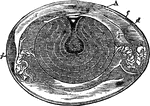
Hen's Egg
" Fig 110 - Hens egg, nat. size, in section; from Owen, after A. Thompson. A, cicatricle or "tread,"…
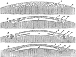
Egg Germination
"Further development of hen's egg; after Haeckel: A, the mulberry mass of cleavage cells, b, same as…