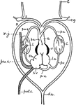
"Diagram of the heart and branchial arches in the Frog. c.g., carotid gland; l., lungs; l.a., left auricle; r.a., right auricle...a, aorta; au., auricle; c, carotid; c.v., cardinal veins (right and left); d.a., dorsal arteries; j, jugular veins; d.c., ductus Cuvieri; s.v., sinus venosus; v, ventricle." -Galloway, 1915

Frog Heart

"Diagram of the heart and branchial arches in the Frog. c.g., carotid gland; l., lungs; l.a., left auricle;…
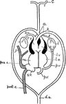
"Diagram of the heart and branchial arches in a Reptile...a, aorta; au., auricle; c, carotid; c.v., cardinal veins (right and left); d.a., dorsal arteries; j, jugular veins; d.c., ductus Cuvieri; s.v., sinus venosus; v, ventricle." -Galloway, 1915

Reptile Heart

"Diagram of the heart and branchial arches in a Reptile...a, aorta; au., auricle; c, carotid; c.v.,…
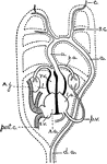
"Diagram of the heart and the branchial arches in Mammals. A dotted outline of the arches of the Fish is drawn for ready comparison. The auricles are represented in a posterior position...a, aorta; au., auricle; c, carotid; d.a., dorsal arteries; j, jugular veins; s.v., sinus venosus; v, ventricle." -Galloway, 1915

Mammal Heart

"Diagram of the heart and the branchial arches in Mammals. A dotted outline of the arches of the Fish…
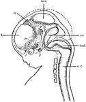
"Diagram of head and brain of human foetus six weeks old (heavy boundaries). The dotted line indicates the outline of the brain of a foetus three months old. Note thee great growth of the hemisphere (h). cer, cerebellum; med, medulla oblongata; mes, mesencephalon; p, pituitary body; pr, prosencephalon; s.c., spinal cord; th, thalamencephalon; 1, olfactory nerve; 2, optic nerve." -Galloway, 1915

Human Fetus

"Diagram of head and brain of human foetus six weeks old (heavy boundaries). The dotted line indicates…

"Diagram of a cross-section of the spinal cord through the roots of spinal nerves. c, central canal; d.f., dorsal fissure; d.r., dorsal root of spinal nerve arising from the dorsal horn of the gray matter (g); gn., ganglion on the dorsal root; n, spinal nerve; v.f., ventral fissure; v.r., ventral root of the spinal nerve, arising from the ventral horn of the gray matter; w., white matter." -Galloway, 1915

Spinal Cord

"Diagram of a cross-section of the spinal cord through the roots of spinal nerves. c, central canal;…

"Diagram of the principal vessels in the circulation of a Fish, lateral view. a, aorta; au., auricle; br.a., afferent branchial arteries; br.e., efferent branchial arteries; bu., bulbus (or conus) arteriosus; c, carotid; c.a., caudal artery; c.v.r., right cardinal vein; c.v.l., left cardinal vein; d.a., dorsal artery; d.c., ductus Cuvieri; g, gills; h.v., hepatic vein; h.p., hepatic portal vein; i, intestine; j, jugular vein; k, kidney; l, liver; r.p., renal portal vein; s.v., sinus venosus; v, ventricle." -Galloway, 1915

Fish Circulation Vessels

"Diagram of the principal vessels in the circulation of a Fish, lateral view. a, aorta; au., auricle;…

"Diagram of the female genital organs of a Bird. c, cloaca; i, intestine; k, kidney; o, ovary with ova of different size; od., oviduct; o.f., funnel of the oviduct; o.o., opening of the oviduct into the cloaca; u, ureter; u.o., opening of ureter into the cloaca. Only one ovary and oviduct are fully developed in the Birds." -Galloway, 1915

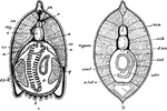
Female Bird Genital Organs

"Diagram of the female genital organs of a Bird. c, cloaca; i, intestine; k, kidney; o, ovary with ova…

"Diagram of the urino-genital organs of a male Bird. ad., adrenal body; c, cloaca; i, intestine; k, kidney; t, testis; u, ureter; u.o., opening of ureter into the cloaca; v.d., vas deferens; v.d.o., opening of the vas deferens; v.s., vesicula seminalis." -Galloway, 1915

Male Bird Genital Organs

"Diagram of the urino-genital organs of a male Bird. ad., adrenal body; c, cloaca; i, intestine; k,…
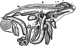
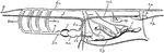
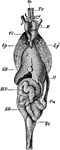
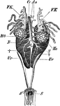
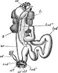
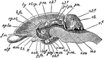
"General view of the viscera of a male frog, from the right side. a, stomach; b, urinary bladder; c, small intestine; cl, cloacal aperture; d, large intestine; e, liver; f, bile duct; g, gall bladder; h, spleen; i, lung; k, larynx; l, fat body; m, testis; n, ureter; o, kidney; p, pancreas; s, cerebral hemisphere; sp, spinal cord; t, tongue; u, auricle; ur, urostyle; v, ventricle; v.s, vesicula seminalis; w, optic lobe; x, cerebellum; y, Eustachian recess; z, nasal sac." -Parker, 1900

Frog Viscera

"General view of the viscera of a male frog, from the right side. a, stomach; b, urinary bladder; c,…

"Transverse section through the wall of a cylinder (parallel with the course of the canals), showing one incurrent canal (IC), and one radial (R) throughout their length; sp, triradiate spicules; sp', oxeote spicules of dermal cortex (dc); sp'', tetraradiate spicules of gastral cortex (gc); ec, ectoderm; en, endoderm; pm, pore membrane; pp, prosopyles; ap, apopyle; di, diaphragm; exc, excurrent passage; PG, paragastric cavity; em, early embryo; em, late embryo. The arrows indicate the course of the water through the sponge." -Parker, 1900

Sycon Gelatinosum

"Transverse section through the wall of a cylinder (parallel with the course of the canals), showing…

"Vertical section of a fresh-water sponge (Spongilla), showing the arrangement of the canal-system. C, ciliated chambers; DP, dermal pores; Ex, excurrent canals; GO, openings of the excurrent canals; PG, paragastric cavity; SD, subdermal cavities; O, osculum." -Parker, 1900

Spongilla

"Vertical section of a fresh-water sponge (Spongilla), showing the arrangement of the canal-system.…
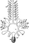
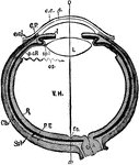
"Ambulacral system of a starfish. a, ampullae; ap, Polian vesicles; c, circular canal; m, madreporite; m', madreporic canal; p, tube-feet; r, radial vessels; r', branches to ampullae." -Parker, 1900

Ambulacral System

"Ambulacral system of a starfish. a, ampullae; ap, Polian vesicles; c, circular canal; m, madreporite;…
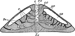
"Transverse section of thorax of crayfish, diagrammatic. abm, ventral abdominal muscles; bf, leg; bm, ventral nerve-cord; d, intestine; dbm, dorsal muscles of abdomen; ep, wall of thorax; h, heart; k, gills; kd, gill-cover; l, liver; ov, ovary; pc. pericardial sinus; sa. sn, sternal artery; vs, ventral sinus. The arrow shows the direction of the blood-current." -Parker, 1900

Crayfish Thorax

"Transverse section of thorax of crayfish, diagrammatic. abm, ventral abdominal muscles; bf, leg; bm,…

"Nervous system of Astacus fluviatilis. bg, sub-oesphageal ganglion; cs, commissural ganglion; g, brain; s, visceral nerve; sc, oesophageal connective; y, post-oesophageal commissure; IV-VIII, thoracic ganglia; 1-6, abdominal ganglia." -Parker, 1900

Crayfish Nervous System

"Nervous system of Astacus fluviatilis. bg, sub-oesphageal ganglion; cs, commissural ganglion; g, brain;…
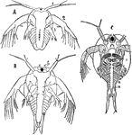
"Three stages in the development of Apus. fs, frontal sensory organ; L, digestive gland; s, carapace; 1-4, cephalic appendages; I-XIII, body segments and appendages." -Parker, 1900

Apus Stages

"Three stages in the development of Apus. fs, frontal sensory organ; L, digestive gland; s, carapace;…

"Periplaneta. General view of the nervous system. abd. 6, sixth abdominal ganglion; ant, antennary nerve; br, brain; conn, oesophageal connective; inf, sub-oesophageal ganglion; opt, optic nerve; thor. 1, thor. 2, thor. 3, first, second, and third thoracic ganglia." -Parker, 1900

Cockroach Nervous System

"Periplaneta. General view of the nervous system. abd. 6, sixth abdominal ganglion; ant, antennary nerve;…

"Patella vulgata, seen from the ventral side. f, foot; g.l, circlet of gill lamellae; m. e, edge of the mantle; mu, attachment muscle; sl, slits in the attachment muscle; sh, shell; v, efferent branchial vessel; v', aorta; ve, smaller vessels." -Parker, 1900

Limpet

"Patella vulgata, seen from the ventral side. f, foot; g.l, circlet of gill lamellae; m. e, edge of…

"Sepia officinalis, enteric canal. a, anus; b. d, one of the bile ducts; b. m, buccal mass; c, caecum; i, ink-sac; i. d, ink-duct; j, jaws; l, l, liver lobes; oe, oesophagus; p, pancreatic appendages1; r, rectum; s. g, salivary glands; st, stomach." -Parker, 1900

Cuttlefish Enteric Canal

"Sepia officinalis, enteric canal. a, anus; b. d, one of the bile ducts; b. m, buccal mass; c, caecum;…

"Oral surface of a male (A) and female (B) Nautilus pompilius in an expanded state. a, shell; b, external annular lobe carrying 19 tentacles on each side, and anteriorly enlarged to form the hood; c, right and left inner lobes, each carrying 12 tentacles in the female, and divided in the male into two parts; d, posterior inner lobe; e, oral cone; f, tentacles of the outer annular lobe projecting from their sheaths; g, two anterior tentacles of this lobe belonging to the hood; i, superior, k, inferior, ophthalmic tentacle; l, eye; n, lamellated organ on the posterior inner lobe of the female; o, funnel; p, spadix; q, antispadix." -Parker, 1900

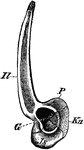
Nautilus Oral Surface

"Oral surface of a male (A) and female (B) Nautilus pompilius in an expanded state. a, shell; b, external…

"Amphioxus lanceolatus. A, transverse section of the pharyngeal region. a, dorsal aorta; b, atrium; c, notochord; co, coelom; e, endostyle; g, gonad; kb, branchial lamellae; kd, pharynx; l, liver; my, myomere; n, nephridium; r, neuron or dorsal nerve tube; sn, spinal nerves; sp, gill-slits. B, transverse section of the intestinal region; atr, atrium; coel, coelom; d. ao, dorsal aorta; int, intestine; myom, myomere; nch, notochord; neu, neuron; s. int. v, subintestinal vein." -Parker, 1900

Lancelet Section

"Amphioxus lanceolatus. A, transverse section of the pharyngeal region. a, dorsal aorta; b, atrium;…

"Lacerta agilis. General view of the viscera in their naturaal relations. Bl, urinary bladder; Ci, post-caval vein; ED, rectum; GB, gall-bladder; H, heart; Lg, Lg', the lungs; M, stomach; MD, small intestine; Oe, oesophagus; Pn, pancreas; Tr, trachea." -Parker, 1900

Sand Lizard Viscera

"Lacerta agilis. General view of the viscera in their naturaal relations. Bl, urinary bladder; Ci, post-caval…

"Diagrammatic horizontal section of the eye of man. c, cornea; ch. choroid (dotted); C. P, ciliary processes; e. c, epithelium of cornea; e. cj, conjunctiva; f. c, yellow spot; I, iris; L, lens; ON, optic nerve; OS, ora serrata; o-x, optic axis; p. c. R, anterior non-visual portion of retina; P. E, pigmented epithelium (black); R, retina; sp. l, suspensory ligament; Scl, sclerotic; V. H, vitreous body." -Parker, 1900

Human Eye

"Diagrammatic horizontal section of the eye of man. c, cornea; ch. choroid (dotted); C. P, ciliary processes;…
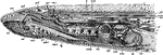
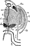
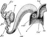
The dissection of a female sea lamprey or Petromyzon marinus. ad, anterior dorsal cartilage; an, annular cartilage; au, auricle; bd, bile-duct; bp', basal plate; br, brain; cd, cardinal vein; d.ao, dorsal aorta; dm, dorsal muscles; ea, external gill aperture; ft, fibrous tissue; gr, groove canal; hp, basal plate to fontanelle; hv, hepatic vein; i, integument; i.ju; inferior jugular vein; int, intestine; ju, jugular; lg, lingual cartilage; lr, liver; m, buccal vavity; mv, median ventral cartilage; my, spinal cord; na, olfactory sac; na', pituitary pouch; na'', nasal aperture; na.c, nasal capsule; nc, notochord; n.ca, spinal canal; oc, cranial roof; oes, gullet; o.f, buccal funnel; ov, ovary; pc, pericardium; pd, posterior dorsal cartilage; pmt, protractor muscle of tongue; rmt; retractor of tongue; rt, respiratory tube; ss, lymph sinus; sv, sinus venosus; t, tongue; v, ventricle; v.ao, ventral aorta; vl, velum; vm, ventral muscles. -Parker, 1900

Lamprey Anatomy

The dissection of a female sea lamprey or Petromyzon marinus. ad, anterior dorsal cartilage; an, annular…
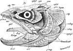
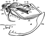
"Salmo fario, the entire skull, from the left side. art, articular; branchiost, branchiostegal rays; dent, dentary; epiot, epiotic; eth, supra-ethnoid; fr, frontal; hyom, hyomandibular; intop, inter-opercular; pal, palantine; par, parietal; pmx, pre-maxilla; praop, pre-opercular; pt, pterygoid; pter, pterotic; Quad, quadrate; socc, supra-occipital; sphot, sphenotic; subop, sub-opercular; sympl, symplectic; Zunge, basi-hyal." -Parker, 1900

Fish Skull

"Salmo fario, the entire skull, from the left side. art, articular; branchiost, branchiostegal rays;…

"Rana esculenta. The shoulder girdle from the ventral aspect. Co, coracoid; Co', epicoracoid; Cl, clavicle; G, glenoid cavity; Ep, episternum; Fe, fenestra between procoracoid and coracoid; KC, cartilage separating scapula and clavicle; Kn, xiphisternum; m, junction of epicoracoids; S, scapula; St, sternum." -Parker, 1900

Edible Frog Shoulder Girdle

"Rana esculenta. The shoulder girdle from the ventral aspect. Co, coracoid; Co', epicoracoid; Cl, clavicle;…

"Rana esculenta. Pelvic girdle from the right side. G, acetabulum; Il, P, ilium; Is, ischium; Kn, pubis." -Parker, 1900

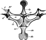
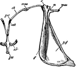
Edible Frog Pelvic Girdle

"Rana esculenta. Pelvic girdle from the right side. G, acetabulum; Il, P, ilium; Is, ischium; Kn, pubis."…
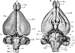
"Rana esculenta. The brain. A, from above; B, from below. ch. opt, optic chiasma; HH, cerebellum; Hyp, pituitary body; Inf, infundibulum; L. ol, olfactory lobe; Med, spinal cord; MH, mid-brain; NH, medulla oblongata; Th. opt, optic thalamus; Tr. opt, optic tract; VH, cerebral hemisphere; ZH, diencephalon; I-X, cerebral nerves; XII. (1), hypoglossal (first spinal) nerve." -Parker, 1900

Edible Frog Brain

"Rana esculenta. The brain. A, from above; B, from below. ch. opt, optic chiasma; HH, cerebellum; Hyp,…

"Rana esculenta. Urinogenital organs of the male. Ao, dorsal aorta; Cl, cloaca; Cv, post-caval vein; FK, fat bodies; HO, testes; N, kidneys; S, apertures of ureters into cloaca; Ur, ureters." -Parker, 1900

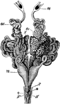
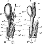
Male Edible Frog Genital Organs

"Rana esculenta. Urinogenital organs of the male. Ao, dorsal aorta; Cl, cloaca; Cv, post-caval vein;…

"Rana esculenta. Urinogenital organs of the female. N, kidneys; Od, oviduct; Ot, its coelomic aperture; Ov, left ovary (the right is removed); P, cloacal aperture of oviduct; S.S', cloacal apertures of ureters; Ut, uterine dilatation of oviduct." -Parker, 1900

Female Edible Frog Genital Organs

"Rana esculenta. Urinogenital organs of the female. N, kidneys; Od, oviduct; Ot, its coelomic aperture;…

"Skeleton of crocodile. C, causal region; D, thoracic region of spinal column; F, fibula; Fe, femur; H, humerus; I, ischium; L, lumbar region; R, radius; Ri, ribs; S, sacrum; Sc, scapula; Sta, abdominal ribs; T, tibia; U, ulna." -Parker, 1900

Crocodile Skeleton

"Skeleton of crocodile. C, causal region; D, thoracic region of spinal column; F, fibula; Fe, femur;…

"Cistudo lutaria. Skeleton seen from below; the plastron has been removed and is represented on one side. C, costal plate; Co, coracoid; e, entoplastron; Ep, epiplastron; F, fibula; Fe, femur; H, humerus; Il, ilium; Is, ischium; M, marginal plates; Nu, nuchal plate; Pb, pubis; Pro, pro-coracoid; Py, pygal plates; R, radius; Sc, scapula; T, tibia; U, ulna." -Parker, 1900

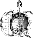
Marsh Turtle Skeleton

"Cistudo lutaria. Skeleton seen from below; the plastron has been removed and is represented on one…

"Chelone midas. Transverse section of skeleton. C, costal plate; C', centrum; M, marginal plate; P, lateral element of plastron; R, rib; V, expanded neural plate." -Parker, 1900

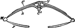
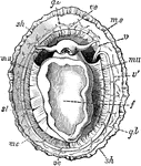
Green Turtle Skeleton

"Chelone midas. Transverse section of skeleton. C, costal plate; C', centrum; M, marginal plate; P,…

"A, lateral view of skull of rattlesnake (Crotalus). B. O, basi-occipital; B. S, basi-sphenoid; E. O, exoccipital; F. O, fossa ovalis; La, conjoined lacrymal and pre-fontal; L. f, articulation between lacrymal and frontal; Mn, mandible; Mx, maxilla; Na, nasal; Pl, palatine; Pmx, pre-maxilla; P. Sph, pre-sphenoid; Pt, pterygoid; Qu, quadrate; Sq, squamosal; II. V, foramina of exit of the second and fifth cranial nerves. B, transverse section at point lettered B in Fig. .A; T, trabeculae." -Parker, 1900

Rattlesnake Skull

"A, lateral view of skull of rattlesnake (Crotalus). B. O, basi-occipital; B. S, basi-sphenoid; E. O,…

"Heart of monitor (Varanus) dissected to show the cavity of the ventricle and the vessels leading out from it. A. A', auricles; Ao, dorsal aorta; Ap. Ap', pulmonary arteries; Asc, subclavian artery; Ca. Ca', carotids; RA. RA, roots of dorsal aorta; Trca, innominate trunk; V, ventricle; +, right aortic arch; *, left aortic arch." -Parker, 1900

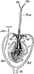
Monitor Lizard Heart

"Heart of monitor (Varanus) dissected to show the cavity of the ventricle and the vessels leading out…

"Brain of alligator, from above. B. ol., olfactory bulb; G. p, epiphysis; HH, cerebellum; Med, spinal cord; MX, optic lobes; NH, medulla oblongata; VH, cerebral hemispheres; I-XI, cranial nerves; 1, 2, first and second spinal nerves." -Parker, 1900

Alligator Brain

"Brain of alligator, from above. B. ol., olfactory bulb; G. p, epiphysis; HH, cerebellum; Med, spinal…

"Poison apparatus of rattlesnake. A, eye; Gc, poison-duct entering the poison-fang at +; Km, muscles of mastication partly cut through at *; Mc, masseter or constrictor muscle; Mc', continuation of the constrictor muscle to the lower jaw; N, nasal opening; S, fibrous poison-sac; z, tongue; za, opening of the poison-duct; zf, pouch of mucous membrane enclosing the poison-fangs." -Parker, 1900

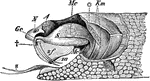
Rattlesnake Poison Apparatus

"Poison apparatus of rattlesnake. A, eye; Gc, poison-duct entering the poison-fang at +; Km, muscles…

"Pterylosis of Columba livia. A, ventral; B, dorsal. al. pt, alar pteryla or wing-tract; c. pt, cephalic pteryla or head-tract; cd. pt, caudal pteryla or tail-tract; cr. pt, crural pteryla; cr. apt, cervical apterium or neck-space; fm. pt, femoral pteryla; hu. pt, humeral pteryla; lat. apt, lateral apterium; sp. pt, spinal pteryla; v. apt, ventral apterium; v. pt, ventral pteryla." -Parker, 1900

Rock Pigeon Feather Tracts

"Pterylosis of Columba livia. A, ventral; B, dorsal. al. pt, alar pteryla or wing-tract; c. pt, cephalic…
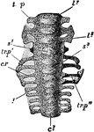
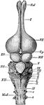
"Columba livia. Sacrum of a nestling (about fourteen days old), ventral aspect. c1, centrum of first sacral vertebra; c1, centrum of fifth caudal; c. r, first sacral rib; l1, centrum of first lumbar; l3, of third lumbar; s1, of fourth lumbar; s3, of sixth lumbar; tr. p, transverse process of first lumbar; tr. p', of fifth lumbar; tr. p'', of first sacral." -Parker, 1900

Rock Pigeon Sacrum

"Columba livia. Sacrum of a nestling (about fourteen days old), ventral aspect. c1, centrum of first…

"Columba livia. Skull of young specimen. A, dorsal; B, ventral; C, left side. al. s, alisphenoid; au, angular; ar, articular; b. o, basi-occipital; d, dentary; e. o, ex-occipital; eu, aperture of Eustachian tube; f. m, foramen magnum; fr, frontal; i.o. s, inter-orbital septum; ju, jugal; lc, lachrymal; lb. s, lambdoidal suture; m. eth, mesethmoid; mx, maxilla; mx. p, maxillo-palatine process; na, na', na'', nasal; o. c, occipital condyle; or. fr, orbital plate of frontal; pa, parietal; pa. s, parasphenoid (rostrum); pl, palatine; p. mx, premaxilla; pt, pterygoid; qu, quadrate; s. an, supra-angular; s. o, supra-occipital; sq, squamosal; ty, tympanic cavity; IL-XII, foramina for cerebral nerves." -Parker, 1900

Rock Pigeon Skull

"Columba livia. Skull of young specimen. A, dorsal; B, ventral; C, left side. al. s, alisphenoid; au,…
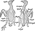
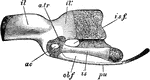
"Columba livia. Left manus of a nestling. The cartilaginous parts are dotted. cp. 1, radiale; cp. 2, ulnare; mcp. 1, 2, 3, metacarpals; ph. 1, phalanx of first digit; ph. 2, ph. 2', phalanges of second digit; ph. 3, phalanx of third digit; ra, radius; ul, ulna." -Parker, 1900

Rock Pigeon Manus

"Columba livia. Left manus of a nestling. The cartilaginous parts are dotted. cp. 1, radiale; cp. 2,…

"Columba livia. Left innominate of a nestling. The cartilage is dotted. ac, acetabulum; a. tr, anti-trochanter; il, pre-acetabular; and il', post-acetabular portion of ilium; is, ischium; i s. f, ischiatic foramen; ob. f, obturator notch; pu, pubis." -Parker, 1900

Rock Pigeon Innominate

"Columba livia. Left innominate of a nestling. The cartilage is dotted. ac, acetabulum; a. tr, anti-trochanter;…

"Columba livia. Part of left foot of an unhatched embryo. The cartilage is dotted. mtl. 2, second; mtl. 3, third; and mtl. 4, fourth metatarsal; ti, tibia; tl. 1, proximal tarsal cartilage; tl. 2, distal tarsal cartilage." -Parker, 1900

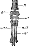
Rock Pigeon Embryo Foot

"Columba livia. Part of left foot of an unhatched embryo. The cartilage is dotted. mtl. 2, second; mtl.…
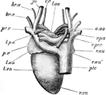
"A, heart of the pigeon, dorsal aspect. a. ao, arch of aorta; br. a, brachial artery; br. v, bachial vein; c. c, common carotid; ju, jugular; l. au, left auricle; l. p. a, left pulmonary artery; l. vn, left ventricle; pc. v, left pre-caval; ptc, post-caval; p. v, pulmonary veins; r. au, r. au', right auricle; r. p. a, right pulmonary artery; r. prc, right pre-caval; r.r vn, right ventricle." -Parker, 1900

Pigeon Heart

"A, heart of the pigeon, dorsal aspect. a. ao, arch of aorta; br. a, brachial artery; br. v, bachial…

"Columba livia. The brain; A, from above; B, from below; C, from the left side. cb, cerebellum; c. h, cerebral hemispheres; f, flocculus; inf, infundibulum; m. o, medulla oblongata; o. l, optic lobes; o. t, optic tracts; pn, pineal body; II-XIII, cerebral nerves; sp. 1, first spinal nerve." -Parker, 1900

Rock Pigeon Brain

"Columba livia. The brain; A, from above; B, from below; C, from the left side. cb, cerebellum; c. h,…

"Columba livia. Male urino-genital organs. adr, adrenal; cl. 2, urodaeum; cl. 3, proctodaeum; k, kidney; ts, tetis, that of the right side displaced; ur, ureter; ur', aperture of ureter; vd, vas deferens; vd', its cloacal aperture; v. s, vesicula seminalis." -Parker, 1900

Male Rock Pigeon Genitalia

"Columba livia. Male urino-genital organs. adr, adrenal; cl. 2, urodaeum; cl. 3, proctodaeum; k, kidney;…

"Columba livia. Female urino-genital organs. cl. 2, urodaeum; cl. 3, proctodaeum; k, kidney; l. od, left oviduct; l. od', its cloacal aperture; l. od'', its coelomic funnel; l. od''', its coelomic aperture; ov, ovarty; r. od, right oviduct; r. od', its cloacal aperture; ur, ureter; ur', its cloacal aperture." -Parker, 1900

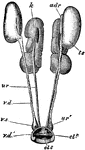
Female Rock Pigeon Genitalia

"Columba livia. Female urino-genital organs. cl. 2, urodaeum; cl. 3, proctodaeum; k, kidney; l. od,…

"Lepus cuniculus. Shoulder-girdle with anterior end of sternum of young specimen. a, acromion; af, pre-scapular fossa; c, coracoid; cl, ossified clavicle; ma, meta-cromion; mss, meso-scapular segment; ost, pre-sternum; pc, pre-coracoid; pf, post-scapular fossa; sr, sternal ribs." -Parker, 1900

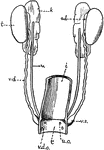
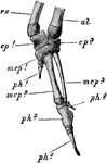
Rabbit Shoulder Girdle

"Lepus cuniculus. Shoulder-girdle with anterior end of sternum of young specimen. a, acromion; af, pre-scapular…
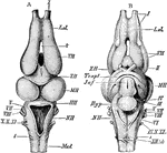
"Lepus cuniculus. Brain. A, dorsal view; B, ventral; b. o, olfactory lobe; cb', median lobe of cerebellum (vermis); cb'', lateral lobe of cerebellum; cr, cauro-cerebri; ep, epiphysis; f. b, parencephala; f. p, longitudinal fissure; h. b, hind-brain; hp, hypophysis; m. b, mid-brain (corpora quadrigemina); md, medulla oblongata; p.v, pons Varolii; i-xii, cranial nerves." -Parker, 1900

Rabbit Brain

"Lepus cuniculus. Brain. A, dorsal view; B, ventral; b. o, olfactory lobe; cb', median lobe of cerebellum…

"Lepus cuniculus. Longitudinal vertical sectioin of the brain. cb, cerebellum, showing arbor vitae; c. c, crus cerebri; ch1, parencephalon; ch2, temporal lobe; c. ma, corpus mammillare; f. m, foramen of Monro; inf, infundibulum; ly, lyra; m. o, medulla oblongata; o. ch, optic chiasma; olf, olfactory lobe; pty, pituitary body; vl. ip, velum interpositum; v. vn, valve of Vieussens; II, optic nerve." -Parker, 1900

Rabbit Brain

"Lepus cuniculus. Longitudinal vertical sectioin of the brain. cb, cerebellum, showing arbor vitae;…

"Lepus cuniculus. The urogenital organs. A, of male; B, of female, from the left side. The kidneys and proximal ends of the ureters, and in B, the ovaries. Fallopian tubes and uteri are not shown. an, anus; bl, urinary bladder; c. c, corpus cavernosum; c. s, corpus spongiosum; c. gl, perineal gland; p. gl', aperture of its duct on the perineal space; pr, anterior, pr', posterior, and pr', lateral lobes of prostate; rct, rectum; r. gl, rectal gland; u. g. a, urino-genital aperture; u. m, uterus masculinus; ur, ureter; va, vagina; vb, vestibule; v. d, vas deferens." -Parker, 1900

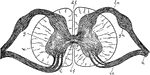
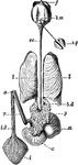
Rabbit Urogenital Organs

"Lepus cuniculus. The urogenital organs. A, of male; B, of female, from the left side. The kidneys and…
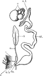
"Lepus cuniculus. The anterior end of the vagina, with the right uterus. Fallopian tube, and ovary. Part of the ventral wall of the vagina is removed, and the proximal end of the left uterus is shown in longitudinal section fl. t, Fallopian tube; fl. t', its peritoneal aperture; l. ut, left uterus; l. ut', left os uteri; r. ut, right uterus; r. ut', right os uteri; s, vaginal sepum; va, vagina." -Parker, 1900

Rabbit Vagina

"Lepus cuniculus. The anterior end of the vagina, with the right uterus. Fallopian tube, and ovary.…

The skeleton of a bat's wing.

Bat Wing

The skeleton of a bat's wing.

in early development of race, the Negro type were found in Africa.

Early Races, Negro

in early development of race, the Negro type were found in Africa.

in early development of race, the Mongolian type consistent of Kalmucks, Chinese, and Amerindians.

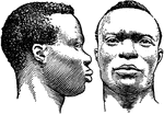
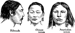
Early Races, Mongolian

in early development of race, the Mongolian type consistent of Kalmucks, Chinese, and Amerindians.

in early development of race, the Caucasian type consisted of Mediterranean men (Jews of Algiers), Mediterranean woman (Berber) and Nordics (Englishmen).

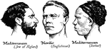
Early Races, Caucasian

in early development of race, the Caucasian type consisted of Mediterranean men (Jews of Algiers), Mediterranean…

Early developments of racial types, a tomb paint from an Egyptian tomb.

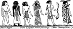
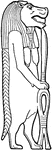
Racial Types From Egyptian Paintings

Early developments of racial types, a tomb paint from an Egyptian tomb.

Perhaps the earliest people to form real cities in the western region of the world, were a people of mysterious origin called Sumerians. This drawing is a very early Sumerian stone carving showing Sumerian warriors in phalanx.

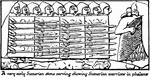
Stone Carvings of Sumerian Warriors

Perhaps the earliest people to form real cities in the western region of the world, were a people of…
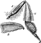
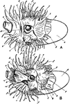
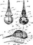
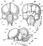
An early figure of the Egyptian hippopotamus goddess.

Egyptian Hippopotamus Goddess

An early figure of the Egyptian hippopotamus goddess.

"A, right hind leg of a honey-bee (seen fron behind and within); B, the tibia. ti, seen from the outside, showing the collecting basket formed of stiff hairs." -Bergen, 1896

Bee Leg

"A, right hind leg of a honey-bee (seen fron behind and within); B, the tibia. ti, seen from the outside,…

Singular warfare of the American Indians. Caption below illustration: "I no longer hesitated; I took my aim; discharged ,y piece; and the animal was instantly stretched before me with a groan, which I conceived to be that of a human creature. I went up to it, and judge my astonishment when I found I had killed an Indian."

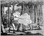
American Indians Warfare

Singular warfare of the American Indians. Caption below illustration: "I no longer hesitated; I took…