The hyoid series is composed of five distinct pieces- a body, or hyoid bone proper, two cornua or horns and two cornicula or lesser horns. Shown us the left aspect of the hyoid series of bones in a horse. Labels: a, proximal, end of left cornu; b, distal end of right cornu; c, proximal end of corniculum; d, hyoid bone. The left heel process points downwards and backwards, the spur process in the opposite direction.

Hyoid Series of Bones in a Horse

The hyoid series is composed of five distinct pieces- a body, or hyoid bone proper, two cornua or horns…

Front aspect of the right carpus of a horse. Labels: 1, cuneiform; 2, lunar; 3, scaphoid; 4, trapezium; 5, unciform; 6, magnum; 7, trapezoid; 8, small metacarpals, the large one lying between and before them. The uppermost bone is the distal end of the radius.

Carpus of a Horse

Front aspect of the right carpus of a horse. Labels: 1, cuneiform; 2, lunar; 3, scaphoid; 4, trapezium;…

External view of bones of right carpus, metacarpus, and digit of a horse. 1, distal end of radius; 2, groove for extensor tendons; 3, scaphoid; 4, lunar; 5, cuneiform;6, trapezium; 7, magnum; 8, unciform; 9, great metacarpal; 10, external small metacarpal (splint); 11, sesamoids; 12, proximal phalanx; 13, median phalanx; 5, distal phalanx; 16, basilar process.

Horse Leg

External view of bones of right carpus, metacarpus, and digit of a horse. 1, distal end of radius; 2,…

Generic development of the horse's foot. A, Foot of Eohippus; B, That of Orohippus; C, That of Hipparion; D, that of horse. The digits are numbered, when present, I. to V.

Evolution of Horse Foot

Generic development of the horse's foot. A, Foot of Eohippus; B, That of Orohippus; C, That of Hipparion;…

Posterior view of phalanges of a horse disarticulated. Labels: a, os suffraginis; b, os coronae; C, os pedis; D, os naviculare, or third sesamoid; 1, glenoid cavities joined by the groove, 2; 3, rough surface for V-shaped sesamoidean ligament; 4, condyles articulating with cavities, 5; 6, condyles of coronae articulating with glenoid cavities of pedal bone, 7; 8, pyramidal process; 9, basilar process; 10, retrosal processes; 11, articular surfaces between navicular and pedal bones; 12, surface, continues with glenoid cavities, 7; 13, posterior edge; 14, extremities for lateral ligaments of navicular bone.

Phalanges of a Horse

Posterior view of phalanges of a horse disarticulated. Labels: a, os suffraginis; b, os coronae; C,…

Left posterolateral view of a horse's pelvis. 1, anterior iliac spine; 2, Posterior iliac spine. The iliac crest is the border joining 1 and 2. 3, Iliac shaft. 4, The acetabulum, the large rough ridge above is the sciatic spine; 6, inferior ischiatic spine, posterior to which is the tuberosity.

Pelvis of a Horse

Left posterolateral view of a horse's pelvis. 1, anterior iliac spine; 2, Posterior iliac spine. The…

Posterior view of a left femur of a horse. Labels: 1, head; 2, trochanter major; 3, trochanter minor; 4, trochanter internus; 5, notch for the round ligament; 6, trochanteric fossa; 7, 8, places for attachment for the laterla ligaments; 11, the condlyes, separated by the intercondyloid groove; 10, supracondyloid fossae.

Femur of a Horse

Posterior view of a left femur of a horse. Labels: 1, head; 2, trochanter major; 3, trochanter minor;…

Bones of left tarsus of a horse, seen from in front and outside. Labels: 1, calcaneum; 2, astragalus; 3, cuneiforme magnum; 4, cuneiforme medium; 5, cuboid.

Tarsus of a Horse

Bones of left tarsus of a horse, seen from in front and outside. Labels: 1, calcaneum; 2, astragalus;…

External view of bones of left tarsus, metatarsus, and digit of a horse. Labels: 1, distal end of tibia; 2, calcaneum; 3, astragalus; 4, cuboid; 5, cuneiforme magnum; 6, cuneiforme medium; 7, great metatarsal; 8, external small metatarsal (splint); 9, proximal phalanx; 10, sesamoids; 11, median phalanx; 12, distal phlanx; 14, third sesamoid; 15, basilar process.

Horse Leg

External view of bones of left tarsus, metatarsus, and digit of a horse. Labels: 1, distal end of tibia;…

Posterior view of left metatarsus of a horse. 1, Large metatarsal bone; 2, Internal small metatarsal bone; 3, external, small metatarsal bone; 4, partial origin of suspensory and check ligaments; 5, nutrient foramen; 6, articular ridge joining the condyles.

Metatarsus of a Horse

Posterior view of left metatarsus of a horse. 1, Large metatarsal bone; 2, Internal small metatarsal…
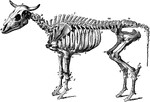
The skeleton of an ox. Axial Skeleton. The skull. Cranial Bones- occipital, 1: b, parietal, 2; a, frontal, 2; c, temporal, 2; sphenoid, 1; ethmoid, 1; auditory ossicles, 8. Facial Bones- h, nasal, 2; e, lachrymal, 2; d, malar, 2; f, maxilla, 2; g, premaxilla, 1; i, inferior maxilla, 2; palatine, 2; pterygoid, 2; vomer, 1; turbinals, 4; hyoid series, 7. Teeth: incisors, 6; canines, 2; molars, 24. The trunk- k, cervical vertebrae, 7; l, dorsal vertebrae, 13; m, lumbar vertebrae, 6; n, sacrum ( five segments), 1; o, coccygal vertebrae (variable), 20; p, ribs, 26; * sternum (seven sternebrae), 1. Appendicular skeleton. Pectoral limb. t, scapula, 2; u, humerus, 2; v, radius, 2; w, ulna, 2. Carpus- x, trapezium, 2; y, cuneiform, 2; z, lunar; a', scaphoid, 2; b', unciform, 2; c', magnum, 2. Metacarpus: d', large bone, 2; e', small bone, 4; f', large sesamoids, 8. Digit- g', proximal phalanges, 4; h', median phalages, 4; i', distal phalanges, 4; k', small sesamoids, 4. Pelvic Limb. Pelvis- q, ilium, 2; s, ischium, 2; r, pubis, 2. The limb- l', femur, 2; m', patella, 2; n', tibia, 2. Tarsus-o', maleolar, 2; p', calcaneum, 2; q', astragalus, 2; r', cubocuneiform, 2; s', cuneiforme medium, 2; t', cuneiforme parvum, 2. Metatarsus: large bone, 2; u', small bone, 2. Large sesamoids, 8. Digit: proximal phalanges, 4; median phalanges, 4; distal phalanges, 4; small sesamoids, 4. Visceral skeleton. Bones of the heart, 2. The separate bones of the ruminant skeleton, as here considered are 251.

Ox Skeleton

The skeleton of an ox. Axial Skeleton. The skull. Cranial Bones- occipital, 1: b, parietal, 2; a, frontal,…

Skeleton of the hog. Axial skeleton. The skull. Cranial bones- a, occipital, 1; b, parietal, 2; d, frontal, 2; c, temporal, 2; sphenoid, 1; ethmoid, 1; auditory ossicles, 8. Facial Bones- g, nasal, 2; h, os rostri, 1; f, lachrymal, 2; e, malar, 2; i, maxilla, 2; k, premaxilla, 2; l, inferior maxilla, 1; palatine, 2; 2, pterygoid, 2; vomer, 1; turbinals, 4; hyoid series, 5. Teeth: incisors, 12; canines, 4; molars, 28. The trunk- m, cervicle vertebre (variable), 18; r, ribs, 28; * sternum (seven sternebrae), 1. Appendicular Skeleton. Pectoral Limb. v, scapula, 2; w, humerus, 2; x, radius, 2; y, ulna, 2. carpus: z, trapezium, 2; a', cuneiform, 2; b', lunar, 2; c', scaphoid, 2; d', unciform, 2; e', magnum, 2; f', trapezoid, 2; g', pisiform, phalanges, 8; l', distal phalanges, 8; l", small sesamoids, 8. Pelvic Limb. Pelvis: s, ilium, 2; t, pubis, 2; u, ischium, 2. The limb-m', femur, 2; n', patella, 2; o', tibia, 2; p' fibula, 2. Tarsus- q' calcaneum, 3; r', astragalus, 2; s', cuboid, 2; t', cuneiforme magnum, 2; u', ecto-cuneiforme, 2; v' w', meso and endo cuneiformes, 4. Metatatarsus: large bones, 8; x', small bones 2; y', large sesamoids, 16. Digit: proximal phalanges, 8; median phalanges, 8; distal phalanges, 8; small sesamoids, 8. Visceral Skeleton- None. The separate bones of the skeleton of the hog, as here considered, are 234.

Skeleton of a Hog

Skeleton of the hog. Axial skeleton. The skull. Cranial bones- a, occipital, 1; b, parietal, 2; d, frontal,…
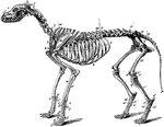
The skeleton of the dog. Axial skeleton. The skull. Cranial bones- a, occipital, 1; b, parietal, 2; d, frontal, 2; c, temporal, 2; sphenoid, 1; ethmoid, 2; auditory ossicles, 8. Facial Bones- f, nasal, 2; e, lachrymal, 2; d, malar, 2; h, maxilla, 2; g, premaxilla, 2. Teeth: incisors, 12; canines, 4; molars, 26. The trunk- l, cervical vertebrae, 7; m, dorsal vertebrea, 13; n, lumbar vertebrae, 7; o, sacrum (three segments), 1; p, coccygeal phalanges (variable), 20; t, ribs, 26; * sternum (eight sternebrae), 1. Appendicular Skeleton. Pectoral Limb. u, scapula, 2; v, humerus, 2; w, radius, 2; x, ulna. Carpus: y, trapezium, 2; z cuneiform, 2; a', scaphoid, 2; b', unciform, 2; c', magnum, 2; d', trapezoid, 2; e', pisiform,2; metacarpal bones, 10; h', anterior sesamoids, 10; g' posterior sesamoids, 20. Digits- i' proximal phalanges, 10; k', median phalanges, 8; l', distal phalanges, 10; small sesamoids wanting. Pelvic Limb. Pelvis: q, ilium, 2; r, pubis, 2; s, ischium, 2. The limb-m', femur, 2; o', fabellae, 4; n', patella, 2; q', tibia, 2; p', tibial sesamoid, 2; r', fibula, 2. Tarsus: s', calcaneum, 2; t', astragalus, 2; u', cuboid, 2; v', superior cuneiform, 2; w', ectocuneiforme, 2; x', mesocuneiforme, 2; y', edocuneiforme, 2. Metatarsus: large bones, 8; z',small bones, 2; anterior sesamoids, 8; posterior sesamoids, 16. Digit: proximal phalanges, 8; median phalanges, 8; distal phalanges, 8; small sesamoids wanting. Visceral Skeleton. Os Penis, 1; Rudimentary clavicle (inconstant), 2. The bones of the Carnivora Skeleton, thus considered, are 345.

Skeleton of a Dog

The skeleton of the dog. Axial skeleton. The skull. Cranial bones- a, occipital, 1; b, parietal, 2;…
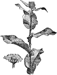
Elecampane, also called Horse-heal (Inula helenium) or Marchalan (in Welsh), is a perennial composite plant common in many parts of Great Britain, and ranges throughout central and Southern Europe, and in Asia as far eastwards as the Himalayas. It is a rather rigid herb, the stem of which attains a height of from 3 to 5 feet; the leaves are large and toothed, the lower ones stalked, the rest embracing the stem; the flowers are yellow, 2 inches broad, and have many rays, each three-notched at the extremity. The root is thick, branching and mucilaginous, and has a warm, bitter taste and a camphoraceous odor.

Elecampane

Elecampane, also called Horse-heal (Inula helenium) or Marchalan (in Welsh), is a perennial composite…

Seahorses are a genus (Hippocampus) of fish belonging to the family Syngnathidae, which also includes pipefish and leafy sea dragons. There are over 32 species of seahorse, mainly found in shallow tropical and temperate waters throughout the world. They prefer to live in sheltered areas such as sea grass beds, coral reefs, or mangroves.

Seahorse

Seahorses are a genus (Hippocampus) of fish belonging to the family Syngnathidae, which also includes…
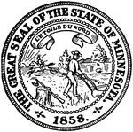
The Great Seal of the State of Minnesota. The seal depicts a farmer plowing as a Native American rides by horseback. Above, the state motto reads, 'L'etoile du nord' meaning "Star of the North."

Seal of Minnesota

The Great Seal of the State of Minnesota. The seal depicts a farmer plowing as a Native American rides…

The Great Seal of the State of New Jersey. The seal features Liberty and Ceres holding a shield with three plows representing agriculture. The state motto in the ribbon below reads, "Liberty and Prosperity."

Seal of New Jersey

The Great Seal of the State of New Jersey. The seal features Liberty and Ceres holding a shield with…
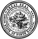
The Great Seal of the State of North Dakota. The seal depicts a tree, wheat, a plow, and a Native American on horseback hunting a buffalo. Above is the state motto, "Liberty and Union Now and Forever, One and Inseparable."

Seal of North Dakota

The Great Seal of the State of North Dakota. The seal depicts a tree, wheat, a plow, and a Native American…
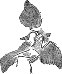
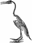
An illustration of a bird skeleton.

Bird Skeleton

An illustration of a bird skeleton.

An illustration of the skeletal fossil of Archaeopteryx, the earliest and most primitive bird known. Archaeopteryx lived in the late Jurassic Period around 155–150 million years ago, in what is now southern Germany during a time when Europe was an archipelago of islands in a shallow warm tropical sea, much closer to the equator than it is now.

Archaeopteryx Fossil

An illustration of the skeletal fossil of Archaeopteryx, the earliest and most primitive bird known.…
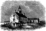
The Sackett's Harbor lighthouse was erected on Horse Island during the War of 1812.

Lighthouse on Horse Island

The Sackett's Harbor lighthouse was erected on Horse Island during the War of 1812.

An illustration of an Ichthyosaurus skeleton. Ichthyosaurus is an extinct genus of ichthyosaur from the Early Jurassic (Hettangian - Sinemurian) of Europe (Belgium, England and Germany). It is among the best known ichthyosaur genera, with the Order Ichthyosauria being named after it. Ichthyosaurus was the first complete fossil to be discovered in the early 1800's by Mary Anning in England.

Archaeopteryx Skeleton

An illustration of an Ichthyosaurus skeleton. Ichthyosaurus is an extinct genus of ichthyosaur from…
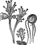
An illustration of a small jellyfish. Jellyfish are free-swimming members of the phylum Cnidaria. ellyfish don't have specialized digestive, osmoregulatory, central nervous, respiratory, or circulatory systems. They digest using the gastrodermal lining of the gastrovascular cavity, where nutrients are absorbed. They do not need a respiratory system since their skin is thin enough that the body is oxygenated by diffusion. They have limited control over movement and mostly free-float, but can use the hydrostatic skeleton of the water pouch to accomplish vertical movement through pulsations of the disc-like body.

Jellyfish

An illustration of a small jellyfish. Jellyfish are free-swimming members of the phylum Cnidaria. ellyfish…
An illustration of the skeleton of a kangaroo's right hind foot.

Kangaroo Foot

An illustration of the skeleton of a kangaroo's right hind foot.

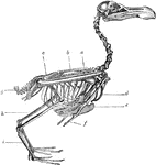
The skeleton of a bird. Labels: a, radius and ulna; b, dorsal vertebrae; c, sacrum and pelvis; g, ploughshare bone; h, tibia; i, metatarsus; d, furculum; e, carina of the sternum; f, distal phalanx of pectoral limb.

Skeleton of a Bird

The skeleton of a bird. Labels: a, radius and ulna; b, dorsal vertebrae; c, sacrum and pelvis; g, ploughshare…
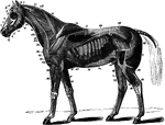
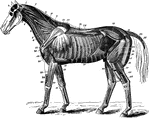
The superficial muscles of the horse with the panniculus and tunica abdominalis removed. Labels: 1, abducens; 2, retrahentes muscles; 2' attollens maximus; 2", attollens anticus; 3, temporalis; 4, nasalis longus; 5, orbicularis palpebrarum; 6, levator labii superioris alaeque nasi; 7, dilatator naris lateralis; 8, orbicularis oris; 9, zygomaticus; 9', buccinator; 10, depressor labii inferioris; 11, masseter; 12, levator humeri; 13, trapezius cervicalis; 14, trapezius dorsalis; 15, latissimus dorsi; 18, pextoralis parvus; 19, pectoralis magnus; 20, sterno-maxillaris; 23, subscapulo-hyoideus; 24, antea-spinatus; 25, teres externus; 26, postea-spinatus; 30, caput medium; 31, caput magnum; 33, extemsor metacarpi magnus; 34, humeralis obliquus; 35, extensor peis; 36, flexor metacarpi externus and medius; 37, flexor metacarpi internus; 46, cervical serratus magnus; 47, dorsal serratus magnus; 48, rhomboideus longus; 49, superficialis costarum; 50, splenius; 52, intercostales; 54, tensor fasciae latae; 55, triceps abductor femoris; 56, gluteus externus; 61, biceps rotator tibialis; 62, rectus femoris; 63, vastus externus; 65, gastrocnemius; 68, flexor pedis perforans; 69, peroneous; 70, extensor pedis; 71, flexor metatarsi; 72, flexor pedis accessorius; 74, obliquus abdominis externus.

Superficial Muscles of the Horse

The superficial muscles of the horse with the panniculus and tunica abdominalis removed. Labels: 1,…
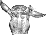
Muscles of the external ear- posterior view. Labels: a, inferior and b, superior layer of the scuto-auricularis internus; c, attollens posticus; d, retrahens externus; e, retrahens medius; f, retrahens internus; g, mastoido-auricularis.

Muscles of the External Ear of a Horse

Muscles of the external ear- posterior view. Labels: a, inferior and b, superior layer of the scuto-auricularis…
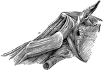
The neck muscles of a horse-lateral view. Labels: a, oblique capitis posticus; b, obliquus capitis anticus; c, complexus minor; d, rectus capitis posticus major; e, rectus capitis posticus minor.

Neck Muscles of a Horse

The neck muscles of a horse-lateral view. Labels: a, oblique capitis posticus; b, obliquus capitis anticus;…
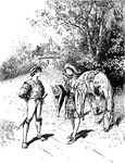
Two men in renaissance clothing having a conversation on the side of the road. The man to the left is noted to be a jester for a king. The caption reads, "'What wilt thou that a fool can do for thee?' he asked again."

Two Men Talking

Two men in renaissance clothing having a conversation on the side of the road. The man to the left is…

An illustration of a Diplodocus skeleton and the caudal vertebrae (A and B). Diplodocus is a genus of diplodocid sauropod dinosaur whose fossils were first discovered in 1877 by S. W. Williston. It lived in what is now western North America at the end of the Jurassic Period. Diplodocus is one of the more common dinosaur fossils found in the Upper Morrison Formation, a sequence of shallow marine and alluvial sediments deposited about 150 to 147 million years ago, in what is now termed the Kimmeridgian and Tithonian stages.

Diplodocus

An illustration of a Diplodocus skeleton and the caudal vertebrae (A and B). Diplodocus is a genus of…
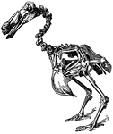
Illustration of a dodo bird skeleton. The dodo (Raphus cucullatus) was a flightless bird endemic to the Indian Ocean island of Mauritius. Related to pigeons and doves, it stood about a meter tall, weighing about 20 kilograms (44 lb), living on fruit and nesting on the ground.

Dodo Skeleton

Illustration of a dodo bird skeleton. The dodo (Raphus cucullatus) was a flightless bird endemic to…
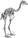
An illustration of the skeleton of a pezophaps solitarius, part of the dodo family.

Pezophaps Solitarius (Skeleton)

An illustration of the skeleton of a pezophaps solitarius, part of the dodo family.
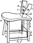
"Sewing-horse. In saddlery, a sewing-clamp with its supports. a, seat; b, legs; c, c', clamping-jaws, c' hinged to c at d; e, strap fastened to c' passing through c, and attached by chain f to the foot-lever g, the latter pivoted at h; i, spring which opens the jaws when not pulled together by e; k, ratch which g engages to hold he jaws together." -Whitney, 1911

Sewing Horse

"Sewing-horse. In saddlery, a sewing-clamp with its supports. a, seat; b, legs; c, c', clamping-jaws,…
The deep muscles of the horse. Labels: 1, temporalis; 1', stylo-maxillaris; 2, rectus capitis anticus major; 3, sterno-thyro-hyoideus; 4, sterno-maxillaris; 5, trachea; 6, scalenus; 7, splenius; 8, funicular part of ligamentum nuchae; 9, rhomboideus longus; 10, cervical serratus magnus; 15, costal serratus magnus; 11, cartilage of prolongation; 12, rhomboideus brevis; 13, transversalis costarum; 14, longissimus dorsi; 15, serratus magnus; 16, external intercostarum; 17, internal intercostals; 18, rectus abdominis; 19, pexctoralis magnus; 20, postea-spinatus minor; 21, flexor brachii; 22, humeralis obliquus; 22', capus parvum (of treceps extensor brachii); 23, extensor suffraginis; 24, extensormetacarpimagnusdivided; 25, extensor metacarpo obliquus; 25', tendon; 26, 28, flexor pedis perfoans and perforatus; 29, obliquus abdominis internus; 30, gluteus maximus; 31, erector coccygis; 32, curvator coccygia; 33, depressor coccygis; 34, rectus femoris; 35, vastus externus; 36, part covered by triceps abductor; 37, biceps rotator tibialis; 38, gastrocnemius; 39, plantaris; 40, flexor pedis perforans; 41, peroneus; 42, flexor metatarsi; 43, extensor pedis. (cut across)

Neck Muscles of the Horse

The deep muscles of the horse. Labels: 1, temporalis; 1', stylo-maxillaris; 2, rectus capitis anticus…
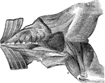
Muscles of the sublumbar and internal deep femoral regions-seen from below. Labels: a, quadratus lumborum; b, psoas parvus; c, psoas magnus; d, e, iliacus; f, pectineus; g, adductor brevis; h, adductor longus; i, adductor magnus; k, vastus internuus; l, rectus femoris.

Muscles From the Femoral Region of the Horse

Muscles of the sublumbar and internal deep femoral regions-seen from below. Labels: a, quadratus lumborum;…
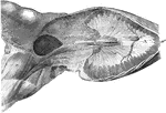
The diaphragm, and superficial muscles of the internal femoral region-viewed from below. A, the diaphragm; a, the tendinous center; a', the fleshy periphery; a", the crura; a"', the hiatus aorticus; a"", the foramen sinistrum; a""', the foramen dextrum; b, the sartorius; c, the gracilis.

Muscles From the Femoral Region of the Horse

The diaphragm, and superficial muscles of the internal femoral region-viewed from below. A, the diaphragm;…

Muscles of the anterior limb-external view. Labels: a, antea-spinatus; b, postea-spinatus; c, teres externus; d, caput magnum; e, caput medium; f, flexor brachii; g, extensor metacarpi magnus; h, extensor metacarpi obliquus; i, flexor metacarpi externus; k, extensor pedis; k', its tendon; k", fibrous band from the suspensory ligament; l, extensor suffraginis; l', its tendon.

Horse Leg Muscles

Muscles of the anterior limb-external view. Labels: a, antea-spinatus; b, postea-spinatus; c, teres…

Muscles of the anterior limb-internal view. Labels: a, subscapularis; b, teres internus; c, coracohumeralis; d, scapulo-ulnaris; e, flexor metacarpi internus; f, flexor metacarpi medius; g, caput parvum.

Horse Leg Muscles

Muscles of the anterior limb-internal view. Labels: a, subscapularis; b, teres internus; c, coracohumeralis;…

External view of the muscles of the anterior limb-showing the deeper ones of the upper region. Labels: a, posteaspinatus minor; b, deep portion of the postea-spinatus; c, flexor brachii divided and the upper part raised; c', its tendon, which fits the cicipital groove; d, humeralis obliquus; e, anconeus; f, extensor pedis; g, Thiernesse's muscles; h, Phillips' muscle; h', its tendon.

Horse Leg Muscles

External view of the muscles of the anterior limb-showing the deeper ones of the upper region. Labels:…

Internal view of the deep muscles of the anterior limb. Labels: a, caput parvum of triceps extensor brachii; b, flexor pedis perforatus; b' its tendon; b", slips to the phalanges; c, flexor pedis perforans; c', radialis accesorius; c', ulnaris accesorius; c"', inferior check ligament; d, perforans tendon leaving the sheath of perforatus tendon.

Horse Leg Muscles

Internal view of the deep muscles of the anterior limb. Labels: a, caput parvum of triceps extensor…
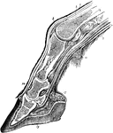
Longitudinal section through the digit of a horse. Labels: 1, skin; 2, extensor pedis tendon; 3, synovial capsule of patern joint; 4, metacarpus; 5, perforatus tendon; 6 (omitted in cut), perforans tendon; 7, synovial sheath; 8, projecting synovial cul-de-sac; 9, inner aspect of sesamoid bone; 10, the fetlock; 11, the y-ligament; 12, the v-ligament; 12, proximal phalanx; 14, terminal portion of extensor tendon; 15, median phalanx; 16, third sesamoid or navicular bone; 17, the sensitive frog; 18, distal phalanx; 19, the hoof; 20, sensitive laminae.

Section Through the Horse Leg and Hoof

Longitudinal section through the digit of a horse. Labels: 1, skin; 2, extensor pedis tendon; 3, synovial…
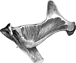
Deep-seated muscles of the gluteal region. Labels: a, deep anterior portion of gluteus maximus; b, gluteus internus; c, common tendon of obturator internus and pyriformis; d, gemellus anticus and posticus; e, ischiofemoralis.

Muscles from Gluteal Region of a Horse

Deep-seated muscles of the gluteal region. Labels: a, deep anterior portion of gluteus maximus; b, gluteus…
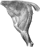
The muscles of the thigh and haunch- left side; the external fascia being removed. Labels: a, tensor fasciae latae; b, gluteus externus; c, gluteus maximus; d, triceps abductor femoris; e, biceps rotator tibialis; f, part of adductor magnus.

Thigh and Haunch Muscles of a Horse

The muscles of the thigh and haunch- left side; the external fascia being removed. Labels: a, tensor…

Anterior tibial group of muscles of the right limb, seen from before and the outside. Labels: a, flexor metatarsi; a', tendonto the cuboid; and a", temdom to the metatarsus, from its funicular portion; a"', tendon of its fleshy portion, sending a straight slip to the metatarsus, and an oblique one to the cuneiform bone; b, extensor pedis; b', its tendon; c, peroneus; c', point where its tendon joins that of the extensor pedis; d, extensor pedis brevis; e, superior, and f, inferior thick portions of the annular ligament.

Horse Leg Muscles

Anterior tibial group of muscles of the right limb, seen from before and the outside. Labels: a, flexor…

Tendons and ligaments of the left anterior extremity of ox, viewed from external side. Labels: a, flexor perforatus tendon; a', its superficial; a", its deep branch; b, interosseus metacarpeus; c, flexor perforans tendon; d-d", e-e"', superior suspensory ligament and attachments; d, anterior division; e', e", branches forming superior ligaments of small claw; e'". inferior ligament of small claw; f, common annular ligament; g, g', superior and inferior special annular ligaments; h, interdigital ligament; i, i', tendon of the extensor proprius externus.

Tendons and Ligaments of Ox Leg

Tendons and ligaments of the left anterior extremity of ox, viewed from external side. Labels: a, flexor…
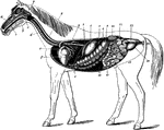
The digestive apparatus of the horse. Labels: a, mouth; 2, pharynx; 3, esophagus; 4, diaphragm; 5, spleen; 6, stomach (left sac); 7, duodenum; 8, liver (upper extremity); 9, great colon; 10, caecum; 11, small intestine; 12, floating colon; 13, rectum; 14, anus; 15, left kidney and ureter; 16, bladder; 17, urethra; a, hard palate; b, tongue; c, soft palate; d, trachea; e, pulmonary artery (divided); f, heart; g, posterior aorta.

Digestive Apparatus of the Horse

The digestive apparatus of the horse. Labels: a, mouth; 2, pharynx; 3, esophagus; 4, diaphragm; 5, spleen;…

Right infero-lateral view of the muscles of the maxillary space, the ramus and hyoid cornu are cut away. Labels: a, lingualis; b, hyo-glossus longus; c, ho-glossus brevis; d, genio-hyo-glossus; e, genio-hyoideus; f, stylo-hyoideus; g, hyoideus magnus; h, levator palati; i, tensor palati; k, pharyngostaphylinus; l, pterygo-pharyngeus; m, kerato-pharyngeus; n, hyo-pharyngeus; o, thyro-pharyngeus; p, crico-pharyngeus; q, esophagus.

Muscles of the Maxillary Space of a Horse

Right infero-lateral view of the muscles of the maxillary space, the ramus and hyoid cornu are cut away.…

Parotid and molar glands of the left side. Labels: a, parotid gland; b, Steno's duct; c, superior molar glands; d, inferior molar gland.

Parotid and Molar Glands of a Horse

Parotid and molar glands of the left side. Labels: a, parotid gland; b, Steno's duct; c, superior molar…

Right infero-lateral view of the head; the maxillary ramus, cheek, parotid gland, and upper lip being removed. Labels: a, submaxillary gland; b, Wharton's duct; c, sublingual gland; d, its excretory orifices; e, labial glands; f, their excretory orifices; g, papilla, with orifice of Steno's duct; h, orifices of the superior, and i, those of the inferior molar glands; k, the hard palate; l, the soft palate; m, the tongue, with its papillae; n, pillars of the tongue and soft palate; o, fraenumlinguae.

Horse Head

Right infero-lateral view of the head; the maxillary ramus, cheek, parotid gland, and upper lip being…
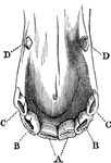
Incisor and canine teeth of a horse. A, front, B, lateral, and C, corner incisor; D, canine teeth.

Incisor and Canine Horse Teeth

Incisor and canine teeth of a horse. A, front, B, lateral, and C, corner incisor; D, canine teeth.

Incisor tooth of a horse-posterior view. Labels: a, outer layer of enamel; b, inner layer of enamel round the infundibulum; c, dental star; d, the dentine.

Horse Incisor Tooth

Incisor tooth of a horse-posterior view. Labels: a, outer layer of enamel; b, inner layer of enamel…
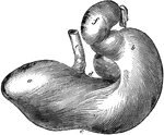
Posterior view of the stomach of a horse. Labels: a, left cul-de-sac; b, right cul-de-sac; c, greater curvature; d, lesser curvature; e, esophagus; f, duodenum.

Horse Stomach

Posterior view of the stomach of a horse. Labels: a, left cul-de-sac; b, right cul-de-sac; c, greater…
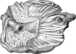
Internal aspect of a horse stomach, opened from below. Labels: a, cuticular mucous membrane; b, villous mucous membrane; c, line of demarcation between the two portions; d, cardiac orifice; e, pyloric orifice and valve.

Horse Stomach

Internal aspect of a horse stomach, opened from below. Labels: a, cuticular mucous membrane; b, villous…
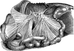
The two mesenteries; the great colon being removed. Labels: a, anterior mesentery; b, mesenteric glands; c, loops of the small intestine; d, colic mesentery; e, loops of the floating colon; f, sphincter ani- internal layer; g, levator ani.

Mesenteries of a Horse

The two mesenteries; the great colon being removed. Labels: a, anterior mesentery; b, mesenteric glands;…
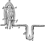
Section of mucous membrane of the small intestine. One the left a villus is seen in seen in section. Labels: a, the epithelial covering; c, the blood-vessels; d, the basement membrane, or subepithelial layer; e, spaces for reception of the chyle; f, origin of a lacteal vessel. On the right is a follicular depression in the mucous membrane, with b, the cells lining it; and c, the subepithelial layer.

Mucous Membrane

Section of mucous membrane of the small intestine. One the left a villus is seen in seen in section.…
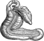
Caecum and great colon of a horse. Labels: a, caecum; b, c, its muscular bands; d, termination of the ileum; e, first; e', second; f, third; f', fourth division of colon; g, pelvic flexure; h, origin of floating colon. The arrows indicate the course of the food through the colon.

Caecum and Colon of Horse

Caecum and great colon of a horse. Labels: a, caecum; b, c, its muscular bands; d, termination of the…
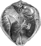
Posterior view of the liver and diaphragm in situ. Labels: a, left lobe; b, right lobe; c, quadrate lobe; d, spigelian lobe; d', spigelian ligament; just above in the right kidney; e, left lateral ligament; f, right lateral ligament; g, broad ligament; h, hepatic duct; i, piece of the duodenum opened to show, k, opening of the ductus pancreaticus minor, and l, Eminence of Vater; m, vena porta; n, posterior vena cava; o, notch for esophagus.

Liver and Diaphragm of a Horse

Posterior view of the liver and diaphragm in situ. Labels: a, left lobe; b, right lobe; c, quadrate…
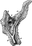
Anterior view of the pancreas. Labels: a, left branch; b, right branch; c, inferior branch; d, duct of Wirsung; e, ductus pancreaticus minor; f, portal vein cut across; g, notch for the great mesenteric artery.

Pancreas of a Horse

Anterior view of the pancreas. Labels: a, left branch; b, right branch; c, inferior branch; d, duct…

Internal aspect of the spleen. Labels: a, superior extremity, or base; b, inferior extremity; c, internal surface; d, the hilus; e, anterior border; f, posterior border; g, suspensory ligament.

Spleen of a Horse

Internal aspect of the spleen. Labels: a, superior extremity, or base; b, inferior extremity; c, internal…
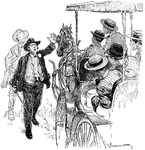
A man waves to an oncoming horse-drawn coach to stop

Stop!

A man waves to an oncoming horse-drawn coach to stop