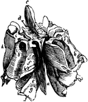
"Dissected fish. a, air bladder; b, urinary bladder; b, urinary bladder; br, brain; c, spinal cord; d and da, dorsal fins; dv, anal fin; e, body of the vertebra; g, gills receiving the blood from the ventral aorta leading out of the heart h; i, intestine; k, kidney; l, liver; m, spines of the vertebra; n, auricle of the heart; ov, ovary; ovd, oviduct; st, stomach; sp, spleen; u, ureter." —Davison, 1906

Dissected Fish

"Dissected fish. a, air bladder; b, urinary bladder; b, urinary bladder; br, brain; c, spinal cord;…

"Frog with the left side cut away and some of the organs pulled downward. a, aorta leading from the ventricle; b, bladder; c, spinal cord surrounded by the vertebral column of bone; d, small intestine; e, opening of the lungs into the pharynx; h, ventricle of the heart; k, kidney; l, lung; liv, liver; n, brain; ov, ovary; ovd, oviduct; p, pancreas; r, rectum; sp, spleen; st, stomach; t, tongue; si, small intestine; u, left auricle; ur, ureter; v, vena cava; vc, body of vertebra." —Davison, 1906

Dissected Frog

"Frog with the left side cut away and some of the organs pulled downward. a, aorta leading from the…
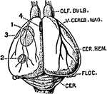
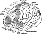
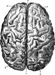
The brain seen from above. 1: Great fissure; 2: Anterior lobes; 3: Posterior lobes.

Brain

The brain seen from above. 1: Great fissure; 2: Anterior lobes; 3: Posterior lobes.

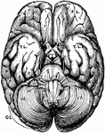
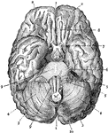
The brain seen from below. 1: Great fissure; 2: Anterior lobes of cerebrum; 3: Posterior lobes of cerebrum; 4: Lobes of cerebellum; 5: Cranial nerves; 6: Auditory nerve; 7: Optic nerve; 8: Olfactory nerve; 9: Main body of medulla oblongata; 10: End of medulla oblongata.

Brain

The brain seen from below. 1: Great fissure; 2: Anterior lobes of cerebrum; 3: Posterior lobes of cerebrum;…
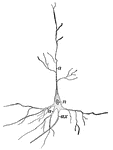
Nerve cell from the brain; <em>a</em>, processes by which it communicates with other cells near by; <em>ax</em>, long process or axis cylinder terminating in the tail end of the spinal cord; <em>n</em>, nucleus.

Nerve Cell

Nerve cell from the brain; a, processes by which it communicates with other cells near by;…

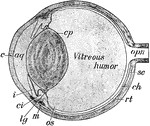
Section of the cat's eye enlarged twice. <em>aq</em>, aqueous humor; <em>c</em>, cornea; <em>ci</em>, cillary processes; <em>cp</em>, capsule surrounding the crystalline lens; <em>ch</em>, choroid coat; <em>i</em>, iris; <em>lg</em>, ligament of the lens; <em>m</em>, muscle to adjust the lens for far and near sight; <em>opn</em>, optic nerve leading to the brain; <em>rt</em>, retina; <em>sc</em>, sclerotic coat.

Cat's Eye

Section of the cat's eye enlarged twice. aq, aqueous humor; c, cornea; ci,…

Diagram of the organs of a Nemertine. <em>m</em>, mouth; <em>div</em>, intestinal diverticula; <em>a</em>, anus; <em>ov</em>, ovaries; <em>n</em>, nephridia; <em>br</em>, brain-lobes; <em>ln</em>, longitudinal nerve stems.

Nemertine

Diagram of the organs of a Nemertine. m, mouth; div, intestinal diverticula; a,…

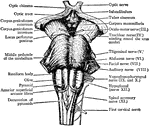
Front view of the medulla, pons and mesencephalon of a full-time human foetus.

Brain

Front view of the medulla, pons and mesencephalon of a full-time human foetus.

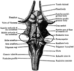
Back view of the medulla, pons and mesencephalon of a full-time human foetus

Brain

Back view of the medulla, pons and mesencephalon of a full-time human foetus

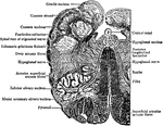
Transverse section through the human medulla in the lower olivary region

Brain

Transverse section through the human medulla in the lower olivary region

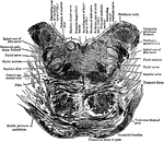
Section through the lower part of the human pons varolii imediately above the medulla

Brain

Section through the lower part of the human pons varolii imediately above the medulla

Mesial section through the Corpus Callosum, the Mesencephalon, the Pons, Medulla and Cerebellum. Showing the third and fourth ventricles joined by the aqueduct of Sylvius

Brain

Mesial section through the Corpus Callosum, the Mesencephalon, the Pons, Medulla and Cerebellum. Showing…
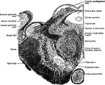
Transverse section through a cerebellar folium.

Brain

Transverse section through a cerebellar folium.

Transverse section through the human mesencephalon at the level of the superior quadrigeminal body

Brain

Transverse section through the human mesencephalon at the level of the superior quadrigeminal body

Gyri and Sulci, on the outer surface of the cerebral hemisphere

Brain

Gyri and Sulci, on the outer surface of the cerebral hemisphere

Orbital surface of the left frontal lobe and the island of Reil; the tip of the temporo-sphenoidal lobe has been removed to display the latter.

Brain

Orbital surface of the left frontal lobe and the island of Reil; the tip of the temporo-sphenoidal lobe…
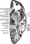
The Gyri and Sulci on the Mesial Aspect of the Cerebral Hemisphere

Brain

The Gyri and Sulci on the Mesial Aspect of the Cerebral Hemisphere

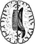
To show the Right Ventricle and the left half of the Corpus Callosum

Brain

To show the Right Ventricle and the left half of the Corpus Callosum

A deeper dissection of the Lateral Ventricle, and of the Velum Interpositum

Brain

A deeper dissection of the Lateral Ventricle, and of the Velum Interpositum

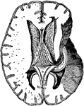
Horizontal section through the Right Cerebral Hemisphere at the Level of the Widest Part of the Lenticular Nucleus

Brain

Horizontal section through the Right Cerebral Hemisphere at the Level of the Widest Part of the Lenticular…
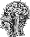
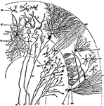
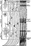
Diagram to illustrate Minute Structure of the Cerebral Cortex

Brain

Diagram to illustrate Minute Structure of the Cerebral Cortex

Brain of Petromyzon marinus (sea lamprey),dorsal view.

Brain of Sea Lamprey

Brain of Petromyzon marinus (sea lamprey),dorsal view.

Brain of Petromyzon marinus (dorsal view).

Brain

Brain of Petromyzon marinus (dorsal view).

Section of the brain of porbeagle shark

Brain

Section of the brain of porbeagle shark

Section of brain of turtle

Brain

Section of brain of turtle

Ventral view of the brain of ornithorynchus

Brain

Ventral view of the brain of ornithorynchus

Dorsal view of the brain of ornithorynchus

Brain

Dorsal view of the brain of ornithorynchus

Mesial view of the brain of ornithorynchus

Brain

Mesial view of the brain of ornithorynchus

Lateral view of the brain of ornithorynchus

Brain

Lateral view of the brain of ornithorynchus

Mesial view of the brain of the tasmanian devil

Brain

Mesial view of the brain of the tasmanian devil

Dorsal view of the brain of a ratel

Brain

Dorsal view of the brain of a ratel

Lateral view of the brain of a ratel

Brain

Lateral view of the brain of a ratel

Lateral view of cerebral hemisphere of Gorilla

Brain

Lateral view of cerebral hemisphere of Gorilla

Diagram of the topography of the main groups of foci in the motor field of chimpanzee

Brain

Diagram of the topography of the main groups of foci in the motor field of chimpanzee

Section through the Head and Neck on the Median Line. 1. Medulla Oblongata 2. Pons 3. Right lobe of cerebrum 4. Cerebellum in section 5. Blood vessel 6. Corpus Striatum 7. Nasal Passage 8. Nasal bone 9. Soft Palate 10. Hard Palate 11. Tongue 12. Epiglottis 13. Os Hyoides 14. Oesaphagus 15. Spinal 16. Larynx 17. Windpipe

Sectional view of the Head

Section through the Head and Neck on the Median Line. 1. Medulla Oblongata 2. Pons 3. Right lobe of…
Under surface, or base of brain.

Base of Brain

Under surface, or base of brain.

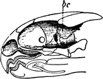
Head of a cuttlefish, showing the brain cavity.

Head of cuttlefish

Head of a cuttlefish, showing the brain cavity.

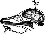
Skull of a Greek tortoise, showing the brain cavity.

Skull of Greek tortoise

Skull of a Greek tortoise, showing the brain cavity.

Skull of <em>corvus corone</em>, or carrion crow, showing the brain cavity.

Crow skull

Skull of corvus corone, or carrion crow, showing the brain cavity.

Skull of a deer, showing the brain cavity.

Deer skull

Skull of a deer, showing the brain cavity.

Skull of a baboon, showing the brain cavity.

Baboon skull

Skull of a baboon, showing the brain cavity.

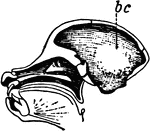
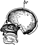
Showing the brain cavity of a human.

Human skull

Showing the brain cavity of a human.

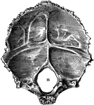
Occipital bone of the human skull, inner surface. It is situated at the back and base of the skull. Labels: 9,9, and 10,10, depression for reception of lobes of brain; 11, foramen magnum.

Occipital Bone of the Human Skull

Occipital bone of the human skull, inner surface. It is situated at the back and base of the skull.…
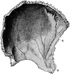
Parietal bone of the human skull, inner surface. The parietal bones form the greater part of the sides and roof of the skull. Labels: A, parietal depression; E, furrow for ramification of arteries.

Parietal Bone of the Human Skull

Parietal bone of the human skull, inner surface. The parietal bones form the greater part of the sides…

Frontal bone of the human skull, outer surface. The frontal bone forms the forehead, roof of the orbital cavities, and the nasal cavity. Labels: 1, frontal eminence; 7, roof of orbital cavitiy; 10, orbital arch.

Frontal Bone of the Human Skull

Frontal bone of the human skull, outer surface. The frontal bone forms the forehead, roof of the orbital…
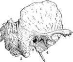
Temporal bone of the human skull. The temporal bones are situated at the sides and base of the skull. Labels: 1, squamous portion; 2, placed below external opening of auditory canal in petrous portion; 3, placed below mastoid portion; 4, placed below glenoid cavity for reception of condyle of lower jaw.

Temporal Bone of the Human Skull

Temporal bone of the human skull. The temporal bones are situated at the sides and base of the skull.…

Sphenoid bone, situated the anterior part of the base of the skull, articulating with all the other cranial bones, which it binds firmly and solidly together. Labels: a, greater wing; b, lesser wing.

Sphenoid Bone of the Human Skull

Sphenoid bone, situated the anterior part of the base of the skull, articulating with all the other…
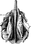
Ethmoid bone, posterior surface. The ethmoid bone is an exceedingly light, spongy bone, placed between the two orbits and at the root of the nose, contributing to form a part of each of these cavities. Label: 2, cribriform, or perforated plate.

Ethmoid Bone of the Human Skull

Ethmoid bone, posterior surface. The ethmoid bone is an exceedingly light, spongy bone, placed between…

Base of brain, spinal cord, and spinal nerves. Labels: V, 5th nerve; VI, 6th nerve; VII, a, facial nerve, b, auditory nerve; VIII, pneumo-gastric nerve; VIII, a, glosso-pharyngeal, b, spinal accessory; IX, hypoglossal; c1-c7, cervical nerve root; d1-d12, dorsal nerve roots; L1-L5, lumbar nerve roots; S4-S5, 4th and 5th sacral nerves; Cocc, coccygeal nerves; B.P., brachial plexus; L.P., lumbar plexus; S.P. sacral plexus; S a, b, c, cervical sympathetic ganglia.

Spinal Cord, Spinal Nerves, and Base of Brain

Base of brain, spinal cord, and spinal nerves. Labels: V, 5th nerve; VI, 6th nerve; VII, a, facial nerve,…
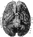
The base of the brain. Labels: 1, longitudinal fissure; 2, 2, anterior lobes of cerebrum; 3, olfactory bulb; 7, optic commissure; 9, 3rd nerve, ; 11, 4th nerve; 13, 5th nerve; 14, crura cerebri, 15, 6th nerve; 16, pons Varolii; 17, 7th nerve; 19, 8th nerve; 20, medulla oblongata; 21, 9th nerve, 23, 10th nerve; 25, 11th nerve; 27, 12th nerve; 28, 29, 30, 31, 32, cerebellum.

Base of Brain

The base of the brain. Labels: 1, longitudinal fissure; 2, 2, anterior lobes of cerebrum; 3, olfactory…

Diagram of nervous system. Labels: a, a, cortex of cerebral hemispheres; b, b, cell body and dendrites of upper motor neuron, situated in cerebral cortex; b', axon of upper motor neuron, branching at its termination near the dendrites of lower motor neuron, situated in the ventral horn of gray matter in the spinal cord; B', axon of lower motor neuron passing to its termination in a voluntary muscle fiber B"; C, cell body and dendrites of supper sensory neuron, situated in the medulla oblongata; C'C', axons of upper sensory neurons, terminating in cortex; c, cell body of lower sensory neuron situated in the dorsal root ganglion; c''', dendrite of lower motor neuron, conducting impulses from the periphery to the central nervous system; c", long axon of lower sensory neuron, conducting impulses toward the brain; c', short axon of lower sensory neuron, conducting impulses direct to ventral horn. (For the sake of simplicity the connection with the cerebellum are omitted.)

Nervous System Diagram

Diagram of nervous system. Labels: a, a, cortex of cerebral hemispheres; b, b, cell body and dendrites…

Diagrammatic longitudinal section of the body. Labels: a, the neural tube, with its upper enlargement in the skull cavity at a'; N, the spinal cord; N', the brain; ee, vertebrae forming the solid partition between the dorsal and ventral cavities; b, the pleural, and c, the abdominal division of the ventral cavity, separated from one another by the diaphragm, d; i, the nasal, and o, the mouth chamber, opening behind into the pharynx, from which one tube leads to the lungs, l, and another to the stomach, f; h, the heart; k, a kidney; s, the sympathetic nervous chain. From the stomach, f, the intestinal tube leads through the abdominal cavity to the posterior opening of the alimentary canal.

Longitudinal Section of Body

Diagrammatic longitudinal section of the body. Labels: a, the neural tube, with its upper enlargement…
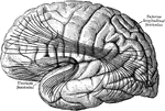
The most important association tracts of the brain. The fibers are projected upon the external surface of the hemisphere.

Cerebral Cortex

The most important association tracts of the brain. The fibers are projected upon the external surface…
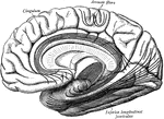
The most important association tracts of the brain. The fibers are projected upon the mesial (medial) surface of the hemisphere.

Cerebral Cortex

The most important association tracts of the brain. The fibers are projected upon the mesial (medial)…
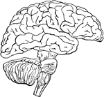
Diagram illustrating the general relationships of the parts of the brain. Labels: A, fore-brain; b, mid-brain; B, cerebellum; C, pons Varolii; D, medulla oblongata; B,c,and D together constitute the hind-brain.

Brain

Diagram illustrating the general relationships of the parts of the brain. Labels: A, fore-brain; b,…
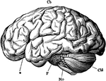
The brain from the left side. Labels: Cb, the cerebral hemispheres forming the main bulk of the fore-brain; Cbl, the cerebellum; Mo, the medulla oblongata; P, the pons Varolii; *, the fissure of Sylvius.

Brain

The brain from the left side. Labels: Cb, the cerebral hemispheres forming the main bulk of the fore-brain;…
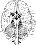
The base of the brain. The cerebral hemispheres are seen overlapping all the rest. Labels: I, olfactory lobes; II, optic tract passing to the optic commissure from which the optic nerves proceed; III, the third nerve or motor oculi; IV, the fourth nerve or patheticus; V, the fifth nerve or trigeminalis; VI, the sixth nerve or abducens; VII, the seventh or facial nerve or portio dura; VIII, the auditory nerve or portio mollis; IX, the ninth or glosso-pharyngeal; X, the tenth or pneumogastric or vagus; XI, the spinal accessory; XII, the hypoglassal; ncI, the first cervical spinal nerve.

The Base of the Brain

The base of the brain. The cerebral hemispheres are seen overlapping all the rest. Labels: I, olfactory…
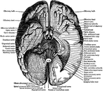
View of the under surface of the brain, with the lower portion of the temporal and occipital lobes, and the cerebellum on the left side removed, to show the origins of the cranial nerves.

Under Surface of the Brain

View of the under surface of the brain, with the lower portion of the temporal and occipital lobes,…
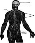
View of the nervous system of man, showing the nerve centers (brain and spinal cord) giving off nerves to supply the whole of the body.

The Nervous System

View of the nervous system of man, showing the nerve centers (brain and spinal cord) giving off nerves…
The ethmoid bone of the skull, that separates the nasal cavity from the brain.

The Ethmoid Bone

The ethmoid bone of the skull, that separates the nasal cavity from the brain.