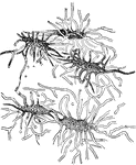
Bone points and bone needles. Crafted during the Reindeer age.

Reindeer age articles, Bone points and needles

Bone points and bone needles. Crafted during the Reindeer age.
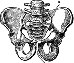
A bony structure located at the bottom of the spine. The human sacrum forms the back part of the pelvis, is roughly triangular in shape, consists of five united vertebrae, and from its solidity it is well adapted to serve as the keystone of the pelvic arch.

Pelvic Bone, Male

A bony structure located at the bottom of the spine. The human sacrum forms the back part of the pelvis,…

Labels: T, thyroid cartilage: C, cricoid cartilage; Tr, trachea; H, hyoid bone; E, epiglottis; I, joint of thyroid cartilage.

Side View of the Larynx

Labels: T, thyroid cartilage: C, cricoid cartilage; Tr, trachea; H, hyoid bone; E, epiglottis; I, joint…
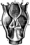
Labels: T, thyroid cartilage: C, cricoid cartilage; Tr, trachea; H, hyoid bone; E, epiglottis; I, joint of thyroid cartilage; Ar, arytenoid cartilages.

Back View of the Larynx

Labels: T, thyroid cartilage: C, cricoid cartilage; Tr, trachea; H, hyoid bone; E, epiglottis; I, joint…
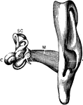
The auditory apparatus with the surrounding bone removed. Labels: M, external auditory canal; SC, semicircular canals; C, cochlea.

The Auditory Apparatus

The auditory apparatus with the surrounding bone removed. Labels: M, external auditory canal; SC, semicircular…

The chief organs of the body from the side. Labels: a, arch of the aorta or main artery of the trunk; c, large intestine; d, diaphragm; e, throat; g, lung; k, kidney; l, spinal cord within the back bone; m, main artery divided to go to h legs; n, pancreas; o, gullet; s, spleen; tr, wind pipe; t, main vein of body; ap, appendix.

View of Organs from the Side

The chief organs of the body from the side. Labels: a, arch of the aorta or main artery of the trunk;…

The thigh bone cut through the middle. Labels: b, hard bone; h and d, spongy bone; ma, marrow.

Femur

The thigh bone cut through the middle. Labels: b, hard bone; h and d, spongy bone; ma, marrow.
The human skeleton.

The Human Skeleton

The human skeleton.

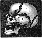
A diagram of the bones of the head. Label: 1, frontal lobe; 2, parietal bone; 3, temporal bone; 4, occipital bone; 5, nasal bone; 6, malar bone; 7, upper jaw; 8, os unguis; 9, lower jaw.

Bones of the Head

A diagram of the bones of the head. Label: 1, frontal lobe; 2, parietal bone; 3, temporal bone; 4, occipital…

The upper surface of the bones of the foot. Labels: 1, the surface of the astragulus or ankle bones, where it unites with the tibia. 2, The body of the astragulus. 3, Calcis or heel-bone. 4, The scaphoid. 5,6,7, The cuneiform. 8, The cuboid. 9,The metatarsal bones. 10,11, The phalanges of the great toe. 12, 13, 14, The phalanges of the other toes.

Bones of the Foot

The upper surface of the bones of the foot. Labels: 1, the surface of the astragulus or ankle bones,…

A diagram of the relative position of the bone, cartilage, and synovial membrane. Labels: 1,The extremities of two bones to form a joint. 2, The cartilage that covers the end of the bone. 3, The synovial membrane, which covers the cartilage of both bones and is then doubled back from one to the other, represented by the dotted lines.

Position of the Bone, Cartilage, and Synovial Membranes

A diagram of the relative position of the bone, cartilage, and synovial membrane. Labels: 1,The extremities…
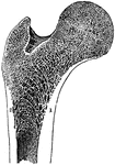
The longitudinal section of the extremity of the femur, exhibiting the arrangement of the spongy substance. Labels: 1, 2 Positions in which the compact substance appears to resolve itself into a series of arches.

Longitudinal Section of the Femur

The longitudinal section of the extremity of the femur, exhibiting the arrangement of the spongy substance.…
A skeleton of a cow shown to illustrate the internal skeleton which all vertebrates share. Labels: 1, Frontal bone of the head. 2, Upper jaw (superior maxillary). 3. Lower jaw (inferior maxillary). 4, Cervical vertebrae. 5, Dorsal vertebrae. 6, Lumbar vertebrae. 7 Sacral vertebrae. 8, Caudal vertebrae. 9; Scapula. 10, Humerus. 11, Radius and ulna. 12, Carpus. 13, Metacarpus. 14, Phalanges (toes). 15, Femur. 16, Tibia. 17, Tarsus. 18, Metatarsus. 19, Phalanges.

Skeleton of a Cow

A skeleton of a cow shown to illustrate the internal skeleton which all vertebrates share. Labels: 1,…
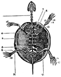
A skeleton of a tortoise The ribs of which are expanded, forming the dorsal part of its shell. Labels: 1, cervical; 2, dorsal; 3, lumbar vertebra; 4, scapula; 5, clavicle; 6, coracoid bone; 13, humerus; 14, ulna; 15, radius, 16, carpus. 17, phalanges (fingers); 7, femur; 8, tibia; 9, fibula; 10, tarsus. 11, metatarsus, 12, phalanges (toes).

Skeleton of a Tortoise

A skeleton of a tortoise The ribs of which are expanded, forming the dorsal part of its shell. Labels:…

The skeleton of a haddock. In some species such as the haddock, there is a modified form of the coracoid bone, free at its lower extremities, which may, perhaps, subserve the purpose of the clavicle of the higher animals.

Skeleton of a Haddock

The skeleton of a haddock. In some species such as the haddock, there is a modified form of the coracoid…

The metacarpal and phalangeal bones of the fingers, with their tendons and ligaments. Labels: 1, metacarpal bone, 2. tendon of the superficial flexor. 3, Tendon of the deep flexor passing through a perforation (*) of the superficial flexor.

Metacarpal and Phalangeal Bones of the Fingers

The metacarpal and phalangeal bones of the fingers, with their tendons and ligaments. Labels: 1, metacarpal…

A side view of the cartilages of the larynx. Labels: *, The front side of the thyroid cartilage. 1, The os hyoides (bone at the base of the tongue). 2, The ligament that connects the hyoid bone and thyroid cartilage. 3, 4, 5, The thyroid cartilage. 6, The cricoid cartilage. 7, The trachea.

A Side View of the Cartilages of the Larynx

A side view of the cartilages of the larynx. Labels: *, The front side of the thyroid cartilage. 1,…

A section of the end of a finger and nail. Labels: 4, Section of the last bone of the finger. 4, Fat, forming the cushion at the end of the finger. 2, The nail. 1, The cuticle continued under and around the root of the nail (3).

A Section of the End of the Finger and Nail

A section of the end of a finger and nail. Labels: 4, Section of the last bone of the finger. 4, Fat,…
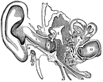
A view of all the parts of the ear, Labels: 1, The tube that leads to the internal ear. 2, The membrana tympani. 3, 4, 5, The bones of the ear. 7, The central part of the labyrinth, names the vestibule. 8, 9, 10, The semicircular canals. 11, 12, The channels of the cochlea. 13, The auditory nerve. 14, The channel from the middle ear to the throat (Eustachian tube). 15, A nerve. 16, A process. 17, The seventh pair of nerves (facial). 18, A process of the temporal bone.

Parts of the Ear

A view of all the parts of the ear, Labels: 1, The tube that leads to the internal ear. 2, The membrana…
A femur sawed lengthwise.

A Femur

A femur sawed lengthwise.

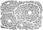
A thin slice of bone, highly magnified, showing the lacunae, the tiny tubs (canaliculi) radiating from them, and four Huversian canals, three seen crosswise and one lengthwise.

A Magnified View of a Bone

A thin slice of bone, highly magnified, showing the lacunae, the tiny tubs (canaliculi) radiating from…
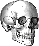
The human skull. Labels: 1, frontal lobe; 2, parietal lobe; 3, temporal lobe; 4, the sphenoid bone; 5, ethmoid bone; 6, superior maxillary (upper jaw) bone; 7, malar bone; 8, lachrymal bone; 9, nasal bone; 10, inferior maxillary (lower jaw) bone.

The Skull

The human skull. Labels: 1, frontal lobe; 2, parietal lobe; 3, temporal lobe; 4, the sphenoid bone;…
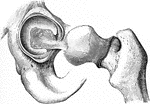
The femur articulates with the hip bone by a ball-and-socket joint. The cup is deep, thus affording less play and greater strength.

The Mechanism of the Hip Joint

The femur articulates with the hip bone by a ball-and-socket joint. The cup is deep, thus affording…
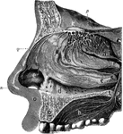
The olfactory system. Labels: a, b, c, d, interior of the nose, which is lined by a mucous membrane; n, the nose; e, the wing of the nose; q, the nose bones; o, the upper lip; g, section of the upper jawbone; h, the upper part of the mouth, or hard palate; m, frontal bone of the skull; k, the ganglion or bulb of the olfactory nerve in the skull, from which are seen the branches of the nerve passing in all direction.

Olfactory System

The olfactory system. Labels: a, b, c, d, interior of the nose, which is lined by a mucous membrane;…

Red marrow occupies the spaces in the cancellous tissue; it is highly vascular, and this maintains the nutrition of the spongy bone, the interstices of which it fills. Shown are the cells of the red marrow of the the guinea pig. Labels; a, a large cell, the nucleus of which appears to be partly divided into three by constrictions; b, a cell, the nucleus of which shows an appearance of being constricted into a number of smaller nuclei; c, so called giant cell, or myeloplaxe, with many nuclei; d, a smaller myeloplaxe, with three nuclei; e-i, proper cells of the marrow.

Cells of the Red Marrow

Red marrow occupies the spaces in the cancellous tissue; it is highly vascular, and this maintains the…
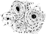
The bone contains a multitude of small irregular spaces, approximately fusiform in shape, called lacunae, with very minute canals leading from them and anastomosing with similar little prolongations from the other lacunae. Shown is a transverse section of compact bony tissue of a humerus. Three of the Haversian canals are seen, with their concentric rings; also the lacunae, with the canaliculi were filled with debris in grinding down the section, and therefore appear black in the figure, which represents the object as viewed with transmitted light. The Haversian systems are so closely packed in this section that scarcely any interstitial lamellae are visible.

Microscopic Structure of Bone

The bone contains a multitude of small irregular spaces, approximately fusiform in shape, called lacunae,…
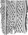
The bone contains a multitude of small irregular spaces, approximately fusiform in shape, called lacunae, with very minute canals leading from them and anastomosing with similar little prolongations from the other lacunae. Shown is a longitudinal section from the human ulna, showing Haversian canal, lacunae, and canaliculi.

Microscopic Structure of Bone

The bone contains a multitude of small irregular spaces, approximately fusiform in shape, called lacunae,…
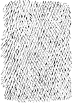
Bone corpuscles with their processes as seen in a thin section of human bone.

Bone Corpuscles

Bone corpuscles with their processes as seen in a thin section of human bone.

Shown is a thin layer peeled off from a soft bone, which is intended to represent the reticular structure of a lamella.

Reticular Structure of a Lamellae

Shown is a thin layer peeled off from a soft bone, which is intended to represent the reticular structure…
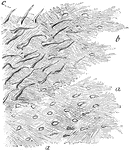
Lamellae torn off from a decalcified human parietal bone at some depth from the surface. Labels: a, lamellae, showing reticular fibers; b, darker part, where several lamellae are superposed; perforating fibers. Apertures through which perforating fibers has passed, are seen especially in the lower part of the figure.

Lamellae

Lamellae torn off from a decalcified human parietal bone at some depth from the surface. Labels: a,…
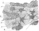
Osteoblasts from the parietal bone of a human embryo, thirteen weeks old. Labels: a, bony septa with cells of the lacunae; b, layers of osteoblasts; c, the latter in transition to bone corpuscles.

Osteoblasts

Osteoblasts from the parietal bone of a human embryo, thirteen weeks old. Labels: a, bony septa with…
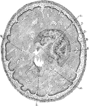
Transverse section through the tibia of a fetal kitten. Labels: P, Periosteum. O, Osteogenetic layer of periosteum showing the osteoblasts newly formed bones. B, the periosteal bone deposited in successive layers beneath the periosteum and ensheathing E, the spongy endochondral bone; represented as more deeply shaded. Within the trabeculae of endochondral spongy bone are seen the medulla, with V, veins. In the lower half of the figure the endochondral spongy bone has been completely absorbed.

Formation of Compact Bone in a Kitten

Transverse section through the tibia of a fetal kitten. Labels: P, Periosteum. O, Osteogenetic layer…
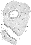
Transverse section of femur of a human embryo about eleven weeks old. Labels: a, rudimentary Haversian canal in cross sections; b, in longitudinal section; c, osteoblast; d, newly formed osseous substance of a lighter color; e, that of greater age; f, lacunae with their cells; g, a cell still united to an osteoblast.

Formation of Compact Bone in a Human

Transverse section of femur of a human embryo about eleven weeks old. Labels: a, rudimentary Haversian…

Section of a portion of the dentine and cement from the middle of the root of an incisor tooth. Labels: a, dental tubuli ramifying and terminating, some of them in the interglobular spaces b and c, which somewhat resemble bone lacunae; d, inner layer of the cement with numerous closely set canaliculi; e, outer later of cement; f, lacunae; g, canaliculi.

Dentine and Cement

Section of a portion of the dentine and cement from the middle of the root of an incisor tooth. Labels:…

The first step in the development of teeth consists in a downward growth from the Rete Malpighi or the deeper layer of stratified epithelium of the mucous membrane of the mouth, which first becomes thickened in the neighborhood of the maxillae now in the course of formation. This development is shown in a section of the upper jaw of a fetal sheep. A- 1, common enamel germ dipping down into the mucous membrane; 2, palantine process of jaw; 3, Reta Malpighi. B- Section similar to A, but passing through one of the special enamel germs here becoming flask-shaped; c, d, epithelium of mouth; f, neck; f', body of special enamel organ; p, papilla; s, dental sac forming; f p, the enamel germ of permanent tooth; m, bone of jaw; v, vessel cut across.

Development of Teeth

The first step in the development of teeth consists in a downward growth from the Rete Malpighi or the…

Outline showing the general form of the larynx, trachea, and bronchi, as seen from front. Labels: h, the great cornu of the hyoid bone; e,epiglottis; t, superior and inferior cornu of the thyroid cartilage; c, middle of the cricoid cartilage; tr, the trachea, showing sixteen cartilaginous rings; b, the right bronchus; b1, the left bronchus.

Front View of Respiratory Apparatus

Outline showing the general form of the larynx, trachea, and bronchi, as seen from front. Labels: h,…

Outline showing the general form of the larynx, trachea, and bronchi, as seen from behind. Labels: h, the great cornu of the hyoid bone; e,epiglottis; t, superior cornu of the thyroid cartilage ; t1, inferior cornu of the thyroid cartilage; a, points to the back of both the arytenoid cartilages, which are surmounted by the cornicula; c, the middle ridge on the back of the cricoid cartilage; tr, the posterior membranous part of the trachea; b, b1, right and left bronchi.

Back View of Respiratory Apparatus

Outline showing the general form of the larynx, trachea, and bronchi, as seen from behind. Labels: h,…

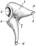
The hammer-bone or malleus, seen from the front. 1, the head; 2, neck; 3, short process; 4, long process.
Malleus

The hammer-bone or malleus, seen from the front. 1, the head; 2, neck; 3, short process; 4, long process.

The incus, or anvil-bone. Labels: 1, body; 2, ridged articulation from the malleus; 4, processus brevis, with 5, rough articular surface from ligament of incus; 6, processus magnus, with articulating surface for stapes; 7, nutrient foramen.

Incus

The incus, or anvil-bone. Labels: 1, body; 2, ridged articulation from the malleus; 4, processus brevis,…
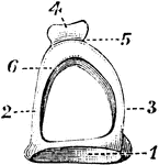
The stapes on stirrup-bone. 1, base; 2 and 3, arch; 4, head of bone, which articulates with orbicular process of the incus; 5, constricted part of the neck; 6, one of the crura.

Stapes on Stirrup-Bone

The stapes on stirrup-bone. 1, base; 2 and 3, arch; 4, head of bone, which articulates with orbicular…
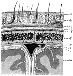
Diagram showing the layers of the scalp and membranes of the brain in section. Labels: a, skin; b, subcutaneous tissue with hair roots and vessels; c, epicranius; d, subepicranial layer; e, pericranium; fm parietal bone; g, dura mater; k, arachnoid; l, pia mater; m, cortex; n, in subdural space near a Pacchionian body projecting within the superior longitudinal sinus.

Layers of the Scalp and Membrane of the Brain

Diagram showing the layers of the scalp and membranes of the brain in section. Labels: a, skin; b, subcutaneous…
The occipital bone at birth.

Occipital Bone at Birth

The occipital bone at birth.

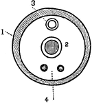
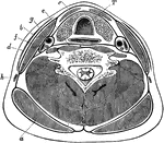
Transverse section through the lower part of the neck, to show the arrangement of the cervical fascia. Labels: a, Trapezius; b, sternomastoid; c, depressors of hyoid bone; d, platysma; e, anterior spinal muscles; f, scalenus anticus; g, carotid artery; h, external jugular vein; i, posterior spinal muscles; T, trachea, with gullet behind and thyroid body in front.

Transverse Section of the Neck

Transverse section through the lower part of the neck, to show the arrangement of the cervical fascia.…
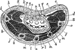
Horizontal section of the hand through the middle of the thenar and hypothenar eminences. Labels: a, metacarpal bone; b, first dorsal interosseous; c, palmaris brevis; d, abductor quinti digiti; e, flexor brevis min. dig.; f, opponens min. dig.; g, flexor brevis poll.; h, abductor brevis poll.; i, opponens poll.; j, adductor poll.; k, flexor long. poll.; l, dorsal interossei; m, palmar interossei; n, flexor sublimis; o, flexor profundus; p, superfic. volae; q, median nerve, and (on inner side) ulnar artery nerve; r, deep volar arch; 1, palmar fascia; 2, outer septum; 3, inner septum; 4, deep fascia of palm.

Horizontal Section of the Hand

Horizontal section of the hand through the middle of the thenar and hypothenar eminences. Labels: a,…
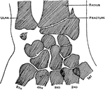
Showing the situation of Colle's fracture of the radius, with fracture of the styloid of the ulna. The usual position of a fraction of the scaphoid is also indicated.

Colle's Fraction of the Radius

Showing the situation of Colle's fracture of the radius, with fracture of the styloid of the ulna. The…
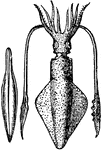
"The Common Calmar or Squid. They propel themselves backward through the water with great velocity, driving back the water by means of their funnel."

Loligo Vulgaris, with its pen, or internal bone (Lamarck)

"The Common Calmar or Squid. They propel themselves backward through the water with great velocity,…

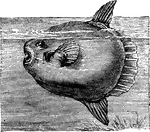
"A species with which we do not often meet with in this country."

Sunfish (Orthagoriscus Mola)

"A species with which we do not often meet with in this country."

"The Trunk-fish, or Ostracion, is without scales, but covered with regular, osseous plates, which are so jointed the one to the other that the body is, as it were, inclosed in a kind of box or long coffer."

The Trunkfish (Ostracion)

"The Trunk-fish, or Ostracion, is without scales, but covered with regular, osseous plates, which are…
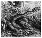
"The Viperine Snakes have a long, perforated, erectile fang on the maxillary bone, which is extremely short and bears no teeth."

Fer-de-Lance

"The Viperine Snakes have a long, perforated, erectile fang on the maxillary bone, which is extremely…
Back view of the femur.

Back View of the Femur

Back view of the femur.

The longitudinal section of a long bone.

Structure of a Long Bone

The longitudinal section of a long bone.

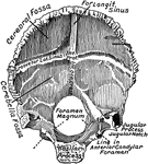
The occipital bone, viewed from above.

Occipital Bone

The occipital bone, viewed from above.

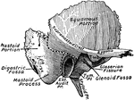
The outer surface of the temporal bone. The dotted lines indicate the lines of suture between squamous, mastoid, or tympanic portions.

Temporal Bone

The outer surface of the temporal bone. The dotted lines indicate the lines of suture between squamous,…

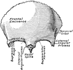
The frontal bone, seen from in front.

Frontal Bone

The frontal bone, seen from in front.

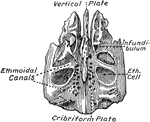
The ethmoid bone, seen from above.

Ethmoid Bone

The ethmoid bone, seen from above.

The sphenoid bone, viewed from above.

Sphenoid Bone

The sphenoid bone, viewed from above.

Nasal bone viewed from before.

Nasal Bone

Nasal bone viewed from before.

The external surface of right inferior turbinate bone.

Turbinate Bone

The external surface of right inferior turbinate bone.

The inner surface of the right lachrymal bone.

Lachrymal Bone

The inner surface of the right lachrymal bone.

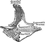
Right malar bone.

Malar Bone

Right malar bone.