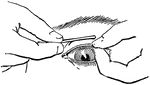
Everted eyelid
"Showing how the upper eyelid may be everted with a pencil or penholder." — Blaisedell, 1904
Muscles of the Eyes
"The eye is moved about by six muscles. The back ends of these muscles are attached to the eye sockets.…
White fibrous tissue
"The connective tissue with white fibers sometimes forms a very thin sheet, as in the delicate covering…
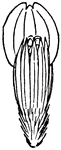
Longitudinal seciont of a fingernail
"A, last bone of finger; B, true skin on the dorsal surface of finger; C,…
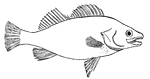
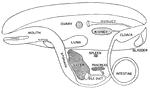
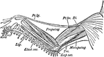
Fish
Outline of a fish, showing the "paired" and "median" fins. (p) one of the pectoral fins; (v) one of…
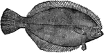
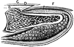
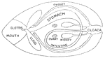
Flounder
Flounder keep near the bottom, swimming on one side, and the two eyes are both on the side that is uppermost.
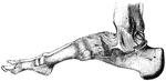
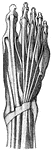
Ligaments of the Foot and Ankle
"The bones are fastened together, kept in place, and their movements limited, by tough and strong bands,…
Bones of the Foot
"The foot is built in the form of a half-dome or half-arch. This is to afford a broad, strong support…
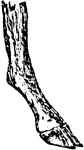
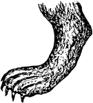
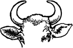
Compressed Foot
An extreme form of a compressed foot, typically seen in the deer and ox. It is useful for land transit.
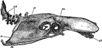
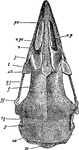
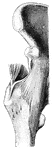
Fowl Skull
The skull of an adult fowl. Here the temporal fossa is bridged over by the junction of the post-frontal…
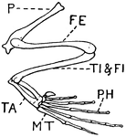
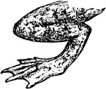
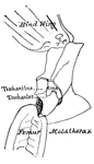
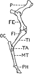
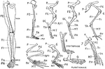
Leg of Frog
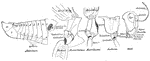
This illustration shows the leg of a frog. P. Pelvis, FE. Femur, TI. Tibia, FI. Fibula, TA. Tarsus,…
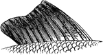
Gar
The gar fish has a cylindrical body covered by rhomboidal bony scales that are coated with enamel, making…
Gland
"Diagram to show the working parts of a gland. v and a are blood tubes with thin-walled branches around…
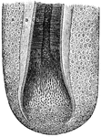
Sweat gland
"The convoluted gland is seen surrounded by fat cells and may be traced through the true skin to its…
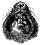
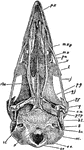
Plains Pocket Gopher
"Under Side of Head of Geomys bursarius, showing entrance of external cheek-pouches and sulcate superior…
Hair with ringworm
"A Piece of Hair from the Scalp infested with a Mold which produces Ringworm. Ringworm may occur anywhere…
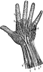
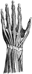
Hand
1. Nerves of the skin 2. Tendons 3. Arteries of the palm of the hand 4. Elbow nerve 5. Elbow artery…
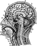
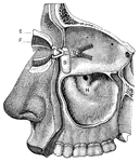
Sectional view of the Head
Section through the Head and Neck on the Median Line. 1. Medulla Oblongata 2. Pons 3. Right lobe of…
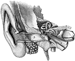
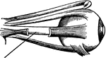
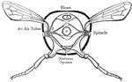
General view of organ of hearing
"A, pinna; B, cavity of the concha, showing the openings of a great number of sebaceous…
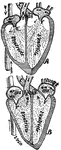
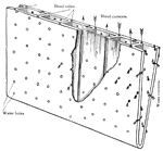
Beating heart
"Diagram of the rush of blood when the heart beats. The valves v open above are closed below while the…
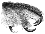
Powerful Ligament at the Hip Joint
"The bones are fastened together, kept in place, and their movements limited, by tough and strong bands,…
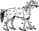
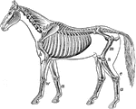
Horse
The anatomy of a horse. 1, ears; 2, forelock; 3, forehead; 4, eyes; 5, eye-pits; 6, nose; 7, nostril;…
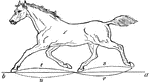
Trotting Horse
"Horse in the act of trotting. In this, as in all the other paces, the body of the horse is levered…
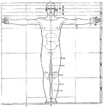
Proportions of human figure
"The proportions of the human figure. As handed down to us by Vitruvius and described by Joseph Bonomi."…
Cross-section of a human hair
"Cross-section of One Half of a Human Hair. A hair is made up of horny cells of the outer layer of the…
Human Leg (Front View), and Comparative Diagrams showing Modifications of the Leg
This illustration shows a human leg (front view), and comparative diagrams showing modifications of…