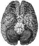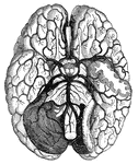
<em>A</em>, anterior lobe of the cerebellum; <em>B</em>, olfactory nerve; <em>C</em>, portion of the posterior lobe; <em>D</em>, optic chiasm; <em>E</em>, optic tract; <em>H, M,</em> hemispheres of the cerebellum; <em>K</em>, portion of the occipital lobe; <em>N</em>, medulla oblongata; <em>R</em>, pons Varolii."

Base of the brain

A, anterior lobe of the cerebellum; B, olfactory nerve; C, portion of the…

"Arteries and their Branches at the Base of the Brain." &mdash; Blaisedell, 1904

Blood vessels of the brain

"Arteries and their Branches at the Base of the Brain." — Blaisedell, 1904
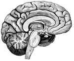
"<em>A</em>,frontal love of the cerebrum; <em>B</em>, parietal lobe; <em>C</em>, parieto-occipital lobe; <em>D</em>, occcipital lobe; <em>E</em>, cerebellum; <em>F</em>; arbor vitae; <em>H</em>, pons Varolii; <em>K</em>, medulla oblongata." — Blaisedell, 1904

Left Half of the Brain

"A,frontal love of the cerebrum; B, parietal lobe; C, parieto-occipital lobe;…
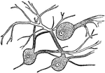
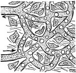
"Wherever nerve cells are abundant, the nerve tissue has a gray color; in other places, it looks white. Most of the gray matter of the brain is on the surface." — Blaisedell, 1904

Nerve Cells of the Brain

"Wherever nerve cells are abundant, the nerve tissue has a gray color; in other places, it looks white.…
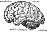
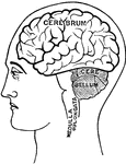
"The brain seen from the side, showing the three principal divisions." &mdash; Ritchie, 1918

Side view of the brain

"The brain seen from the side, showing the three principal divisions." — Ritchie, 1918
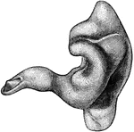
"A Cast of the External Auditory Canal. The auditory canal is a passage in the solid potion of the temporal bone. It is lined by skin on which there are fine hairs, and a set of glands secreting earwax, which serves to moisten the parts, catch particles fo dust, and keep away small insects." &mdash; Blaisedell, 1904

External auditory canal

"A Cast of the External Auditory Canal. The auditory canal is a passage in the solid potion of the temporal…

"Blood plasma passing out of capillary to feed the body cells." &mdash;Davison, 1910

Capillaries

"Blood plasma passing out of capillary to feed the body cells." —Davison, 1910

"Showing (1) cartilage with martrix and cells; (2) cartilage with matrix containing cells and white fibers." &mdash; Blaisedell, 1904

Longitudinal section of cartilage

"Showing (1) cartilage with martrix and cells; (2) cartilage with matrix containing cells and white…
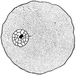
"In a general what we may describe a cell as a tiny mass of jelly in which floats another still smaller mass of slightly different composition, called the <em>nucleus</em> of the larger one."

Cell

"In a general what we may describe a cell as a tiny mass of jelly in which floats another still smaller…

"A, columnar cells found lining various parts of the intestines (called <em>columnar epthelium</em>); B, cells of a fusiform or spindle shape found in the loose tissue under the skin and in other parts (called <em>connective-tissue cells</em>); C, cell having many processes or projections - such are found in some kinds of connective tissue; D, primitive cells composed of protoplasm with nucleus, and having no cell wall." &mdash; Blaisedell, 1904

Various forms of cells

"A, columnar cells found lining various parts of the intestines (called columnar epthelium);…

"A, a, Cephalic plate; b, Tergum of segment bearing first pair of legs (d); c, Tip of palpognath; e, Antenna; f, toxicognath; g, Last pair of appendages, enlarged and directed backwards." (Britannica, 1910)

Centipede

"A, a, Cephalic plate; b, Tergum of segment bearing first pair of legs (d); c, Tip of palpognath; e,…

"Brain and spinal cord, with the thirty-one pairs of spinal nerves." — Tracy, 1888

Central Nervous System

"Brain and spinal cord, with the thirty-one pairs of spinal nerves." — Tracy, 1888
"Diagram showing the position of the nervous centers in the head." &mdash; Tracy, 1888

Cerebellum

"Diagram showing the position of the nervous centers in the head." — Tracy, 1888

"The Upper Surface of the Cerebrum. Showing its division into two hemispheres, and also of the convolutions." &mdash; Blaisedell, 1904

Cerebrum

"The Upper Surface of the Cerebrum. Showing its division into two hemispheres, and also of the convolutions."…
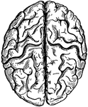
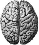
"The cerebrum seen from above, showing the hemispheres." — Ritchie, 1918

Top View of the Cerebrum

"The cerebrum seen from above, showing the hemispheres." — Ritchie, 1918

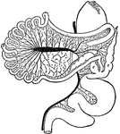
cc. Colic caeca, d. Duodenum. g. Glandular patch, l.l. Meckel's tract, l.i. Hind-gut, p.v. Cut root of portal vein, r.v. Rectal vein, s. Proventriculus, y. Meckel's diverticulum.

Intestinal Tract of Chauna Chavaria

cc. Colic caeca, d. Duodenum. g. Glandular patch, l.l. Meckel's tract, l.i. Hind-gut, p.v. Cut root…

"<em>A</em>, a muscle which aids in pushing the food down the esophagus; <em>B</em>, esophagus; <em>C</em>, section of the right bronchus; <em>D</em>, two right pulmonary veins; <em>E</em>, great azygos vein crossing the esophagus to empty into the superior vena cava, <em>F</em>, thoracic duct; <em>H</em>, thoracic aorta; <em>K</em>, lower portion of the esophagus passing through the diaphragm; <em>L</em>, diaphragm as it appears in sectional view, enveloping the heart; <em>M</em>, inferior vena cava, passing through the diaphragm and emptying into the right auricle; <em>N</em>, right auricle; <em>O</em>, section of the right branch of the pulminary artery; <em>P</em>, aorta; <em>R</em>, superior vena cava; <em>S</em>, trachea." &mdash; Blaisedell, 1904

Lateral section of the chest

"A, a muscle which aids in pushing the food down the esophagus; B, esophagus; C,…
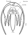
The sternum of a chick three days old.

Chick Sternum

The sternum of a chick three days old.

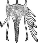
A ripe chick's head, 1.25 inches long.

Chick's Head

A ripe chick's head, 1.25 inches long.

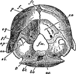
The end view of the skull of a chicken three weeks old.

Chicken Skull

The end view of the skull of a chicken three weeks old.

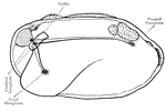
Internal structures of the body of a clam.

Clam

Internal structures of the body of a clam.

Internal structures of the body of a clam, side view.

Clam

Internal structures of the body of a clam, side view.

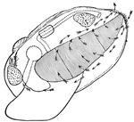
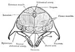
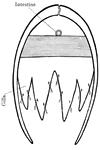
Cross section of the body of a clam, through the heart. Arrows indicate water current through the gills.
Clam

Cross section of the body of a clam, through the heart. Arrows indicate water current through the gills.

Cross section of the body of a clam, through the posterior adductor muscles. Arrows indicate water current through the gills.

Clam

Cross section of the body of a clam, through the posterior adductor muscles. Arrows indicate water current…
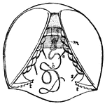
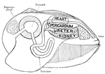
Digestive and excretory organs of a clam.

Clam

Digestive and excretory organs of a clam.

The nervous system of a clam.

Clam

The nervous system of a clam.

Young clam, still within the egg membrane. m, adductor muscle; t, hooks by which it attaches itself to the gills or fins of fishes; b, byssus; s, sense organs.

Clam

Young clam, still within the egg membrane. m, adductor muscle; t, hooks by which it attaches itself…
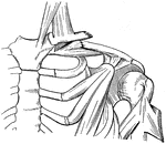
"When a bone is broken, blood trickles out between the incjured parts, and afterwards gives place to a sticky, watery fluid, which gradually becomes thicker, like a syrup or jelly. This is slowly replaced by a new bone structure and forms a kind of cement to gold together the broken ends." — Blaisedell, 1904

Broken Clavicle

"When a bone is broken, blood trickles out between the incjured parts, and afterwards gives place to…
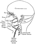
A typical example of a spiny-rayed fish is the codfish.

Codfish

A typical example of a spiny-rayed fish is the codfish.

"The cranial nerves are thus arranged in pairs: 1, olfactory nerves, special nerves of smell; 2, optic nerves, passing to each eyeball, devoted to sight; 3, 4, and 6 control the muscles fo the eyes; 5, trifacial in three branches, which proceed mainly to the face, partly sensory and partly motor; 7, facial nerves, controlling the facial muscles; 8, auditory, or nerves of hearing; 9, glossopharyngeal nerves, partly sensory and partly motor: each nerve contains two roots, one a nerve of taste, the other a motor nerve, which controls the muscles engaged in swallowing; 10, pneumogastric nerves; 11, spinal accessory nerves, supplying some of the muscles of the neck and back; 12, hypoglossal nerves, controlling the movements of the tongue in speech and swallowing." — Blaisedell, 1904

Distribution of the Cranial Nerves

"The cranial nerves are thus arranged in pairs: 1, olfactory nerves, special nerves of smell; 2, optic…

Cross-section of abdomen of crayfish.

Crayfish

Cross-section of abdomen of crayfish.

Longitudinal-section of a crayfish, showing digestive, circulatory, reproductive, excretory, and nervous systems.

Crayfish

Longitudinal-section of a crayfish, showing digestive, circulatory, reproductive, excretory, and nervous…
Nervous system of crayfish.

Crayfish

Nervous system of crayfish.

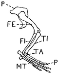
This illustration shows the leg of a crocodile.
P. Pelvis, FE. Femur, TI. Tibia, FI. Fibula, TA. Tarsus, MT. Metatarsus, PH. Phalanges, OC. Os Calcis.

Leg of Crocodile

This illustration shows the leg of a crocodile. P. Pelvis, FE. Femur, TI. Tibia, FI. Fibula, TA. Tarsus,…

"Cast or mold of the interior of the left ventricle of the heart of a deer. Shows that the left ventricular cavity in conical and spiral in its nature."&mdash;Pettigrew, 1874

Heart of a Deer

"Cast or mold of the interior of the left ventricle of the heart of a deer. Shows that the left ventricular…
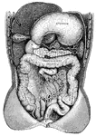
"Showing the Relations of the Stomach, Liver, Intestines, Spleen, and other Organs of the Abdomen. <em>A</em>duodenum</em>; <em>B</em>, upper end of the small intestine; <em>C</em>, lower end of the small intestine; <em>D</em>, caecum; <em>E</em>, bladder. The liver and stomach are drawn up and portions of the lower intesting have been cut away." &mdash; Blaisedell, 1904

Digestive system

"Showing the Relations of the Stomach, Liver, Intestines, Spleen, and other Organs of the Abdomen. Aduodenum;…
A restored skeleton of <em>Uintathcrium (Dinoceras) mirabile.</em>

Dinoceras Skeleton

A restored skeleton of Uintathcrium (Dinoceras) mirabile.

This illustration shows the leg of a dog. This leg is digitigrade. Animals with digitigrade legs walk on their toes (digits).
P. Pelvis, FE. Femur, TI. Tibia, FI. Fibula, TA. Tarsus, MT. Metatarsus, PH. Phalanges, OC. Os Calcis

Leg of Dog

This illustration shows the leg of a dog. This leg is digitigrade. Animals with digitigrade legs walk…
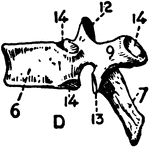
Side view of the dorsal vertebra.

Dorsal vertebra

Side view of the dorsal vertebra.

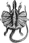
"The Red-Throated Dragon shows a large membranous expansion (b b) situated between the anterior (d d) and posterior extremities, and supported by the ribs. The dragon by this arrangement can take extensive leaps with perfect safety."&mdash;pettigrew, 1874

Red-Throated Dragon

"The Red-Throated Dragon shows a large membranous expansion (b b) situated between the anterior (d d)…
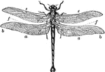
An insect characterized by large multifaceted eyes, two pairs of strong transparent wings, and an elongated body.

Dragonfly

An insect characterized by large multifaceted eyes, two pairs of strong transparent wings, and an elongated…
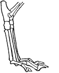
The skeleton of a dugong, one of four living species in the order Sirenia.

Skeleton of the Dugong

The skeleton of a dugong, one of four living species in the order Sirenia.

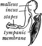
"Across the middle ear a chain of three small bones stretches from the tympanic membrane to the inner ear. These bones are called from their shape the <em>malleus</em> (hammer), the <em>incus</em> (anvil), and the <em>stapes</em> (stirrup)." — Ritchie, 1918

Bones of the Ear

"Across the middle ear a chain of three small bones stretches from the tympanic membrane to the inner…
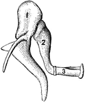
"1, malleus, or hammer; 2, incus, or anvil; 3, stapes, or stirrup." — Blaisedell, 1904

Bones of the Ear

"1, malleus, or hammer; 2, incus, or anvil; 3, stapes, or stirrup." — Blaisedell, 1904

"Many of both sexes wear enormous ear plugs and earrings, some of which are as big around as a napkin. The holes in the lobes of their ears so large that they can carry a cigar in them." &mdash;Carpenter, 1902

Dyak's ear

"Many of both sexes wear enormous ear plugs and earrings, some of which are as big around as a napkin.…

"<em>A</em>, oval window; <em>B, C, D,</em> semicircular canals; * represents the bulging part of each canal; <em>E, F, G,</em> cochlea; <em>H</em>, round window." &mdash; Blaisedell, 1904

Internal ear

"A, oval window; B, C, D, semicircular canals; * represents the bulging part of each…

Side view of internal structures of earthworm.

Earthworm

Side view of internal structures of earthworm.

Dorsal view of internal structures of earthworm after cutting through the dorsal wall lengthwise and turning it outward.

Earthworm

Dorsal view of internal structures of earthworm after cutting through the dorsal wall lengthwise and…
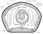
Cross section of earthworm.

Earthworm

Cross section of earthworm.

Cross-section of an egg, showing the different parts.

Cross-section of an egg

Cross-section of an egg, showing the different parts.

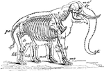
"Skeleton and Outline of African Elephant (Elephas or Loxodon africanus). fr, frontal; ma, mandible; ma', malar; fi, "finger" at end of trunk; C, cervical vertebrae; D, dorsal vertebrae; D, dorsal vertebrae; pe, pelvis; sc, scapula; st, sternum; hu, humerus; ul, ulna; ra, radius; mc, metacarpus; fe, femur; pat, patella; tib, tibia; fib, fibula; met, metatarsus." -Whitney, 1911

African Elephant Skeleton

"Skeleton and Outline of African Elephant (Elephas or Loxodon africanus). fr, frontal; ma, mandible;…

"Shows how the bones of the arm (q), forearm (q'x), and foot (o), are twisted to form an osseous screw."&mdash;Pettigrew, 1875

Anterior Extremity of Elephant

"Shows how the bones of the arm (q), forearm (q'x), and foot (o), are twisted to form an osseous screw."—Pettigrew,…
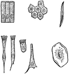
"A, columnar cells of intestine; B, polyhedral cells of the conjuctiva; C, ciliated conical cells of the trachea; D, ciliated cell of frog's mothl E, inverted conical cell of the trachea; F, squamous cell of the cavity of the mouth, seen on its broad surface; G, squamous cell, seen edgeways." &mdash; Blaisedell, 1904

Various kinds of epithelial cells

"A, columnar cells of intestine; B, polyhedral cells of the conjuctiva; C, ciliated conical cells of…

"One of the simplest of the tissues in the body is called the <em>epithelium</em>, and its cells are called epithelial cells. It serves as a lining of various cavities, a covering for the surface of the body, and the essential part of various organs called glands." — Blaisedell, 1904

Cross-Section of the Epithelium

"One of the simplest of the tissues in the body is called the epithelium, and its cells are…

"Diagram illustrating the Manner in which the Image of an Object is inverted on the Retina." — Blaisedell, 1904

Diagram of the Eye

"Diagram illustrating the Manner in which the Image of an Object is inverted on the Retina." — Blaisedell,…

"Section of the human eye." — Blaisedell, 1904

Human eye

"Section of the human eye." — Blaisedell, 1904

"Diagram showing the Change in the Lens during Accomadation. On the right the lens is arranged for distant vision, the cilliary muscle is relaxed, and the ligament <em>D</em> is tense, so flattening by its compression the front of the lens <em>C</em>, on the left the muscle <em>A</em> is acting, and this relaxes the ligament and allows the lens <em>B</em> to become more convex, and so fitted for the vision of near objects." — Blaisedell, 1904

Lens of the eye

"Diagram showing the Change in the Lens during Accomadation. On the right the lens is arranged for distant…
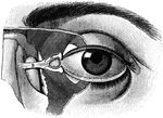
"The Relative Position of the Lachrymal Apparatus, the Eyeball, and the Eyelids. <I>A</em>, lachrymal canals, with the minute orifices represented as two black dots to the right; <em>B</em>, tendon attached to a muscle which surrounds the circumference of the orbit and eyelids; under <em>B</em> is seen the lachrymal sac. The minute openings of the Meibomian glands are seen on the free margins of the eyelids. Below <em>A</em> is seen a small conical elevation, with black dots (the lachrymal papilla, or caruncle)." &mdash; Blaisedell, 1904

Eyeball

"The Relative Position of the Lachrymal Apparatus, the Eyeball, and the Eyelids. A, lachrymal canals,…
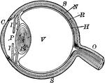
"The most essential parts of human vision are contained in the eyeball, a nearly spherical body, about an inch in diameter, and capable of being turned considerably in its socket by the action of various muscles." — Avery, 1895

Eyeball

"The most essential parts of human vision are contained in the eyeball, a nearly spherical body, about…
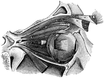
"<em>A</em>, attachment of tendon connected with the four recti muscles; <em>B</em>, external rectus, divided and turned downward, to expose the internal rectus; <em>C</em>, inferior rectus; <em>D</em>, internal rectus; <em>E</em>, superior rectus; <em>F</em>, superior oblique; <em>H</em>, pulley and reflected portion of the superior oblique; <em>K</em>, inferior oblique; <em>L</em> and <em>M</em>, portions of the muscle which raises the upper eyelid; to the right of <em>D</em> and to the left on the same line are seen cut ends of the optic nerve." &mdash; Blaisedell, 1904

Muscles of the eyeball

"A, attachment of tendon connected with the four recti muscles; B, external rectus,…