Clipart tagged: ‘thigh’
Buttock and Thigh
Buttock and back of thigh. Labels: 1, gluteus maximus; 2, gluteus medius; 3, gluteal artery; 4, gluteus…
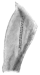
Leg Erased
"Argent, a leg erased at the midst of the thigh gules. ERASED. Signifies any thing torn or plucked off…
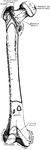
Front of Leg
Front of leg. Labels: 1, external popliteal nerve; 2, anterior tibial artery; 3, musculocutaneous nerve;…
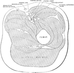
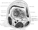
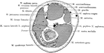
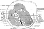
Transverse Section of the Leg
Transverse section of the thigh below the lesser trochanter. The femoral artery, vein, and nerve are…
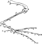
Bones of a Bird's Hind Limb
"Fig 34 - Bones of a bird's hind limb: from a duck, Clangula islandica. A, hip: B, knee: C, heel or…
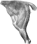
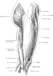
Thigh and Haunch Muscles of a Horse
The muscles of the thigh and haunch- left side; the external fascia being removed. Labels: a, tensor…
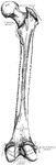
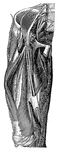
Horse Leg Muscles
Anterior tibial group of muscles of the right limb, seen from before and the outside. Labels: a, flexor…
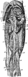
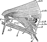
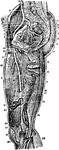
Great Nerve
"A Great Nerve (Crural) and its branches on the Front of the Thigh. The femoral artery with its cut…
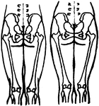
Position of the Pelvic and Thigh Bones in the Male and Female
Diagram showing the position of the pelvic and thigh bones in back view in the male and female.
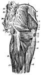
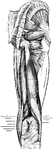
Rear Thigh Muscles
The muscles of the rear thigh. 1, fifth lumbar vertebra; 2, ilio-lumbar ligament; 3, crest of ilium;…
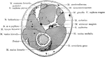
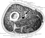
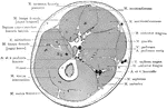
Cross Section Through Thigh Five Inches Above Knee Joint
Section through the lower third of the thigh, five inches above knee joint.
Cross Section Through Thigh Four Inches Above Knee Joint
Section through right thigh, four inches above knee joint.
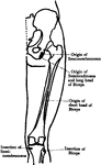
Thigh Muscles
"Some of the Larger Muscles on the back of the Thigh. Powerful tendons at the hip and on the back of…
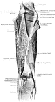
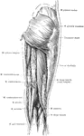
Anterior View of the Superficial Muscles of the Thigh
Superficial muscles of the right thigh, anterior view.
Arteries of the Thigh
Arteries of the thigh. Labels: 1, aorta; 2, common iliac; 3, external iliac; 4, epigastric; 5, circumflex…


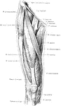
Lateral View of the Superficial Muscles of the Thigh
Superficial muscles of the right thigh, lateral view.
Nerves of the Thigh
Nerves of the thigh. Labels: 1, gangliated cord of sympathetic; 2, third lumbar nerve; 3, branches to…
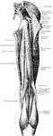
Posterior View of the Superficial Muscles of the Thigh
Superficial muscles of the right thigh, posterior view.