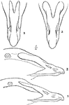
bones of arm
Wing of Bird; h Humerus or bone of upper arm; r and u Radius and Ulna, or bones of the forearm; c Carpus,…
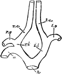
Carotid Arteries of Birds
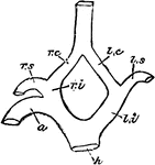
"h, root of aorta; 1, arch of aorta, to the right side; li, left innominate; ri, innominate; ls, left…
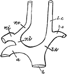
Carotid Arteries of Birds
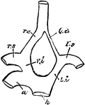
"h, root of aorta; 1, arch of aorta, to the right side; li, left innominate; ri, innominate; ls, left…
Carotid Arteries of Birds
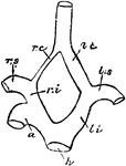
"h, root of aorta; 1, arch of aorta, to the right side; li, left innominate; ri, innominate; ls, left…
Carotid Arteries of Birds
"h, root of aorta; 1, arch of aorta, to the right side; li, left innominate; ri, innominate; ls, left…
Carotid Arteries of Birds
"h, root of aorta; 1, arch of aorta, to the right side; li, left innominate; ri, innominate; ls, left…
Carotid Arteries of Birds
"h, root of aorta; 1, arch of aorta, to the right side; li, left innominate; ri, innominate; ls, left…
Least Auk Adult
"Simorhynchus pusillus. Least Auk. Knob-nosed Auk. Bill small and simple. but stout for its length,…
Least Auk Adult
"Simorhynchus pusillus. Least Auk. Knob-nosed Auk. Bill small and simple. but stout for its length,…
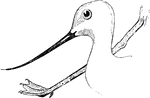
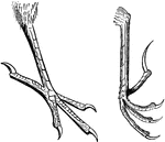
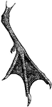
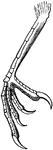
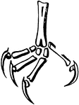
Avocets Head and Foot
"Another small family, characterized by the extreme length of the slender legs, and the extreme slenderness…
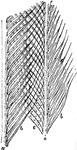
Barbs
"The arrangement shown in fig. 22, where a, a, a, a, are four barbs in transverse section, viewed from…
Single Barbule
"Fig. 21. -A single barbule, baring barbicels and hooklets; magnified; after Nitzsch. ...barbicels (another…
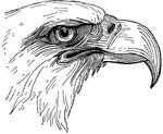
The Beaks and Claws of a Burrowing Owl
"Speotyto. Burrowing Owls. tarsi long, about twice as long as the middle toe without its claw, very…
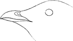
Barn Swallow Bill
Aerial (top) view of Barn Swallow's bill "Hirundo horreorum. Barn Swallow. Bill of moderate size for…
Oriole Bill
"Icterus. Orioles. Bill averaging as long as head (more or less); very acute, sometimes decurved." Elliot…
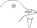
Pelican Bill
"North American White Pelican. Bill and feet ordinarily yellow; much reddened in the breeding season,…
The Parts of a Bird Bill
"Fig. 26 - Parts of a Bill. a, side of upper mandible; b, culmen; c, nasal fossa; d, nostril; e(see…
Typical Passerine Bird Feet
"Fig. Typical passerine feet. The right hand fig. is plectrophanes lapponicus." Elliot Coues, 1884
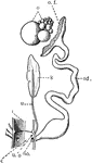
Female Bird Genital Organs
"Diagram of the female genital organs of a Bird. c, cloaca; i, intestine; k, kidney; o, ovary with ova…
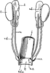
Male Bird Genital Organs
"Diagram of the urino-genital organs of a male Bird. ad., adrenal body; c, cloaca; i, intestine; k,…
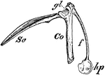
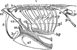
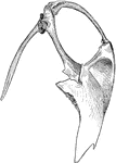
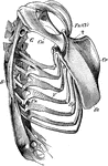
Bird Scapula
"Right shoulder-girdle or scapular arch of fowl, showing hp, the hypoclidium; f, furculum; Co, coracoid;…
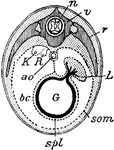
Bird Section
"Diagrammatic section of young bird. n., Spinal cord; v., vertebra; r., rib; L., liver; G., gut; som.…
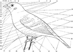
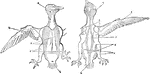
Topography of a Bird
"fig. 25 - Topography of a Bird. 1, forehead (frons). 2, lore. 3, circumocular region. 4, crown (vertex).…
Bluebird Foot
"Scutellate. Foot of bluebird, with laminiplantar and mostly booted tarsus and of the toes." -Whitney,…
Bobolink Epipleurae
"Epipleurae.-- Thorax, scapular arch, and part of pelvic arch of a bobolink (Dolichonyx oryzivorus).…
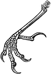
The Foot of a Bridled Foot
"Sterna anaisthetikos. Bridled Tern. The foot of a Bridled Tern; Tarsus .85; middle toe the same, with…
Phalanges of Caprimulgine
"Fig. 41 shows phalanges of caprimulgines foot, where the ratio is 2, 3, 4, 4." Elliot Coues, 1884
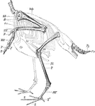
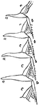
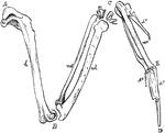
Skeleton of the Limbs and Tail of a Carinate Bird
"Skeleton of the Limbs and Tail of a Carinate Bird. (The skeleton of the body is indicated by dotted…
Scutellate Laminiplanter Tarsus of a Cat-bird
"Figure shows Scutellate laminiplanter tarsus of a cat-bird. A tarsus so disposed as to its podotheca…
Chick Head
"Fig 66 - Head of a chick, second stage, after five days of incubation, section in profile; x6 diameters.…
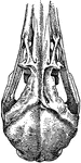
The Skull of a Chick Stage Three
"Skull of chick, third stage, viewed from below, x6 & 2/3 diameters. pn, prenasal cartilage, running…
The Skull of a Chick Stage Two
"Skull of chick, second stage, in profile, brain and membranes removed to show cartilaginous formations,…
Ripe Chick's Skull Profile
"Ripe chick's skull, longitudinal section, vied inside, x 3 diameters; after Parker. px, premaxillary;…
Ripe Chick's Skull
"Ripe chick's skull, longitudinal section, vied inside, x 3 diameters; after parker. In the mandible…
Barn Swallow Claw
Side view of the Barn Swallow's claw. "Hirundo horreorum. Barn Swallow. Tarsi shorter than middle toe…
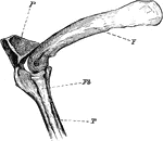
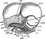
The Knee-joint of a Cormorant
"Phalacrocorax bicristatus. Cormorant. The knee-joint of a Cormorants. F, femur; P, patella; T, tibia;…
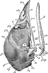
Cormorant Skull
"Phalacrocorax bicristatus. Red-Faced Cormorant. Skull showing sto, occipital style or nuchal bone;…
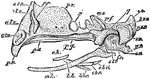
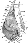
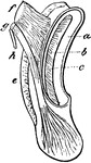
Cormorant Sternum and Shoulder
"Phalacrocorax bicristatus. Red-Faced Cormorant. Sternum and the shoulder from the skeleton of a Cormorant."…
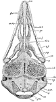
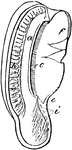
Sandhill Crane Windpipe
"Coiling of the windpipe in the sternum of Grus canadensis. Sandhill Crane." Elliot Coues, 1884
Whooping Crane Windpipe
"Very generally, in cranes and swans, the trachea enters the keel of the sternum, which is excavated…
Curlew Skull
"Schizorhinal skull of curlew (top view), showing the long cleft, a, between upper and lower forks of…
Phalanges of Cypseline Foot
"Fig. 40 Phalanges of Cypseline foot, where the ratio is 2, 3, 3, 3 of Caprimulginae." Elliot Coues,…
Pterylosis of Cypselus Apus
"Fig. 24. - Pterylosis of Cyoselus apus, drawn by Coues after Nitzsch; right hand upper, left hand lower,…
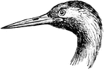
Sea Dove Bill
"Alle. Sea Dove. Size small. Bill very short, stout, and obtuse, as well as high at base, the sides…
White-fronted Dove Details
"Details of Engyptila albifrons (White-fronted Dove); head and foot natural size; wing and tail reduced.
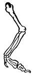
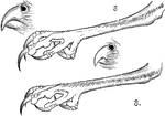
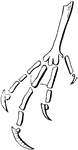
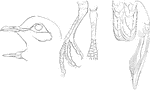
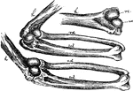
The Bones of the Right Wing of a Duck
"Fig 27. - Bones of the right wing of a duck, Clangula islandica, A, shoulder, omos; B, elbow, ancon;…
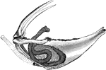
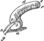
Eagle Cochlea
"Cochlea, X3. a, external, b, internal, cartilaginous prism; c, membranous zone; d, saccular extremity…
A Section of an Eagle's Cochlea
"Section of the cochlea, X3. a, vestibular surface of external cartilaginous prism, extending into d,…
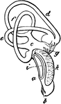
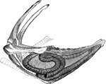
The Inner Ear of a White-tailed Eagle
"Membranous labyrinth of Haliaetus albicilla (White-tailed Eagle), X2. a,b, cochlea; b, its saccular…
Eagle's Ampulal
"Part of the superior vertical semicircular canal, showing its ampulla (which is the dilatation of the…
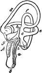
The Inner Ear of an Eagle
"Membranous labyrinth of Haliaetus albicilla (White-tailed Eagle), X2. a,b, cochlea; b, its saccular…
The Bill of an Eider
"Somateri mollissima. Somateri dresseri. Common Eider. Bill gibbous at base of upper mandible; outline…
Mechanism of the Elbow-Joint
"Fig. 28. - Mechanism of elbow-joint. ..., where rc and uc show respectively the size, shape, and position…
Right Eyeball of Bird
"Right Eyeball of Bird, seen from behind, showing the following muscles; a, rectus superior; b, rectus…
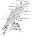
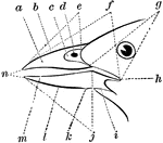
A Labeled Diagram of a Falcon to Show the Nomenclature of the External Parts
"The annexed figure explains the nomenclature of most of the outward of a Bird, but some further explanations…
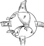
The Skeleton of the Trunk of a Falcon
"Skeleton of the trunk of a Falcon. Ca, coracoid, which articulates with the sternum (St) at ; Cr, keel…
Structure of a Feather
"Fig. - 20 - Two barbs, a, a, of a vane, bearing anterior, b, b, and posterior, c, barbules; enlarged;…