The Human Sensory Systems: Sight ClipArt gallery offers 189 illustrations related to human vision.
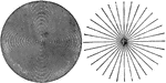
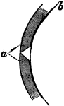
Detection of Astigmatism
Lines for the detection of astigmatism. "The refracting surfaces of the eye acting together are equivalent…
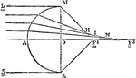
Diagram Illustrating Astigmatism
An illustration depicting an astigmatism. An optical system with astigmatism is one where rays that…
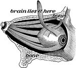
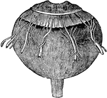
Capsule of Ténon
The capsule of Ténon consists of a thin membrane which envelops the eyeball from the optic nerve…
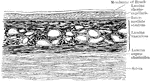
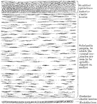
Vertical Section of the Chorioid and Sclera
Vertical section of the chorioid and inner part of the sclera.
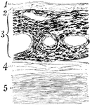
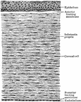
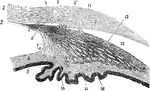
Section Through the Choroid Coat
Section through the choroid coat of the human eye. Labels: 1, elastic membrane, structureless or finely…
Cones and Rods of Retina
A. A cone and two rods from the human retina (modified from Max Schultze); B. Outer part of rod separated…
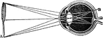
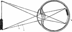
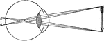
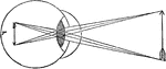
Convergence of Rays in the Aqueous Humor of the Eyeball
The convergence of light rays in the eyeball begins in the aqueous humor is perfected in the crystalline.…
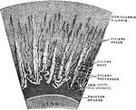
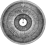
Magnified Corona Ciliaris
A portion of the corona ciliaris magnified. The ciliary processes and the ciliary folds.
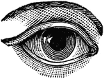

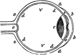
Eye
"Next in order is the aqueous humor, b, e, in the middle of which is the iris, d, c. Behind the pupil…
Eye
"a, sclerotic membrane; b, cornea; d, retina; o, optic nerve; v, vitreous humor." -Comstock 1850
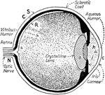
Eye
Section of the eye. 1: Optic nerve; 2: Retina; 3: Vitreous humor; 4: Crystalline lens; 5: Aqueous humor;…
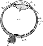
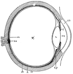
Eye
"Diagram of the eye. C., Cornea; a.h., aqueous humour; c.b., ciliary body; l., lens; I., iris; Sc.,…
Eye
The human eye. Labels: a, crystalline lens; b, retina; c, cornea; d, sclerotic; e, choroid; g, ciliary…
The Eye and its Muscles
The eye and its muscles. Labels: o, the nerve of sight; a, one of the muscles of the eye.
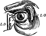
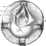
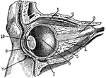
Eye and Lachrymal Gland
Front view of left eye, with eyelid partly removed to show lachrymal gland (tear-producing gland), and…
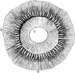
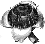
Ciliary Processes of the Eye as Seen from Behind
Ciliary processes, as seen from behind. Labels: 1, posterior surface of the iris, with the sphincter…
Eye Cross-Section
"Cross-section of the eye. Parts: co, cornea; I, iris; aq, anterior chamber of aqueous humor; L, lens;…
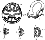
Eye Development
"Development of the eye. 1. Section through first embryonic vesicle, showing outgrowth of optic vesicles…
Eye Diagram
Diagram of the eye. 1: Lines of light from end of arrow; 2: Small, inverted image in the eye.
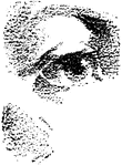
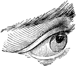
Eye Lid
The lids of the eye close in such a way as to leave a three-cornered canal between them and the surface…
Optical Position and Size of Image Through Lens in Front of Eye
The illustration of putting lenses in front of the eye. The focal point of the image is reflected into…
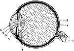
Eye Section
"Section through the left eye, closed. 1, lifting muscle; 2, upper straight muscle; 3, optic nerve;…
A Section of the Eye Seen from Within
A section of the eye seen from within. Labels: 1, The divided edge of the three coats. 2, The pupil.…
A Section of the Eye
A section of the eye. Labels: 1, The sclerotic coat. 2, The cornea. 3, The choroid coat. 6, The iris.…
Choroid Coat of the Eye
The eye, after cutting away the sclerotic coat and cornea, to show the vessels of the choroid coat;…
Ciliary Processes of the Eye
Section through the eye carried through the ciliary processes. Labels: 1, Cornea; 2, membrane of Descemet;…
Cornea too Concave on Eye
"...and the cornea will become too flat, or not suffciently convex, to make the rays of light meet at…
Cornea too Convex on Eye
"If the cornea is too convex, or prominent, the image will be formed before it reaches the retina, for…
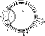
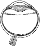
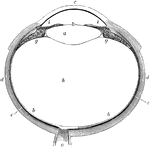
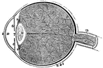
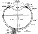
Diagram of the Eye
Plan of the eye seen in section. Labels: A, The Sclerotic Coat; B, The Choroid Coat; C, The Retina;…
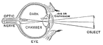
Diagram of the Eye
"Diagram illustrating the Manner in which the Image of an Object is inverted on the Retina." — Blaisedell,…
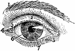
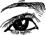
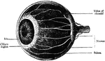
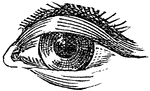
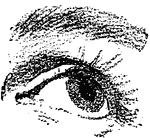
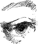
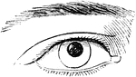
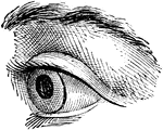
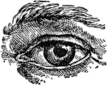
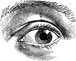
Exterior of Left Human Eye
"Exterior of Left Human Eye. 1, supercilium, or eyebrow; 2, palpebra superior, or upper eyelid; 3, 3,…
Human Eye
c, ciliary nerves going to be distributed in iris; d, smaller ciliary nerve; e, veins known as vasa…
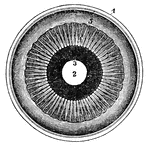
Human Eye
The iris and adjacent structures seen from behind. 1, the divided edge of the three coats, the choroid…