This human anatomy ClipArt gallery offers 825 illustrations of the human skeletal system, including images of both the axial skeleton and the appendicular skeleton. The human axial skeleton includes 80 bones formed by the vertebral column (spine), the thoracic cage (e.g., ribs, sternum), and the skull. The axial skeleton is responsible for the upright position of the body. The human appendicular skeleton is composed of 126 bones formed by the pectoral girdles, the upper and lower limbs, and the pelvic girdle. These bones function in locomotion as well as protection of vital organs.
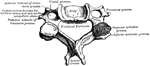
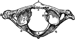
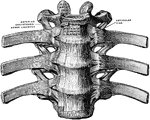
Fifth thoracic vertebra viewed from above.

Fifth Thoracic Vertebra Viewed from Above

Fifth thoracic vertebra viewed from above.

Fourth cervical vertebra viewed from above.

Fourth Cervical Vertebra Viewed from Above

Fourth cervical vertebra viewed from above.

Third thoracic vertebra viewed from above.

Third Thoracic Vertebra Viewed from Above

Third thoracic vertebra viewed from above.

Fourth cervical vertebra viewed from right side.

Fourth Cervical Vertebra Viewed from Side

Fourth cervical vertebra viewed from right side.

Third thoracic vertebra viewed from side.

Third Thoracic Vertebra Viewed from Side

Third thoracic vertebra viewed from side.

Lumbar vertebral, viewed obliquely.

Lumbar Vertebra Viewed Obliquely

Lumbar vertebral, viewed obliquely.

Bony structure of lumbar vertebra.

Bony Structure of Lumbar Vertebra

Bony structure of lumbar vertebra.

A cervical vertebra viewed from above.

Cervical Vertebra

A cervical vertebra viewed from above.

Cervical vertebra.

Cervical Vertebra

Cervical vertebra.

The dorsal vertebra seen from behind, i.e. the end turned from the head. Labels: C, the body; A, neural arch; Fv, the neural ring; Ps, spinous process; Pai, posterior articular process; Pt, transverse process; Fci, articular surfaces on the centrum for articulation with a rib.

Dorsal Vertebra

The dorsal vertebra seen from behind, i.e. the end turned from the head. Labels: C, the body; A, neural…
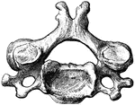
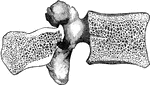
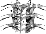
Two dorsal vertebrae viewed from the left side, and in their natural relative positions. Labels: C, the body; A, neural arch; Fv, the neural ring; Ps, spinous process; Pas, anterior articular process; Pai, posterior articular process; Pt, transverse process; Ft, facet for articulation with the tubercle of a rib; Fcs, Fci, articular surfaces on the centrum for articulation with a rib.

Dorsal Vertebra

Two dorsal vertebrae viewed from the left side, and in their natural relative positions. Labels: C,…
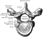
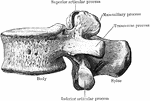
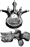
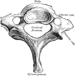
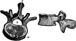
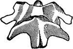
A dorsal vertebra. Labels: 1, the body; 2, face for the head of a rib; 3, superior face of the body; 4, superior half of the intervertebral notch; 5, interior half of the intervertebral notch; 6, spinous process; 7, articular face for the tubercle of a rib; 8, two superior oblique processes; 9, two inferior oblique processes.

Dorsal Vertebra

A dorsal vertebra. Labels: 1, the body; 2, face for the head of a rib; 3, superior face of the body;…

Fourth cervical vertebra from in front.

Front View of Fourth Cervical

Fourth cervical vertebra from in front.

A vertebra of the loins; a, a, their bodies, larger and more spongy than those of the others; b,b,b, the superior; c,c,c the inferior articular processes, strong and deep, the superior concave; the inferior convex; d,d,d, the transverse processes, small and long, serving as levers for the attachment of muscles; e,e, the spinous processes, strong, horizontal, and flattened at the sides; f, the spinal foramen.

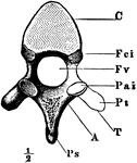
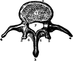
Vertebra of the Loins

A vertebra of the loins; a, a, their bodies, larger and more spongy than those of the others; b,b,b,…
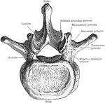
A lumbar vertebra. Labels: 1, face for the intervertebral substance; 2, anterior surface of the body; 3, spinous process; 4, transverse process; 5, oblique process; 6, a portion or the body bridges; 7, the spinal foramen.

Lumbar Vertebra

A lumbar vertebra. Labels: 1, face for the intervertebral substance; 2, anterior surface of the body;…

Lumbar vertebra.

Lumbar Vertebra

Lumbar vertebra.

A vertebra of the neck. Labels: a, body of the bone; b, the spinal process; c, d, the transverse processes double, showing circular holes for the passage of the vertebral artery; e,e,. the superior; f,f, the articular of oblique processes; g, the spinal hole for the spinal marrow. The roots of the articular processes are hallowed out above and below into notches; and these, when the bones are fitted together, form apertures on each side of the spine, through which the nerves pass out from the spinal canal.

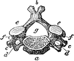
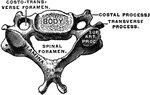
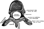
Vertebra of the Neck

A vertebra of the neck. Labels: a, body of the bone; b, the spinal process; c, d, the transverse processes…
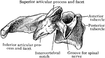
Seventh cervical vertebra.

Seventh Cervical Vertebra

Seventh cervical vertebra.

Fourth cervical vertebra from the side.

Side View of Fourth Cervical

Fourth cervical vertebra from the side.

The atlas, the uppermost vertebra of the spinal column. Labels: 1, anterior tubercle; 2, articular face for the dentata; 3, posterior surface of spinal canal; 4, 4, intervertebral notch; 5, transverse process; 6, foramen for vertebral artery; 7, superior oblique process; 8, tubercle for the transverse ligament.

The Atlas Vertebra

The atlas, the uppermost vertebra of the spinal column. Labels: 1, anterior tubercle; 2, articular face…

A thoracic vertebra, upper surface.

Thoracic Vertebra

A thoracic vertebra, upper surface.

A thoracic vertebra.

Thoracic Vertebra

A thoracic vertebra.

Two views of vertebra. 24 vertebrae make up the spinal column. On the left figure, <em>a</em> is the body of the bone; <em>b</em> is the hole through the vertebra which forms its part of the canal for the spinal marrow; and <em>c</em> is the spinous process. On the right is a side view of the same vertebra.

Two Views of a Vertebra

Two views of vertebra. 24 vertebrae make up the spinal column. On the left figure, a is the…
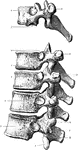
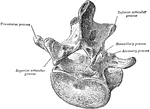
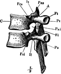
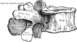
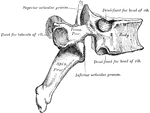
"Section of two lumbar vertebrae, showing ligaments."&mdash;Finley, 1917

Lumbar vertebrae

"Section of two lumbar vertebrae, showing ligaments."—Finley, 1917

Peculiar thoracic vertebrae.

Peculiar Thoracic Vertebrae

Peculiar thoracic vertebrae.

First, ninth, tenth, eleventh, and twelfth thoracic vertebrae from the left side. 1, Inferior articular process with out-turned facet. 2, Single facet for head of XII. rib; no facet on transverse process. 3, Single facet for head of XI. rib; no facet on transverse process. 4, Single or demifacet for head of X. rib. 5, Occasional demifacet for head of X. rib. 6. Demifacet for head of IX. rib. 7, Demifacet for head of II. rib. 8, Single facet o head of II. rib. 9, Facet on transverse process for tuberosity of I. rib. 10, Facet on transverse process for tuberosity of IX. rib. Labels: S, superior tubercles corresponding to mammillary; I, inferior tubercles corresponding to accessory; e, external tubercles corresponding to transverse of lumbar.

Thoracic Vertebrae

First, ninth, tenth, eleventh, and twelfth thoracic vertebrae from the left side. 1, Inferior articular…

The upper two vertebrae of the spinal column, involved in head movement. In performing the rotary motion of turning your head, the skull does not move alone, but it moves together with the first vertebra. The first vertebra in this motion moves on the second around a tooth-like process that stands up from this second vertebra. This tooth works in a smooth notch on the inside of the first vertebra. It is bound very fast in this notch by a strong ligament, so that it may not, in any of the quick motions of the head, be made to press on the delicate spinal marrow which lies against it.

Upper Two Vertebrae

The upper two vertebrae of the spinal column, involved in head movement. In performing the rotary motion…

A vertebral articulation (joint), which are formed by the adjacent surfaces of the bodies of the vertebrae and their processes, and are connected by ligaments.

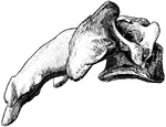
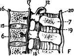
A Vertebral Articulation

A vertebral articulation (joint), which are formed by the adjacent surfaces of the bodies of the vertebrae…

Vertebral column with ligaments, from in front.

Vertebral Column with Ligaments

Vertebral column with ligaments, from in front.

The vertebral column from behind.

Back View of Vertebral Column

The vertebral column from behind.

The spinal column, right lateral view and dorsal view.

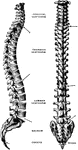
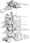
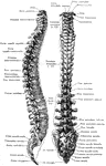
Lateral and Dorsal View of the Vertebral Column

The spinal column, right lateral view and dorsal view.

Lateral and posterior views of the vertebral column.

Lateral and Posterior View of the Vertebral Column

Lateral and posterior views of the vertebral column.

The vertebral column from the left side.

Side View of Vertebral Column

The vertebral column from the left side.

A diagram of a transverse section of a vertebrate. Labels: 1, the walls; 2, digestive organs; 3, circulatory organs; 4, ganglia; 5, spinal cord; 6, spinal column.

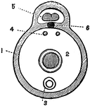
Transverse Section of a Vertebrate

A diagram of a transverse section of a vertebrate. Labels: 1, the walls; 2, digestive organs; 3, circulatory…
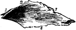
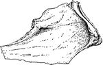
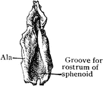
The vomer is a single bone, situated vertically at the back part of the nasal fossae, forming part of the septum of the nose. It is thin, somewhat like a ploughshare in form; but varies in different individuals, being frequently bent to one of the other side; it presents for examination two surfaces and four borders.

Vomer

The vomer is a single bone, situated vertically at the back part of the nasal fossae, forming part of…

Vomer as seen from the right side.

Vomer

Vomer as seen from the right side.

The vomer bone, which forms the posterior-inferior part of the nasal septum.

Vomer Bone

The vomer bone, which forms the posterior-inferior part of the nasal septum.

Vomer from before and above.

Vomer from Before and Above

Vomer from before and above.

Vomer bone, a single bone placed at the back part of the nasal cavity, and forms part of the septum of the nasal fossae.

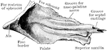
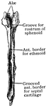
Human Vomer Nasal Bone

Vomer bone, a single bone placed at the back part of the nasal cavity, and forms part of the septum…

Sagittal section of face, a little to the left of the middle line, showing the vomer and its relations.

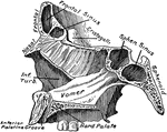
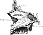
Section of Face Showing Vomer

Sagittal section of face, a little to the left of the middle line, showing the vomer and its relations.

Superior surface of the vomer.

Superior Surface of the Vomer

Superior surface of the vomer.

Bones of the Wrist and Hand. Labels: m, metacarpal bones; p, phalanges; 3, bones of wrist.

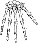
The Human Wrist and Hand Bones

Bones of the Wrist and Hand. Labels: m, metacarpal bones; p, phalanges; 3, bones of wrist.

Section through the wrist joint and carpal bones.

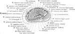
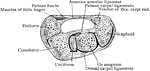
Cross Section Through the Wrist Joint and Carpal Bones

Section through the wrist joint and carpal bones.

Ligaments on anterior aspect of radio carpal, carpal, and carpo metacarpal joints.

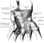
Ligaments of the Wrist

Ligaments on anterior aspect of radio carpal, carpal, and carpo metacarpal joints.

Transverse section through right wrist from above. The flexor tendons have been removed from the canal beneath the annular ligament.

Transverse Section of Wrist

Transverse section through right wrist from above. The flexor tendons have been removed from the canal…