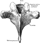
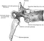
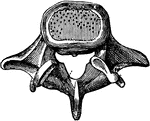
This human anatomy ClipArt gallery offers 825 illustrations of the human skeletal system, including images of both the axial skeleton and the appendicular skeleton. The human axial skeleton includes 80 bones formed by the vertebral column (spine), the thoracic cage (e.g., ribs, sternum), and the skull. The axial skeleton is responsible for the upright position of the body. The human appendicular skeleton is composed of 126 bones formed by the pectoral girdles, the upper and lower limbs, and the pelvic girdle. These bones function in locomotion as well as protection of vital organs.

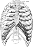
The thorax. Ventral view.

Ventral View of Thorax

The thorax. Ventral view.

Anterior view of the tibia (bone of the leg). Labels: 1, spinous process; 2, surface for condyles of the femur; 3, face for head of the fibula; 4, the head; 5, the tubercle; 6, 6, spine and ankle; 9, tarsal surface; 10, face for lower end of fibula.

Tibia

Anterior view of the tibia (bone of the leg). Labels: 1, spinous process; 2, surface for condyles of…
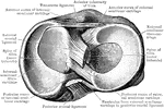
The anterior surface of the tibia and fibula of the right leg.

Tibia and Fibula

The anterior surface of the tibia and fibula of the right leg.

The posterior surface of the tibia and fibula of the right leg.

Tibia and Fibula

The posterior surface of the tibia and fibula of the right leg.

Articular surfaces of tibia and fibula which are opposed to the astragalus.

Tibia and Fibula

Articular surfaces of tibia and fibula which are opposed to the astragalus.

Right tibia from before.

Tibia from Before

Right tibia from before.

Right tibia from behind.

Tibia from Behind

Right tibia from behind.

Head of tibia, with semilunar cartilages. Seen from above.

Head of Tibia Seen from Above

Head of tibia, with semilunar cartilages. Seen from above.

Frontal section of lower end of tibia.

Frontal Section of Lower End of Tibia

Frontal section of lower end of tibia.

Frontal section of upper end of tibia.

Frontal Section of Upper End of Tibia

Frontal section of upper end of tibia.

Upper end of right tibia from above and before.

Upper End of the Tibia

Upper end of right tibia from above and before.

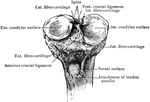
Upper end of tibia, with semilunar cartilages and attached portions of crucial ligamanents.

Upper End of Tibia

Upper end of tibia, with semilunar cartilages and attached portions of crucial ligamanents.

Upper surface of superior extremity of right tibia.

Upper Surface of Tibia

Upper surface of superior extremity of right tibia.

The phalanges of the toes, though more feebly developed, have really the same movements among themselves as those of the fingers.

The Flexibility of the Toes

The phalanges of the toes, though more feebly developed, have really the same movements among themselves…

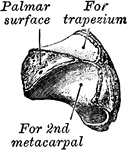
The left trapezium as seen from the front.

Trapezium

The left trapezium as seen from the front.

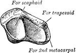
The left trapezium as seen from the above and the mesal side.

Trapezium

The left trapezium as seen from the above and the mesal side.

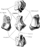
The right trapezium bone.

Trapezium Bone

The right trapezium bone.

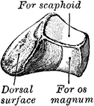
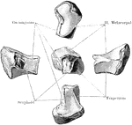
The left trapezoid as seen from the above, the inner side, and behind.

Trapezoid

The left trapezoid as seen from the above, the inner side, and behind.

The left trapezoid as seen from the front, below, and outer side.

Trapezoid

The left trapezoid as seen from the front, below, and outer side.

The right trapezoid bone.

Trapezoid Bone

The right trapezoid bone.

Section through the pelvis at the level of the first sacral vertebra.

Cross Section of the Trunk at First Sacral Vertebra

Section through the pelvis at the level of the first sacral vertebra.
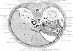
Section through first lumbar vertebra and one inch below ensiform process.

Cross Section of the Trunk through the First Lumbar Vertebra

Section through first lumbar vertebra and one inch below ensiform process.

Back view of the trunk.

Back View of the Bones of the Trunk

Back view of the trunk.

The trunk showing rib, iliac crest, and pubis.

Side View of the Bones of the Trunk

The trunk showing rib, iliac crest, and pubis.

The skeleton of the trunk and the limb arches seen from the front. Labels: c, clavicle; S, scapula; Oc, innominate bone attached to the side of the sacrum dorsally and meeting its fellow at the pubic symphysis in the ventral median line.

Skeleton of Trunk

The skeleton of the trunk and the limb arches seen from the front. Labels: c, clavicle; S, scapula;…

The external surface of right inferior turbinate bone.

Turbinate Bone

The external surface of right inferior turbinate bone.

The turbinated bones are situated one on each side of the outer wall of each nasal fossa. Shown is the external surface of right turbinated bone.

External Surface of Turbinated Bone

The turbinated bones are situated one on each side of the outer wall of each nasal fossa. Shown is the…

The turbinated bones are situated one on each side of the outer wall of each nasal fossa. Shown is the internal surface of right turbinated bone.

Internal Surface of Turbinated Bone

The turbinated bones are situated one on each side of the outer wall of each nasal fossa. Shown is the…

The inner surface (A) and outer surface (B) of the right inferior turbinated bone.

Inferior Turbinated Bone

The inner surface (A) and outer surface (B) of the right inferior turbinated bone.

The eleventh and twelfth ribs have each a single articular facet on the head, which is of rather large size; they have no neck or tuberosity, and are pointed at the extremity.

Twelfth Rib

The eleventh and twelfth ribs have each a single articular facet on the head, which is of rather large…

The plan of development of the ulna for three centuries.

Ulna

The plan of development of the ulna for three centuries.

The ulna bone in the human arm.

Ulna

The ulna bone in the human arm.

The ulna (elbow bone), which is the principal bone of the forearm.

Ulna

The ulna (elbow bone), which is the principal bone of the forearm.

Anterior view of the ulna (bone of the arm) of the left side. Labels: 1, olecranon process; 2, greater sigmoid cavity; 5, external surface; 6, ridge for interosseous ligament; 7, small head for the radius; 8, carpal surface.

Ulna

Anterior view of the ulna (bone of the arm) of the left side. Labels: 1, olecranon process; 2, greater…

The ulna, a bone of the arm.

Ulna

The ulna, a bone of the arm.

The Ulna and Radius. Labels: 1, radius; 2, ulna; o, olecranon process, on the anterior surface of which are seen the large (gs) and the small (ls) cavities for the reception of the lower end of the humerus and of the head of the radius, respectively; h, head of radius.

The Human Ulna and Radius

The Ulna and Radius. Labels: 1, radius; 2, ulna; o, olecranon process, on the anterior surface of which…

Fracture of upper third of ulna, with dislocation of radius forward.

Fractured Ulna with Dislocation of Radius

Fracture of upper third of ulna, with dislocation of radius forward.
Right ulna, inner aspect.

Inner Aspect of Ulna

Right ulna, inner aspect.

Longitudinal section of ulna.

Longitudinal Section of Ulna

Longitudinal section of ulna.

Right ulna, outer aspect.

Outer Aspect of Ulna

Right ulna, outer aspect.

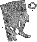
A, transverse section of the ulna, (bone of the arm), natural size, showing the medullary cavity. B, the more deeply shaded part of A magnified 20 diameters.

Transverse Section of Ulna

A, transverse section of the ulna, (bone of the arm), natural size, showing the medullary cavity. B,…

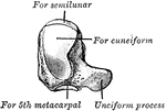
The left unciform seen from the internal surface.

Unciform

The left unciform seen from the internal surface.

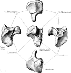
The left unciform seen from the outer surface.

Unciform

The left unciform seen from the outer surface.

The right unciform bone.

Unciform Bone

The right unciform bone.

The upper extremity of the human body. 1: Clavicle; 2: Scapula; 3: Humerus; 4: Ulna; 5: Radius; 6: Carpus; 7: Metacarpal; 8: Phalanges.

Upper Extremity

The upper extremity of the human body. 1: Clavicle; 2: Scapula; 3: Humerus; 4: Ulna; 5: Radius; 6: Carpus;…
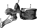
Side view of the vertebra. 1: Body; 2: Processes; 3: Opening for spinal cord.

Vertebra

Side view of the vertebra. 1: Body; 2: Processes; 3: Opening for spinal cord.

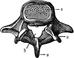
Top view of the vertebra. 1: Body; 2: Processes; 3: Opening for spinal cord.

Vertebra

Top view of the vertebra. 1: Body; 2: Processes; 3: Opening for spinal cord.

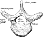
A vertebra of the spinal column seen from above.

Vertebra

A vertebra of the spinal column seen from above.

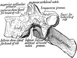
A vertebra of the spine seen from the side.

Vertebra

A vertebra of the spine seen from the side.

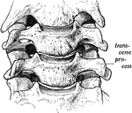
Cervical vertebrae seen from in the front.

Vertebra

Cervical vertebrae seen from in the front.

A vertebra of the spine.

A Vertebra of the Spine

A vertebra of the spine.

A vertebra of the spine.

A Vertebra of the Spine

A vertebra of the spine.

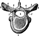
Fourth cervical vertebra from above.

Fourth Cervical from Above

Fourth cervical vertebra from above.

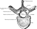
Sixth thoracic vertebra from above.

Sixth Thoracic Vertebra from Above

Sixth thoracic vertebra from above.

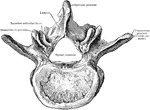
Third lumbar vertebra from above.

Third Lumbar Vertebra From Above

Third lumbar vertebra from above.

Sixth thoracic vertebra from behind.

Sixth Thoracic Vertebra from Behind

Sixth thoracic vertebra from behind.

Sixth thoracic vertebra from side.

Sixth Thoracic Vertebra from Side

Sixth thoracic vertebra from side.

Third lumbar vertebra from behind and the side.

Third Lumbar Vertebra From Behind and the Side

Third lumbar vertebra from behind and the side.

Third lumbar vertebra from the side.

Third Lumbar Vertebra From the Side

Third lumbar vertebra from the side.

A vertebra seen from above.

Vertebra Seen From Above

A vertebra seen from above.