This human anatomy ClipArt gallery offers 825 illustrations of the human skeletal system, including images of both the axial skeleton and the appendicular skeleton. The human axial skeleton includes 80 bones formed by the vertebral column (spine), the thoracic cage (e.g., ribs, sternum), and the skull. The axial skeleton is responsible for the upright position of the body. The human appendicular skeleton is composed of 126 bones formed by the pectoral girdles, the upper and lower limbs, and the pelvic girdle. These bones function in locomotion as well as protection of vital organs.
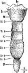
Ventral View of the Sternum
Ventral view of the sternum. Labels: M, manubrium; C, body; P, ensiform cartilage; Icl, notch for the…
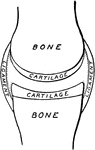
Synovial Membrane
Diagram of articular synovial membranes. The cartilages are represented as drawn apart for the sake…
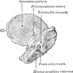
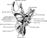
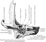
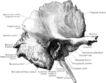
Temporal Bone
The outer surface of the temporal bone. The dotted lines indicate the lines of suture between squamous,…
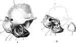
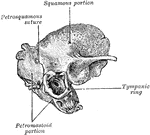
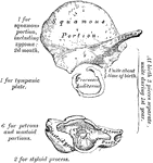
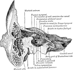
Temporal Bone at Birth
A, The outer surface of the right temporal bone at birth. B, The same with squamozygomatic portion removed.…
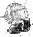
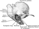
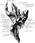
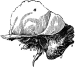
Temporal Bone at Birth
Inner surface of right temporal bone at birth. am squamozygomatic; b, petrosquamosal suture and foramen…
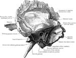
Temporal Bone Cut Open
Right temporal bone cut open to show the anterior surface of the petrous portion.
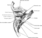
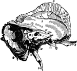
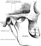
Temporal Bone of the Human Skull
Temporal bone of the human skull. The temporal bones are situated at the sides and base of the skull.…
Anterior Half of Section Through Temporal Bone
The anterior half of a vertical transverse section through the left temporal bone.
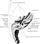
Frontal Section of Temporal Bone
Frontal section of temporal bone, showing the cavities of the outer, middle, and inner ear and the four…
Horizontal Section of Temporal Bone
Horizontal section through left temporal bone showing lower half of section.
Horizontal Section Through Temporal Bone
Horizontal section through right temporal bone, seen from below.
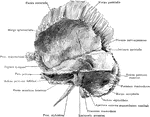
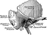
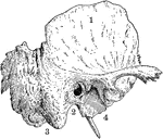
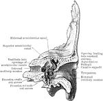
The Mastoid and Petrous Portions of the Temporal Bone
The mastoid and petrous portions of the temporal bone of the skull (ossa temporalia).
Posterior Half of Section Through Temporal Bone
The posterior half of a vertical transverse section through the left temporal bone.
Sagittal Section Through Temporal Bone
Sagittal section through right temporal bone, seen from the outer side.
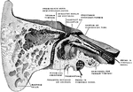
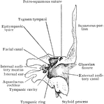
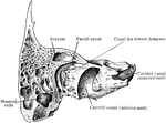
Section Through Temporal Bone
Section through the petrous and mastoid portions of the temporal bone, showing the communication of…
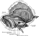
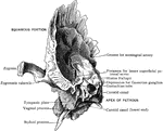
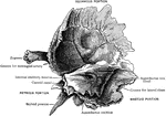
The Squamous Portion of the Temporal Bone
The squamous portion of the temporal bone of the skull (ossa temporalia).
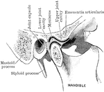
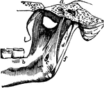
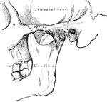
Temporo-mandibular Articulation
The temporo-mandibular articulation, which is a ginglymo-arthroidial joint between the condyle of the…
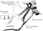
Temporomandibular
The external lateral ligament is a short, thin, and narrow fasciculus, attached, above, to the outer…
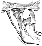
Temporomandibular
The external lateral ligament is a short, thin, and narrow fasciculus, attached, above, to the outer…
Thigh Bone
The thigh bone cut through the middle. Labels: b, hard bone; h, d, spongy bone; ma, marrow.
Thigh Bone and Arm Bone
Here you see the thigh bone on the left and the arm bone on the right. In both, the shaft is hollow,…
Thigh Bone Section
The above cut represents a section of the thigh bone. The extremities (a, w) having a shell or thin…
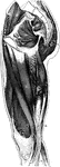
Muscles of the Thigh
Muscles of the thigh. Labels: 46, gluteus maximus; 36, 35, posterior femoral; 33, sartorius; 27, 26,…
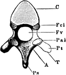

Thoracic Vertebrae
Two thoracic vertebrae seen from behind, i.e., the end turned from the head. Labels: C, the body; A,…
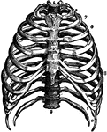
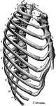
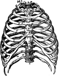
Thorax
The skeleton of the thorax. Labels: a, g, vertebral column; b, first rib; c, clavicle; d, third rib;…
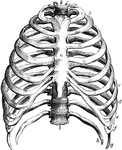
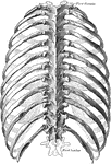
The Thorax
The thorax. Labels: a, the sternum. b to c, the true ribs; d to h, the false ribs; g, h, the floating…
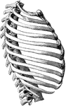
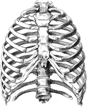
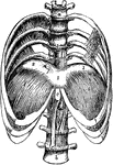
Human Thorax (Chest)
Thorax. The thorax, or chest, is an elongated conical-shaped cage, formed by the sternum and costal…
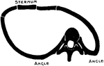
Changes in the Thorax following Scoliosis of the Spine
Showing the changes in the thorax which follow scoliosis of the spine. The convexity of the spinal curvature…
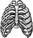
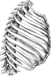
Thorax Skeleton
The skeleton of the thorax. Labels: a, g, vertebral column; b, first rib; c, clavicle; e, seventh rib;…
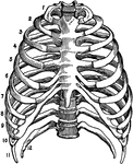
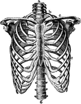
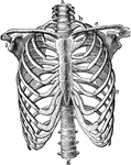
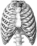
The Bones of the Thorax
Front view of the bones of the thorax, including the ribs, sternum and vertebrae. Labels: 1, first bone…
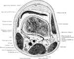
Lower Half of the Thorax
The lower half of the thorax, with four lumbar vertebrae showing the diaphragm from above. Labels: 1,2,3,…
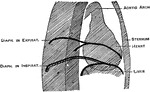
Orthodiagram of the Thorax
Orthodiagram of the thorax. The position of parts is shown in extreme inspiration; the position of the…