This human anatomy ClipArt gallery offers 825 illustrations of the human skeletal system, including images of both the axial skeleton and the appendicular skeleton. The human axial skeleton includes 80 bones formed by the vertebral column (spine), the thoracic cage (e.g., ribs, sternum), and the skull. The axial skeleton is responsible for the upright position of the body. The human appendicular skeleton is composed of 126 bones formed by the pectoral girdles, the upper and lower limbs, and the pelvic girdle. These bones function in locomotion as well as protection of vital organs.
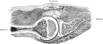
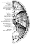
Horizontal Section Through Shoulder Joint
A horizontal section through the left shoulder joint of a boy. Arm abducted to a right angle.
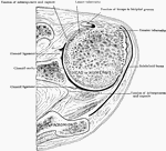
Horizontal Section Through Shoulder Joint
Horizontal section through the right shoulder joint from above.
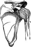
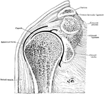
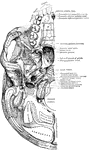
Shoulder-joint and Ligaments of the Scapula
The shoulder joint (articulatio humeri) and the ligaments of the scapula.
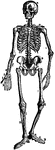
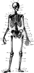
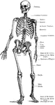
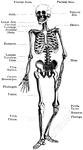
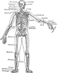
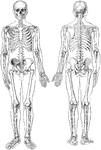
The Human Skeleton
The Human Skeleton. Labels: a, parietal bone; b, frontal; c, cervical vertebrae; d, sternum; e, lumbar…
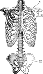
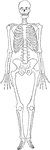
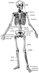
Skeletal Trunk
"The skeleton of the trunk and the limb arches seen from the front. C, clavicle; S, scapula; Oc, innominate…
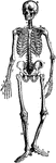
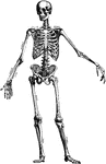
Skeleton
1. Frontal bone 2. Parietal bone 3. Coronal Suture 4. Squamous portion of Temporal bone 5. Mastoid process…
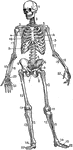
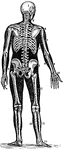
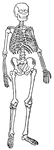
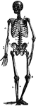
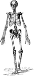
Human skeleton
"The human body, like a great building, has a framework which gives the body its shape and provides…
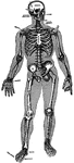
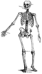
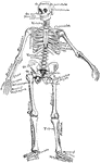
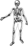
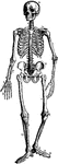
Human skeleton
"There are in all two hundred and six seperate bones in the adult skelton. The teeth are not bones,…
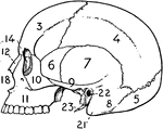
Skull
The outer outline is that of the skull found in the cave of Cromagnon, in France, belogning, as Dawson…
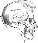
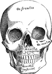
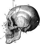
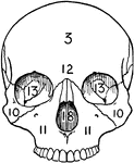
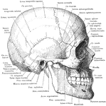
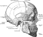
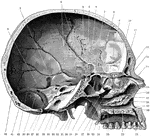
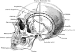
The Skull
A side view of the skull. Labels: O, occipital bone; T, temporal bone; Pr, parietal bone; F, frontal…
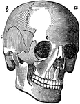
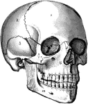
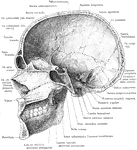
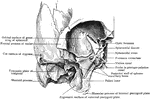
The Skull
The human skull. Labels: 1, frontal lobe; 2, parietal lobe; 3, temporal lobe; 4, the sphenoid bone;…
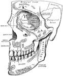
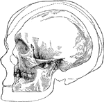
Median Section of the Skull and Mandible
Median section of the skull and mandible, viewed from the left.
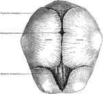
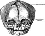
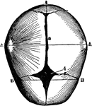
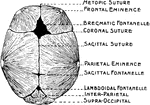
Human Skull at Birth
The skull at birth, superior suerface. The cranial bones of the infant at birth are not fullyformed…
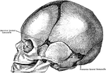
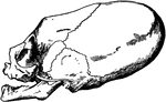
Distorted Skull of Child
Skull of a child of the tribe of Chinook Indians (inhabiting the neighborhood of the Columbia River),…
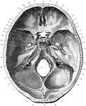
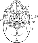
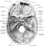
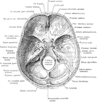
Base of the Skull Seen From Above
Shown is the base of the skull seen from above. Labels: 1, frontal bone; 2, slit for nasal nerve; 3,…
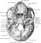
Skull Seen From Side
Shown is the inner aspect of the left half of the skull sagittally divided. Labels: 1, suture between…
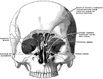
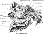
Frontal Section of Skull Showing Nasal Cavity
Front section of skull through plane of outer border of orbits. Arrows pass through communication between…
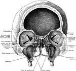
Skull Showing Posterior Wall of Sphenomaxillary Fossa
Portion of right half of skull, showing posterior wall of sphenomaxillary fossa. The superior maxilla,…
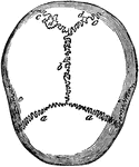
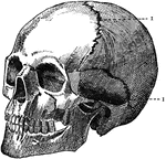
Skull Sutures
Sutures of the skull. Labels: a,a, the coronal suture, from the Latin corona, crown, so called from…